

Книги фарма 2 / Bertram G. Katzung-Basic & Clinical Pharmacology(9th Edition)
.pdfIron crosses the intestinal mucosal cell by active transport. The rate of iron uptake is regulated by mucosal cell iron stores such that more iron is transported when stores are low. Together with iron split from absorbed heme, the newly absorbed iron can be made available for immediate transport from the mucosal cell to the plasma via transferrin or can be stored in the mucosal cell as ferritin, a water-soluble complex consisting of a core crystal of ferric hydroxide covered by a shell of a specialized storage protein called apoferritin. In general, when total body iron stores are high and iron requirements by the body are low, newly absorbed iron is diverted into ferritin in the intestinal mucosal cells rather than being transported to other sites. When iron stores are low or iron requirements are high, however, newly absorbed iron is immediately transported from the mucosal cells to the bone marrow for the production of hemoglobin.
Transport
Iron is transported in the plasma bound to transferrin, a  -globulin that specifically binds ferric iron. The transferrin-ferric iron complex enters maturing erythroid cells by a specific receptor mechanism. Transferrin receptors—integral membrane glycoproteins present in large numbers on proliferating erythroid cells—bind the transferrin-iron complex and internalize the iron, releasing it within the cell. The transferrin and transferrin receptor are then recycled, providing an efficient mechanism for incorporating iron into hemoglobin in developing red blood cells.
Increased erythropoiesis is associated with an increase in the number of transferrin receptors on developing erythroid cells. Iron store depletion and iron deficiency anemia are associated with an increased concentration of serum transferrin.
Storage
Iron binds avidly to a protein, apoferritin, and forms the complex ferritin. Iron is stored, primarily as ferritin, in intestinal mucosal cells and in macrophages in the liver, spleen, and bone. Apoferritin synthesis is regulated by the levels of free iron. When these levels are low, apoferritin synthesis is inhibited and the balance of iron binding shifts toward transferrin. When free iron levels are high, more apoferritin is produced in an effort to safely sequester more iron and protect organs from the toxic effects of excess free iron.
Ferritin is also detectable in plasma. Since the ferritin present in plasma is in equilibrium with storage ferritin in reticuloendothelial tissues, the plasma (or serum) ferritin level can be used to estimate total body iron stores.
Elimination
There is no mechanism for excretion of iron. Small amounts are lost by exfoliation of intestinal mucosal cells into the stool, and trace amounts are excreted in bile, urine, and sweat. These losses account for no more than 1 mg of iron per day. Because the body's ability to increase excretion of iron is so limited, regulation of iron balance must be achieved by changing intestinal absorption and storage of iron, in response to the body's needs.
Clinical Pharmacology
Indications for the Use of Iron
The only clinical indication for the use of iron preparations is the treatment or prevention of iron deficiency anemia. Iron deficiency is commonly seen in populations with increased iron

requirements. These include infants, especially premature infants; children during rapid growth periods; and pregnant and lactating women. Iron deficiency also occurs frequently after gastrectomy and in patients with severe small bowel disease that results in generalized malabsorption. Iron deficiency in these gastrointestinal conditions is due to inadequate iron absorption.
The most common cause of iron deficiency in adults is blood loss. Menstruating women lose about 30 mg of iron with each menstrual period; women with heavy menstrual bleeding may lose much more. Thus, many premenopausal women have low iron stores or even iron deficiency. In men and postmenopausal women, the most common site of blood loss is the gastrointestinal tract. Patients with unexplained iron deficiency anemia should be evaluated for occult gastrointestinal bleeding.
As iron deficiency develops, storage iron decreases and then disappears; next, serum ferritin decreases; and then serum iron decreases and iron-binding capacity increases, resulting in a decrease in iron-binding (transferrin) saturation. Thereafter, anemia begins to develop. Red cell indices (mean corpuscular volume [MCV]: normal = 80–100 fL; mean corpuscular hemoglobin concentration [MCHC]: normal = 32–36 g/dL) are usually low normal when iron deficiency anemia is mild, but cells become progressively more microcytic (low MCV) and hypochromic (low MCHC) as anemia becomes more severe. By the time iron deficiency is diagnosed, serum iron is usually less than 40  g/dL; total iron-binding capacity (TIBC) is greater than 400
  g/dL; ironbinding saturation is less than 10%; and serum ferritin is less than 10 g/L. These laboratory measurements can be used to confirm a diagnosis of iron deficiency anemia in patients who present with signs and symptoms of microcytic anemia.
Treatment
The treatment of iron deficiency anemia consists of administration of oral or parenteral iron preparations. Oral iron corrects the anemia just as rapidly and completely as parenteral iron in most cases if iron absorption from the gastrointestinal tract is normal.
Oral Iron Therapy
A wide variety of oral iron preparations are available. Since ferrous iron is most efficiently absorbed, only ferrous salts should be used. Ferrous sulfate, ferrous gluconate, and ferrous fumarate are all effective and inexpensive and are recommended for the treatment of most patients.
Different iron salts provide different amounts of elemental iron, as shown in Table 33–2. In an irondeficient individual, about 50–100 mg of iron can be incorporated into hemoglobin daily, and about 25% of oral iron given as ferrous salt can be absorbed. Therefore, 200–400 mg of elemental iron should be given daily to correct iron deficiency most rapidly. Patients unable to tolerate such large doses of iron can be given lower daily doses of iron, which results in slower but still complete correction of iron deficiency. Treatment with oral iron should be continued for 3–6 months. This will correct the anemia and replenish iron stores.
Table 33–2. Some Commonly Used Oral Iron Preparations.
Preparation |
Tablet |
Elemental Iron per |
Usual Adult Dosage (Tablets per |
|
Size |
Tablet |
Day) |
|
|
|
|
Ferrous sulfate, |
325 mg |
65 mg |
3–4 |
hydrated |
|
|
|
|
|
|
|

Ferrous sulfate, |
200 mg |
65 mg |
3–4 |
desiccated |
|
|
|
|
|
|
|
Ferrous gluconate |
325 mg |
36 mg |
3–4 |
|
|
|
|
Ferrous fumarate |
200 mg |
66 mg |
3–4 |
|
|
|
|
Ferrous fumarate |
325 mg |
106 mg |
2–3 |
|
|
|
|
Common adverse effects of oral iron therapy include nausea, epigastric discomfort, abdominal cramps, constipation, and diarrhea. These effects are usually dose-related and can often be overcome by lowering the daily dose of iron or by taking the tablets immediately after or with meals. Some patients have less severe gastrointestinal adverse effects with one iron salt than another and benefit from changing preparations. Patients taking oral iron develop black stools; this itself has no clinical significance but may obscure the diagnosis of continued gastrointestinal blood loss.
Parenteral Iron Therapy
Parenteral therapy should be reserved for patients with documented iron deficiency unable to tolerate or absorb oral iron and patients with extensive chronic blood loss who cannot be maintained with oral iron alone. This includes patients with various postgastrectomy conditions and previous small bowel resection, inflammatory bowel disease involving the proximal small bowel, and malabsorption syndromes.
Iron dextran is a stable complex of ferric hydroxide and low-molecular-weight dextran containing 50 mg of elemental iron per milliliter of solution. Iron-sucrose complex and iron sodium gluconate complex are newer, alternative preparations. These agents can be given either by deep intramuscular injection or by intravenous infusion. Adverse effects of parenteral iron therapy include local pain and tissue staining (brown discoloration of the tissues overlying the injection site), headache, light-headedness, fever, arthralgias, nausea and vomiting, back pain, flushing, urticaria, bronchospasm, and, rarely, anaphylaxis and death.
Most adults with iron deficiency anemia require 1–2 g of replacement iron, or 20–40 mL of iron dextran. Most physicians prefer to give the entire dose in a single intravenous infusion in several hundred milliliters of normal saline over 1–2 hours. Intravenous administration eliminates the local pain and tissue staining that often occur with the intramuscular route and allows delivery of the entire dose of iron necessary to correct the iron deficiency at one time. There is no clear evidence that any of the adverse effects, including anaphylaxis, are more likely to occur with intravenous than with intramuscular administration.
Owing to the risk of a hypersensitivity reaction, a small test dose of iron dextran should always be given before full intramuscular or intravenous doses are given. Patients with a strong history of allergy and patients who have previously received parenteral iron are more likely to have hypersensitivity reactions following treatment with parenteral iron dextran.
Clinical Toxicity
Acute Iron Toxicity
Acute iron toxicity is seen almost exclusively in young children who have ingested a number of iron tablets. Although adults are able to tolerate large doses of oral iron without serious consequences, as

few as ten tablets of any of the commonly available oral iron preparations can be lethal in young children. Patients taking oral iron preparations should be instructed to store tablets in child-proof containers out of the reach of children.
Large amounts of oral iron cause necrotizing gastroenteritis, with vomiting, abdominal pain, and bloody diarrhea followed by shock, lethargy, and dyspnea. Subsequently, improvement is often noted, but this may be followed by severe metabolic acidosis, coma, and death. Urgent treatment of acute iron toxicity is necessary, especially in young children. Activated charcoal, a highly effective adsorbent for most toxins, does not bind iron and thus is ineffective. Whole bowel irrigation (see Chapter 59: Management of the Poisoned Patient) should be performed to flush out unabsorbed pills. Deferoxamine, a potent iron-chelating compound, can be given systemically to bind iron that has already been absorbed and to promote its excretion in urine and feces. Appropriate supportive therapy for gastrointestinal bleeding, metabolic acidosis, and shock must also be provided.
Chronic Iron Toxicity
Chronic iron toxicity (iron overload), also known as hemochromatosis, results when excess iron is deposited in the heart, liver, pancreas, and other organs. It can lead to organ failure and death. It most commonly occurs in patients with inherited hemochromatosis, a disorder characterized by excessive iron absorption, and in patients who receive many red cell transfusions over a long period of time.
Chronic iron overload in the absence of anemia is most efficiently treated by intermittent phlebotomy. One unit of blood can be removed every week or so until all of the excess iron is removed. Iron chelation therapy using parenteral deferoxamine is much less efficient as well as more complicated, expensive, and hazardous, but it can be useful for severe iron overload that cannot be managed by phlebotomy.
VItamin B12
Vitamin B12 serves as a cofactor for several essential biochemical reactions in humans. Deficiency of vitamin B12 leads to anemia, gastrointestinal symptoms, and neurologic abnormalities. While deficiency of vitamin B12 due to an inadequate supply in the diet is unusual, deficiency of B12 in adults—especially older adults—due to abnormal absorption of dietary vitamin B12 is a relatively common and easily treated disorder.
Chemistry
Vitamin B12 consists of a porphyrin-like ring with a central cobalt atom attached to a nucleotide. Various organic groups may be covalently bound to the cobalt atom, forming different cobalamins. Deoxyadenosylcobalamin and methylcobalamin are the active forms of the vitamin in humans. Cyanocobalamin and hydroxocobalamin (both available for therapeutic use) and other cobalamins found in food sources are converted to the above active forms. The ultimate source of vitamin B12 is from microbial synthesis; the vitamin is not synthesized by animals or plants. The chief dietary source of vitamin B12 is microbially derived vitamin B12 in meat (especially liver), eggs, and dairy products. Vitamin B12 is sometimes called extrinsic factor to differentiate it from intrinsic factor, a protein normally secreted by the stomach.
Pharmacokinetics
The average diet in the USA contains 5–30  g of vitamin B12 daily, 1–5
  g of which is usually

absorbed. The vitamin is avidly stored, primarily in the liver, with an average adult having a total vitamin B12 storage pool of 3000–5000 g. Only trace amounts of vitamin B12 are normally lost in urine and stool. Since the normal daily requirements of vitamin B12 are only about 2 
 g, it would take about 5 years for all of the stored vitamin B12 to be exhausted and for megaloblastic anemia to develop if B12 absorption stopped. Vitamin B12 in physiologic amounts is absorbed only after it complexes with intrinsic factor, a glycoprotein secreted by the parietal cells of the gastric mucosa. Intrinsic factor combines with the vitamin B12 that is liberated from dietary sources in the stomach and duodenum, and the intrinsic factor-vitamin B12 complex is subsequently absorbed in the distal ileum by a highly specific receptor-mediated transport system. Vitamin B12 deficiency in humans most often results from malabsorption of vitamin B12, due either to lack of intrinsic factor or to loss or malfunction of the specific absorptive mechanism in the distal ileum. Nutritional deficiency is rare but may be seen in strict vegetarians after many years without meat, eggs, or dairy products.
Once absorbed, vitamin B12 is transported to the various cells of the body bound to a plasma glycoprotein, transcobalamin II. Excess vitamin B12 is transported to the liver for storage. Significant amounts of vitamin B12 are excreted in the urine only when very large amounts are given parenterally, overcoming the binding capacities of the transcobalamins (50–100 g).
Pharmacodynamics
Two essential enzymatic reactions in humans require vitamin B12 (Figure 33–1). In one, methylcobalamin serves as an intermediate in the transfer of a methyl group from N 5- methyltetrahydrofolate to methionine (Figure 33–1 A; Figure 33–2, reaction 1). In the absence of vitamin B12, conversion of the major dietary and storage folate, N5-methyltetrahydrofolate, to tetrahydrofolate, the precursor of folate cofactors, cannot occur. As a result, a deficiency of folate cofactors necessary for several biochemical reactions involving the transfer of one-carbon groups develops. In particular, the depletion of tetrahydrofolate prevents synthesis of adequate supplies of the deoxythymidylate (dTMP) and purines required for DNA synthesis in rapidly dividing cells as shown in Figure 33–3, reaction 2. The accumulation of folate as N5-methyltetrahydrofolate and the associated depletion of tetrahydrofolate cofactors in vitamin B12 deficiency have been referred to as the "methylfolate trap." This is the biochemical step whereby vitamin B12 and folic acid metabolism are linked and explains why the megaloblastic anemia of vitamin B12 deficiency can be partially corrected by ingestion of relatively large amounts of folic acid. Folic acid can be reduced to dihydrofolate by the enzyme dihydrofolate reductase (Figure 33–2, reaction 3) and thus serve as a source of the tetrahydrofolate required for synthesis of the purines and dTMP that are needed for DNA synthesis.
Figure 33–1.
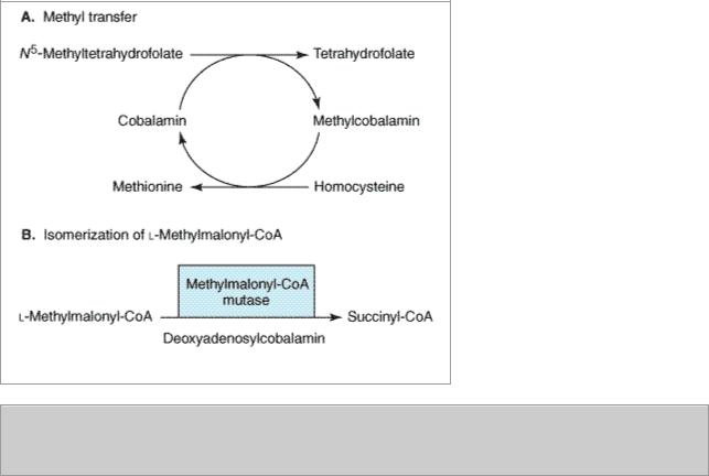
Enzymatic reactions that use vitamin B12. See text for details.
Figure 33–2.

Enzymatic reactions that use folates. Section 1 shows the vitamin B12-dependent reaction that allows most dietary folates to enter the tetrahydrofolate cofactor pool and becomes the "folate trap" in vitamin B12 deficiency. Section 2 shows the dTMP cycle. Section 3 shows the pathway by which folate enters the tetrahydrofolate cofactor pool. Double arrows indicate pathways with more than one intermediate step.
The other enzymatic reaction that requires vitamin B12 is isomerization of methylmalonyl-CoA to succinyl-CoA by the enzyme methylmalonyl-CoA mutase (Figure 33–1 B). In vitamin B12 deficiency, this conversion cannot take place, and the substrate, methylmalonyl-CoA, accumulates. In the past, it was thought that abnormal accumulation of methylmalonyl-CoA causes the neurologic manifestations of vitamin B12 deficiency. However, newer evidence instead implicates the disruption of the methionine synthesis pathway as the cause of neurologic problems. Whatever the biochemical explanation for neurologic damage, the important point is that administration of folic acid in the setting of vitamin B12 deficiency will not prevent neurologic manifestations even though it will largely correct the anemia caused by the vitamin B12 deficiency.
Clinical Pharmacology
Vitamin B12 is used to treat or prevent deficiency. There is no evidence that vitamin B12 injections have any benefit in persons who do not have vitamin B12 deficiency. The most characteristic clinical manifestation of vitamin B12 deficiency is megaloblastic anemia. The typical clinical findings in

megaloblastic anemia are macrocytic anemia (MCV usually > 120 fL), often with associated mild or moderate leukopenia or thrombocytopenia (or both), and a characteristic hypercellular bone marrow with megaloblastic maturation of erythroid and other precursor cells. Vitamin B12 deficiency also causes a neurologic syndrome that usually begins with paresthesias and weakness in peripheral nerves and progresses to spasticity, ataxia, and other central nervous system dysfunctions. A characteristic pathologic feature of the neurologic syndrome is degeneration of myelin sheaths followed by disruption of axons in the dorsal and lateral horns of the spinal cord and in peripheral nerves. Correction of vitamin B12 deficiency arrests the progression of neurologic disease, but it may not fully reverse neurologic symptoms that have been present for several months. Although most patients with neurologic abnormalities caused by vitamin B12 deficiency have full-blown megaloblastic anemias when first seen, occasional patients have few if any hematologic abnormalities.
Once a diagnosis of megaloblastic anemia is made, it must be determined whether vitamin B12 or folic acid deficiency is the cause. (Other causes of megaloblastic anemia are very rare.) This can usually be accomplished by measuring serum levels of the vitamins. The Schilling test, which measures absorption and urinary excretion of radioactively labeled vitamin B12, can be used to further define the mechanism of vitamin B12 malabsorption when this is found to be the cause of the megaloblastic anemia.
The most common causes of vitamin B12 deficiency are pernicious anemia, partial or total gastrectomy, and diseases that affect the distal ileum, such as malabsorption syndromes, inflammatory bowel disease, or small bowel resection.
Pernicious anemia results from defective secretion of intrinsic factor by the gastric mucosal cells. Patients with pernicious anemia have gastric atrophy and fail to secrete intrinsic factor (as well as hydrochloric acid). The Schilling test shows diminished absorption of radioactively labeled vitamin B12, which is corrected when hog intrinsic factor is administered with radioactive B12, since the vitamin can then be normally absorbed.
Vitamin B12 deficiency also occurs when the region of the distal ileum that absorbs the vitamin B12-intrinsic factor complex is damaged, as when the ileum is involved with inflammatory bowel disease, or when the ileum is surgically resected. In these situations, radioactively labeled vitamin B12 is not absorbed in the Schilling test, even when intrinsic factor is added. Other rare causes of vitamin B12 deficiency include bacterial overgrowth of the small bowel, chronic pancreatitis, and thyroid disease. Rare cases of vitamin B12 deficiency in children have been found to be secondary to congenital deficiency of intrinsic factor and congenital selective vitamin B12 malabsorption due to defects of the receptor sites in the distal ileum.
Since almost all cases of vitamin B12 deficiency are caused by malabsorption of the vitamin, parenteral injections of vitamin B12 are required for therapy. For patients with potentially reversible diseases, the underlying disease should be treated after initial treatment with parenteral vitamin B12. Most patients, however, do not have curable deficiency syndromes and require lifelong treatment with vitamin B12 injections.
Vitamin B12 for parenteral injection is available as cyanocobalamin or hydroxocobalamin. Hydroxocobalamin is preferred because it is more highly protein-bound and therefore remains longer in the circulation. Initial therapy should consist of 100–1000 g of vitamin B12 intramuscularly daily or every other day for 1–2 weeks to replenish body stores. Maintenance therapy consists of 100–1000 g intramuscularly once a month for life. If neurologic abnormalities are present, maintenance therapy injections should be given every 1–2 weeks for 6 months before

switching to monthly injections. Oral vitamin B12-intrinsic factor mixtures and liver extracts should not be used to treat vitamin B12 deficiency; however, oral doses of 1000 g of vitamin B12 daily are usually sufficient to treat patients with pernicious anemia who refuse or cannot tolerate the injections.
Folic Acid
Reduced forms of folic acid are required for essential biochemical reactions that provide precursors for the synthesis of amino acids, purines, and DNA. Folate deficiency is not uncommon, even though the deficiency is easily corrected by administration of folic acid. The consequences of folate deficiency go beyond the problem of anemia because folate deficiency is implicated as a cause of congenital malformations in newborns and may play a role in vascular disease (see Folic Acid Supplementation: A Public Health Dilemma).
Chemistry
Folic acid (pteroylglutamic acid) is a compound composed of a heterocycle, p-aminobenzoic acid, and glutamic acid (Figure 33–3). Various numbers of glutamic acid moieties may be attached to the pteroyl portion of the molecule, resulting in monoglutamates, triglutamates, or polyglutamates.
Folic acid can undergo reduction, catalyzed by the enzyme dihydrofolate reductase ("folate reductase"), to give dihydrofolic acid (Figure 33–2, reaction 3). Tetrahydrofolate can subsequently be transformed to folate cofactors possessing one-carbon units attached to the 5-nitrogen, to the 10nitrogen, or to both positions (Figure 33–2). The folate cofactors are interconvertible by various enzymatic reactions and serve the important biochemical function of donating one-carbon units at various levels of oxidation. In most of these, tetrahydrofolate is regenerated and becomes available for reutilization.
Figure 33–3.
The structure and numbering of atoms of folic acid. (Reproduced, with permission, from Murray RK et al: Harper's Biochemistry, 24th ed. McGraw-Hill, 1996)
Pharmacokinetics
The average diet in the USA contains 500–700 g of folates daily, 50–200 
 g of which is usually

absorbed, depending on metabolic requirements (pregnant women may absorb as much as 300–400 g of folic acid daily). Various forms of folic acid are present in a wide variety of plant and animal tissues; the richest sources are yeast, liver, kidney, and green vegetables. Normally, 5–20 mg of folates are stored in the liver and other tissues. Folates are excreted in the urine and stool and are also destroyed by catabolism, so serum levels fall within a few days when intake is diminished. Since body stores of folates are relatively low and daily requirements high, folic acid deficiency and megaloblastic anemia can develop within 1–6 months after the intake of folic acid stops, depending on the patient's nutritional status and the rate of folate utilization.
Unaltered folic acid is readily and completely absorbed in the proximal jejunum. Dietary folates, however, consist primarily of polyglutamate forms of N 5-methyltetrahydrofolate. Before absorption, all but one of the glutamyl residues of the polyglutamates must be hydrolyzed by the enzyme  -1-glutamyl transferase ("conjugase") within the brush border of the intestinal mucosa. The monoglutamate N 5-methyltetrahydrofolate is subsequently transported into the bloodstream by both active and passive transport and is then widely distributed throughout the body. Inside cells, N 5-methyltetrahydrofolate is converted to tetrahydrofolate by the demethylation reaction that requires vitamin B12 (Figure 33–2, reaction 1).
Pharmacodynamics
Tetrahydrofolate cofactors participate in one-carbon transfer reactions. As described above in the section on vitamin B12, one of these essential reactions produces the dTMP needed for DNA synthesis. In this reaction, the enzyme thymidylate synthase catalyzes the transfer of the one-carbon unit of N 5,N 10-methylenetetrahydrofolate to deoxyuridine monophosphate (dUMP) to form dTMP (Figure 33–2, reaction 2). Unlike all of the other enzymatic reactions that utilize folate cofactors, in this reaction the cofactor is oxidized to dihydrofolate, and for each mole of dTMP produced, one mole of tetrahydrofolate is consumed. In rapidly proliferating tissues, considerable amounts of tetrahydrofolate can be consumed in this reaction, and continued DNA synthesis requires continued regeneration of tetrahydrofolate by reduction of dihydrofolate, catalyzed by the enzyme dihydrofolate reductase. The tetrahydrofolate thus produced can then reform the cofactor N 5,N 10-methylenetetrahydrofolate by the action of serine transhydroxymethylase and thus allow for the continued synthesis of dTMP. The combined catalytic activities of dTMP synthase, dihydrofolate reductase, and serine transhydroxymethylase are often referred to as the dTMP synthesis cycle. Enzymes in the dTMP cycle are the targets of two anticancer drugs; methotrexate inhibits dihydrofolate reductase, and a metabolite of 5-fluorouracil inhibits thymidylate synthase (see Chapter 55: Cancer Chemotherapy).
Cofactors of tetrahydrofolate participate in several other essential reactions. As described above, N 5-methy- lenetetrahydrofolate is required for the vitamin B12-dependent reaction that generates methionine from homocysteine (Figure 33–1 A; Figure 33–2, reaction 1). In addition, tetrahydrofolate cofactors donate one-carbon units during the de novo synthesis of essential purines. In these reactions, tetrahydrofolate is regenerated and can reenter the tetrahydrofolate cofactor pool.
Clinical Pharmacology
Folate deficiency results in a megaloblastic anemia that is microscopically indistinguishable from the anemia caused by vitamin B12 deficiency (see above). However, folate deficiency does not cause the characteristic neurologic syndrome seen in vitamin B12 deficiency. In patients with megaloblastic anemia, folate status is assessed with assays for serum folate or for red blood cell folate. Red blood cell folate levels are often of greater diagnostic value than serum levels, since serum folate levels tend to be quite labile and do not necessarily reflect tissue levels.