

clavien_atlas_of_upper_gastrointestinal_and_hepato-pancreato-biliary_surgery2007-10-01_3540200045_springer
.pdfLeft Thoracoabdominal Approach for Carcinoma of the Lower Esophagus and Gastric Cardia |
59 |
|
|
|
|
STEP 4 |
Lymph node removal of the upper abdomen |
|
|
|
|
Lymph nodes of the hepatoduodenal ligament and around the common hepatic artery, left gastric artery and celiac trunk are dissected.
Mobilization of the stomach and transection of the duodenum (see chapter “Total Gastrectomy with Conventional Lymphadenectomy”)
60 |
SECTION 2 |
Esophagus, Stomach and Duodenum |
|
|
|
STEP 5 |
Lower mediastinal lymph node removal |
|
|
|
|
Regarding the left intrathoracic approach, the left pulmonary ligament is divided and the mediastinal pleura is opened. The pleura covering the lower thoracic esophagus is incised, allowing the clearance of loose connective tissue together with the lower thoracic paraesophageal, supradiaphragmatic, posterior mediastinal and intradiaphragmatic lymph nodes.
Left Thoracoabdominal Approach for Carcinoma of the Lower Esophagus and Gastric Cardia |
61 |
|
|
|
|
STEP 6 |
Reconstruction |
|
|
|
|
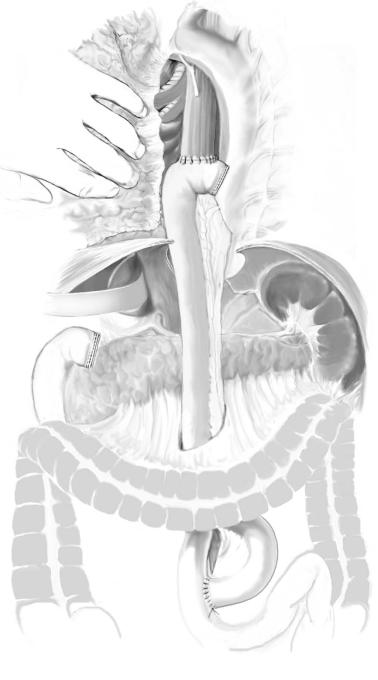
There are several methods of reconstruction according to the tumor location and extension. Roux-Y reconstruction by using an EEA instrument, as shown here, is an option for performing the esophago-jejunostomy.
See chapter on “Subtotal Esophagectomy: Transhiatal Approach” for standard postoperative investigations and complications.

62 |
SECTION 2 |
Esophagus, Stomach and Duodenum |
|
|
|
|
Tricks of the Senior Surgeon |
|
■Kinking of the graft: this is a rare but dangerous complication, due to clinical symptomatic disturbance of the gastrointestinal passage by elongation, which requires surgical intervention and is performed by shortening of the graft.
■If the trachea is injured, use direct suture and pericardial flap.

Subtotal Esophagectomy: Transhiatal Approach
Stefan B. Hosch, Emre F. Yekebas, Jakob R. Izbicki
Introduction
The surgical trauma of the transhiatal approach is less pronounced as compared to a transthoracic approach. On the other hand, the lymphatic clearance is less radical, at least for the mid and upper mediastinum. This is the reason why some surgeons are in favor of the transthoracic approach even for distal adenocarcinoma. Subtotal transhiatal esophagectomy is indicated for benign conditions and for distal carcinoma.
Indications and Contraindications
Indications |
■ |
Adenocarcinoma of distal esophagus (>T1 stage) |
|
■ |
Intraepithelial squamous cell neoplasia |
|
■ |
Poor risk patients |
|
■ |
Extensive stricture (stenosis) due to erosion (chemical burns) unresponsive to |
|
|
nonsurgical treatment including bougienage |
|
■ |
Extensive peptic stricture (stenosis) |
|
■ |
Relapse of megaesophagus after surgical repair of cardiospasm combined with peptic |
|
|
strictures and failure of dilatation |
|
■ |
Extensive benign esophageal tumors (exceptional cases, usually local excision) |
|
■ |
Esophageal rupture or iatrogenic perforation with mediastinitis (primary repair not |
|
|
feasible) |
|
|
Florid gastroduodenal ulcer |
Contraindications |
■ |
|
|
■ |
Infiltration of aorta |
|
■ |
Distant metastasis |
Preoperative Investigation/Preparation for the Procedure
History: |
Previous gastric or colonic surgery |
Risk factors: |
Alcohol, nicotine, gastroesophageal reflux disease (GERD), |
|
Barrett’s esophagus |
Clinical evaluation: |
Recurrent laryngeal nerve status, cervical |
|
lymphadenopathy |
Laboratory tests: |
CEA, liver function tests, coagulation test |
Endoscopy: |
Esophagogastroduodenoscopy with biopsy – |
|
to exclude gastric infiltration |
Colonoscopy |
If colonic interposition is likely |
CT scanning |
Staging |
(thorax + abdomen): |
|
Abdominal ultrasound: |
Staging |
Esophageal endosonography: Staging, r/o aortic infiltration |
|
Bronchoscopy (if tumor is |
r/o bronchial infiltration |
localized in mid-third): |
|
Bowel cleansing |
(If colonic interposition is likely) |
Respiratory therapy |
|
64 |
SECTION 2 |
Esophagus, Stomach and Duodenum |
|
|
|
|
Procedure |
|
|
Access |
|
|
Upper transverse incision with median extension. Alternatively midline laparotomy. |
|
|
|
|
STEP 1 |
Laparotomy and inspection of the stomach, distal esophagus, |
|
liver and regional lymph nodes
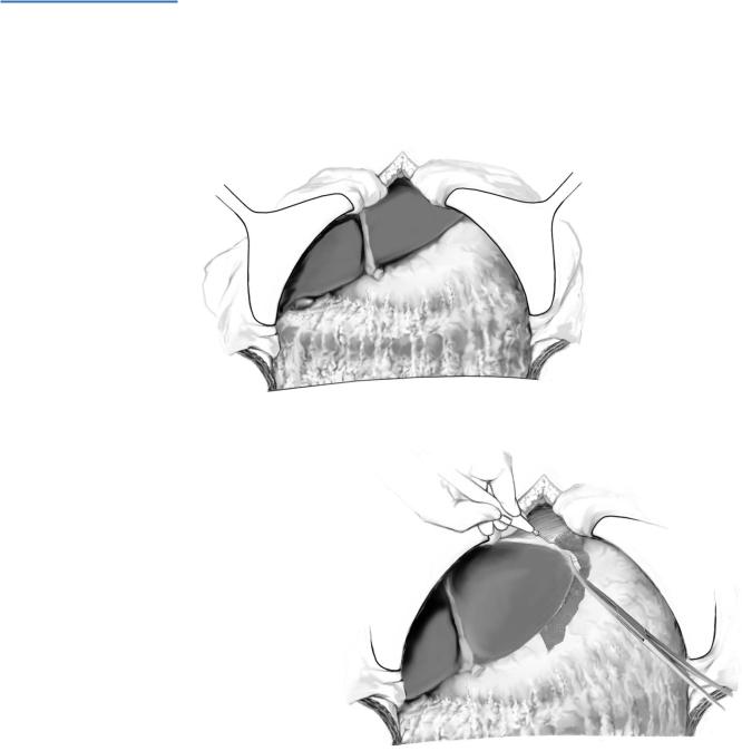
Placement of self-retaining retractor system for exposure of the epigastric region (A). Mobilization of the left lateral liver by transection of the left triangular ligament. To
prevent injury of adjacent structures, a pack is placed under the left lobe of the liver (B).
A
B
Subtotal Esophagectomy: Transhiatal Approach |
65 |
|
|
|
|
STEP 2 |
Preparation and mobilization of the stomach with epigastric lymphadenectomy |
|
including para-aortic lymphatic tissue
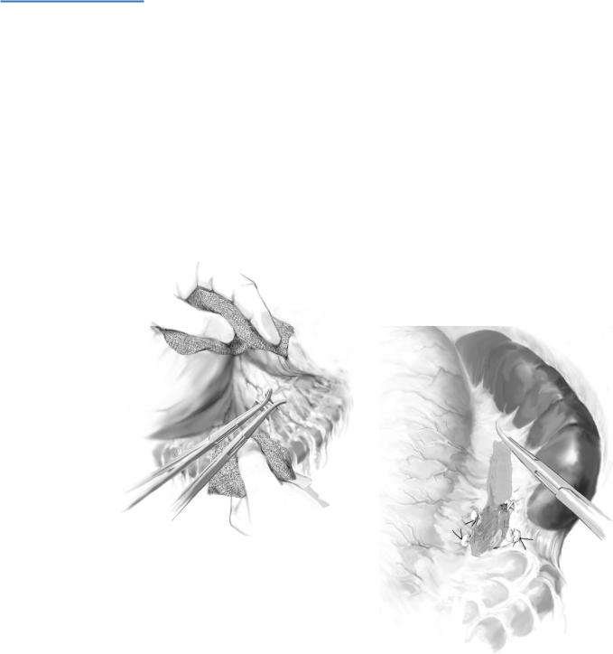
Dissection of the greater curvature is commenced from below, thoroughly sparing the origin of the right gastroepiploic vessels and the arcade between left and right gastroepiploic vessels up to the level of the splenic hilum (A).
Dissection of the greater curvature is continued towards the spleen. The left gastroepiploic artery is transected directly at its origin at the splenic artery. Transection and ligature of the short gastric vessels is performed, thus mobilizing the fundus and the greater curvature completely. For esophageal carcinoma the parietal peritoneum is incised at the upper pancreatic margin and lymphadenectomy is begun along the splenic artery. The flaccid part of the lesser omentum is dissected. The cranial part of the hepatogastric ligament (hepatoesophageal ligament) is dissected from the diaphragm. An accessory left liver artery with strong caliber should be preserved. In this case the left gastric artery has to be diverted distally to the origin of this accessory liver artery (B).
A
B
66 |
SECTION 2 |
Esophagus, Stomach and Duodenum |
|
|
|
STEP 2 (continued) |
Preparation and mobilization of the stomach with epigastric lymphadenectomy |
|
including para-aortic lymphatic tissue
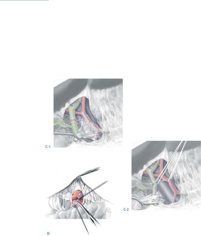
Lymphadenectomy of the hepatoduodenal ligament is performed. Remove all lymphatic tissue around the hepatic artery up to the celiac trunk, of the portal vein and as well as the lymphatic tissue around the common bile duct. Ligature and diversion of the right gastric artery are carried out close to its origin below the pylorus (C-1, C-2).
Transection of the left gastric artery. All lymph nodes along the left gastric artery, the splenic artery, the common hepatic artery, the celiac trunk, and para-aortic lymph nodes are removed (D).
In benign diseases, blunt dissection of the esophagus is performed without lymphadenectomy. The right gastric artery may be ligated below the pylorus.
The blood supply of the gastric tube after preparation is exclusively provided by the right gastroepiploic artery.
Subtotal Esophagectomy: Transhiatal Approach |
67 |
|
|
|
|
STEP 3 |
Mobilization of the abdominal part of the esophagus and incision |
|
|
of the esophageal hiatus |
|
|
|
|
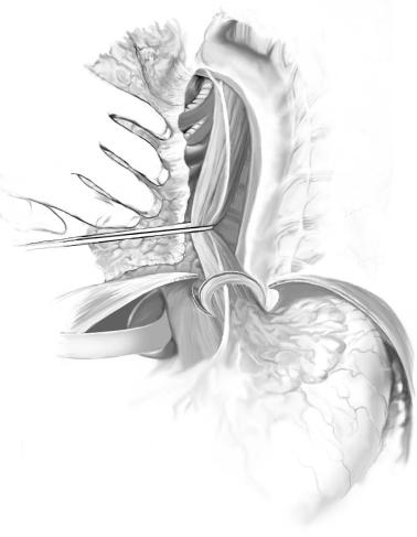
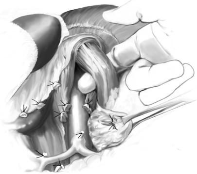
The lymph node dissection is continued along the celiac trunk to the para-aortic region. The lymphatic tissue is transposed to the lesser curvature and is later resected en bloc with the tumor.
For better exposure the diaphragmatic crura are incised with diathermia and the stumps may be ligated. Blunt mobilization of the esophagus is done with the index finger. During this maneuver connective tissue fibers between the esophagus, diaphragmatic crua and abdominal aorta must be removed carefully (A).
The abdominal esophagus is mobilized and pulled caudally with a rubber tube.
A
68 |
SECTION 2 |
Esophagus, Stomach and Duodenum |
|
|
|
STEP 3 (continued) |
Mobilization of the abdominal portion of the esophagus and incision |
|
|
of the esophageal hiatus |
|
|
|
|
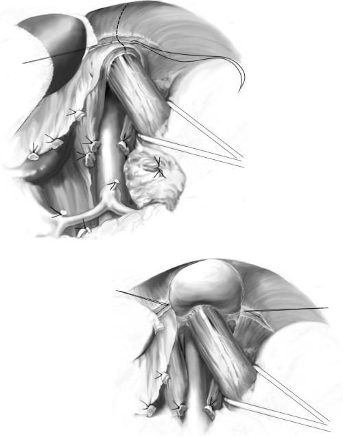
The hiatus is incised ventrally following transection of the left inferior phrenic vein between ligatures (B).
Insertion of retractors. The retrocardial lymphatic tissue is removed en bloc with the specimen (C).
B
C