

clavien_atlas_of_upper_gastrointestinal_and_hepato-pancreato-biliary_surgery2007-10-01_3540200045_springer
.pdfPrinciples of Drainage |
37 |
|
|
Prophylactic Drainage
Drain Orifice
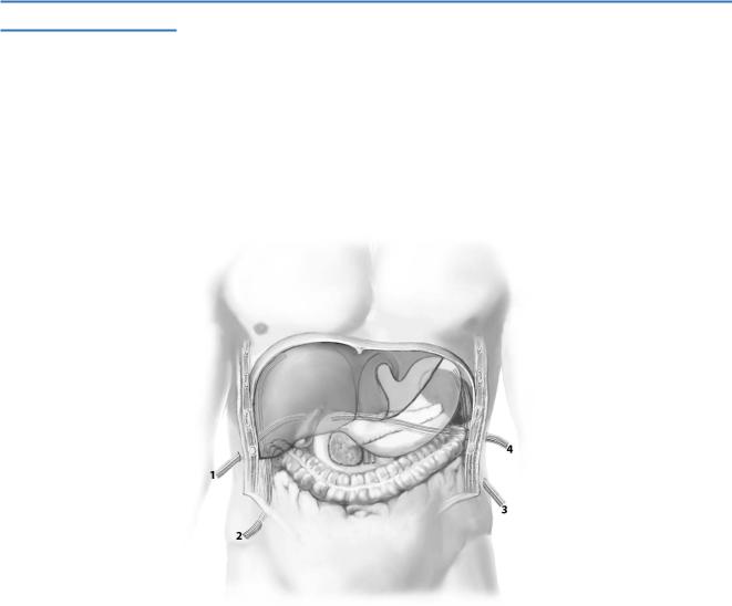
The drain orifice through the skin is created by a penetrating cut with a scalpel (A-1). A Kelly clamp is inserted into the orifice (A-2) and penetrates the abdominal wall diagonally (A-3). The hand serves as protection to prevent bowel injury. This technique creates a tunnel that helps to seal the abdominal cavity after drain removal. After clamping the drain tip, the Kelly clamp and drain are pulled through the abdominal wall from inside outwards (A-4). Others prefer to create the tunnel from inside out and pull the drain into the abdomen. Finally, the drain position
is secured by a non-reactive skin suture, and the drain tube is connected to the suction device.
A-1 |
A-2 |
A-3 |
A-4 |
38 SECTION 1 General Principles
Prophylactic Drains
Prophylactic drainage after upper abdominal operations is used to evacuate intraabdominal fluid that may develop, such as ascites, blood, chyle, bile, pancreatic,
or intestinal juice, that are either harmful/toxic for adjacent tissue or might become infected. Therefore, drains are placed in spaces that tend to accumulate fluid, such
as the subhepatic (1), right subphrenic (2), left subphrenic (3), and parapancreatic (4) spaces.

Principles of Drainage |
39 |
|
|
Anastomosis Drains
Another proposed function of prophylactic drainage is the early detection of anastomotic leakage. If drains are to be used near a high-risk anastomosis, it is important that they are not placed in direct contact with the anastomosis, but, rather, with a safety margin in between to prevent drain-related erosions. This principle is illustrated for a biliodigestive anastomosis, where the drain is placed posterior
to the anastomosis.
Although the routine use of prophylactic drainage has often been considered as a method to prevent complications, there is growing evidence that this practice may
be associated with adverse effects. Retrograde drain infections or drain-related complications are known adverse effects. Several randomized, controlled trials are available investigating the routine use of prophylactic drainage (Table2).
Table 2. Evidence-based recommendations for prophylactic drainage practice
NA not assessed
aOnly one randomized controlled trial in pancreatic cancer
40 |
SECTION 1 |
General Principles |
|
|
|
|
Therapeutic Drainage |
|
Predisposed Spaces for Collections
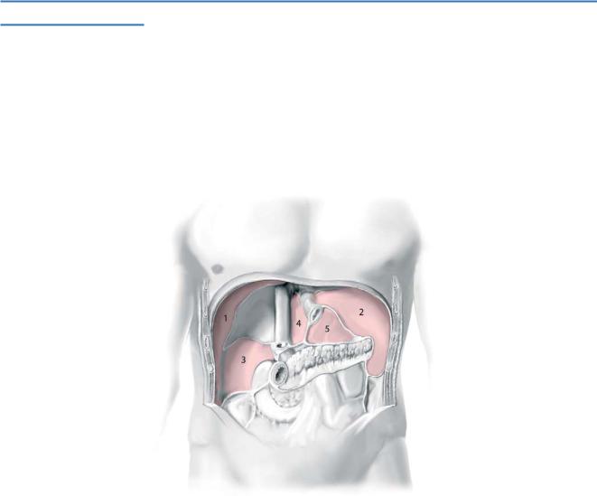
Infected collections, such as abscesses or infected bilomas, are known complications after upper abdominal surgery and require drainage by operative or radiologically guided drain placement. The right subphrenic space (1), left subphrenic space (2), Morison’s pouch (3), left subhepatic space (4), and omental sac (5) are anatomic spaces that predispose to abscess development.
Principles of Drainage |
41 |
|
|
Catheters
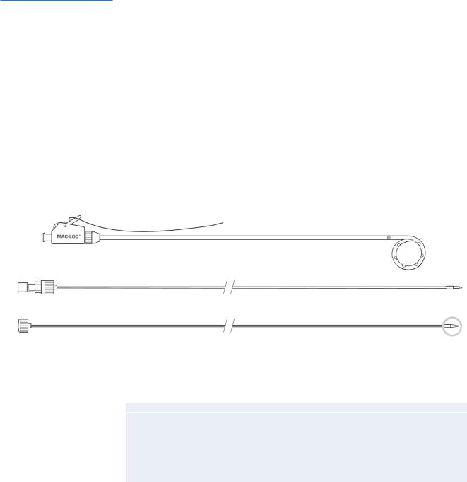
The majority of postoperative collections in the upper abdomen are manageable by means of percutaneous drainage by interventional radiologic techniques using standard aseptic technique and local anesthesia. Thereby, collections are drained percutaneously under ultrasonographic or CT guidance using the Seldinger or trocar techniques. This figure illustrates a typical percutaneous drainage catheter, the MAC-LOC (A-1) that can be inserted by the introduction cannula (A-2) or the trocar stylet (A-3). The catheter has large, oval side ports to increase the drainage capability, as well as a radiopaque band that helps to identify the proximal area of the loop. This type of “self-locking” loop catheter has “memory,” i.e., the loop at the end can be straightened during insertion by introducing a stylet intraluminally. After the catheter is positioned in place, the stylet
is removed, and the loop reforms. This loop prevents displacement of the catheter. Some fluid collections may require surgical drainage with repeated abdominal lavage
and second-look procedures (Table3).
A-1
A-2
A-3
Table 3. Criteria for percutaneous and surgical drainage of infected collections
Percutaneous drainage |
Surgical drainage |
|
|
Unilocular collection/abscess |
Multilocular collections/abscesses |
Low viscosity of drain fluid |
Multiple, non-communicating collections |
Drain route not traversing intra-abdominal |
High viscosity of drain fluid |
organs or thorax |
|
|
Percutaneous drain route traversing intra- |
|
abdominal organs or thorax |
|
|

42 |
SECTION 1 |
General Principles |
|
|
|
|
Tricks of the Senior Surgeon |
|
■Whenever indicated, always use closed drain systems and keep drains as short as possible to minimize the risk of retrograde infections.
■Place drains near but never in direct contact to the anastomotic sutures to prevent drain-induced erosions or drain-induced anastomotic leaks.
■When drains are not productive, do not rely on them! Drains could be occluded or obstructed by adjacent tissue.
■Try to position intraperitoneal drains such that the drain does not rub against or lie in direct contact with blood vessels or hollow organs in an attempt to prevent drain erosions.

SECTION 2
Esophagus, Stomach and Duodenum
Jakob R. Izbicki

Introduction
Jakob R. Izbicki
This section presents the ambitious field of open and laparoscopic surgery in benign and malignant diseases of the esophagus, stomach and duodenum.
Attempts to treat esophageal cancer surgically emerged at the beginning of the twentieth century. Torek successfully resected the thoracic esophagus in 1913, but real progress came as a result of the development of thoracic surgery during and after the Second World War. The concept of extensive lymph node resection in combination with en bloc esophagectomy was proposed by Logan in 1963 but with considerable morbidity and mortality.
However, surgical procedures, preand postoperative management and treatment, and prognosis after surgical treatment have improved considerably in the past 3decades. Expertise of the surgeon and the institution, patient selection, choice and radicality of operation, and preand postoperative care are the most important parameters for outcome.
For this reason, the first eight chapters present a comprehensive survey of the different open and laparoscopic surgical procedures, indications, and choice of operation in esophageal cancer. They give clear guidelines as to how and when to operate with regard to the biological characteristics of the tumor. The focus then turns to benign esophageal diseases such as diverticula, strictures, and achalasia.
The following chapters address the techniques of esophago-gastric, subtotal and total gastric resections in benign and malignant diseases, respectively. Four chapters then outline the open and laparoscopic strategies in the case of palliation such as gastroenterostomy and gastrostomy. Next the laparoscopic procedure is described as the gold standard for gastroesophageal reflux, and then the open approach is covered followed by different laparoscopic techniques for hiatal repair in paraesophageal hernia. Another chapter comprehensively covers the available strategies for morbid obesity, and the last chapter in the section deals with the ambitious pancreas-sparing duodenectomy.
The section has been prepared by experts in their surgical fields, and we hope that it will provide the reader with a comprehensive overview of current surgical standards for the various procedures.

Cervical Esophagectomy
Wolfram T. Knoefel, Rainer Schmelzle
Introduction
Cervical esophagectomy for neoplasia includes removal of the cervical part of the esophagus combined with cervical lymphadenectomy and reconstruction by interposition of a free jejunal transplant with microvascular anastomoses.
Resection of a segment of up to 3cm can be performed with primary anastomosis of the esophagus after adequate mobilization.
Indications and Contraindications
Indications |
■ |
Cervical esophageal neoplasia |
|
■ |
Benign esophageal stricture |
|
|
Local irresectability (infiltration of larynx, trachea or vertebrae) |
Contraindications |
■ |
|
|
■ |
Multifocal lesions |
|
■ |
Distant metastasis |
|
■ |
Florid gastroduodenal ulcer |
|
■ |
Crohn’s disease |
Preoperative Investigation/Preparation for the Procedure
History: |
Risk factors (alcohol, nicotine), previous radiation therapy, |
|
concomitant malignancy, Crohn’s disease |
Clinical evaluation: |
Recurrent laryngeal nerve status, cervical lymphadenopathy |
Endoscopy: |
Esophagogastroduodenoscopy (EGD) to rule out multifocal |
|
lesions and gastroduodenal ulcer |
CT scan or MRI: |
Assessment of stage and resectability (include thorax and |
|
abdomen!) |
Doppler ultrasound: |
Vascular status in the neck |
PET scan: |
Exclude further lesions |
Laboratory test: |
SCC antigen, coagulation parameters |
Avoid: |
Central venous line on the side of anastomoses, wall stents |

48 SECTION 2 Esophagus, Stomach and Duodenum
Procedure
Access
For the cervical part a unilateral or bilateral (U-) incision at the medial margin of the sternocleidomastoid muscle is performed. It extends from the margo inferior of the mandible to the jugulum, where it meets with the contralateral side. For cervicalmediastinal lymph node dissection, the incision is combined with a partial or complete median sternotomy. For harvesting the transplant, a small upper abdominal transverse incision is sufficient.
Exposure
Retraction is performed by hand held retractors of different shapes and sizes according to the anatomy. If available, a self-retaining retraction system will help.