

clavien_atlas_of_upper_gastrointestinal_and_hepato-pancreato-biliary_surgery2007-10-01_3540200045_springer
.pdf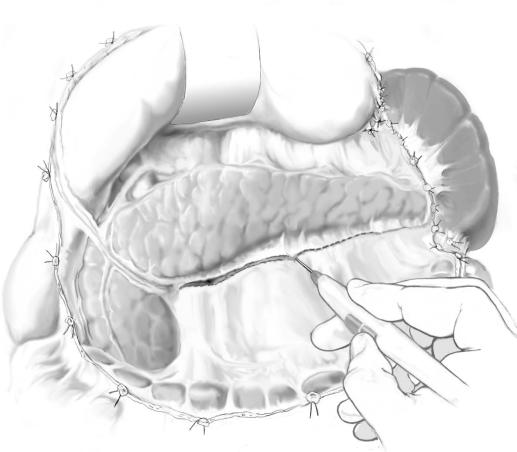
656 |
SECTION 5 |
Portal Hypertension |
|
|
|
STEP 2 |
Mobilization |
|
|
|
|
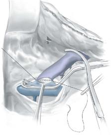
The figure illustrates the initial exposure, which requires mobilization of the greater curve of the stomach from the pylorus to the short gastric vessel. The gastroepiploic vessels should be interrupted and ligated. Access is further enhanced by taking down the splenocolic ligament and mobilizing the splenic flexure of the colon inferiorly. These two moves are also a component of the devascularization that separates the low pressure shunt from the high pressure portal venous system. Once this mobilization has taken place, inferior retractors can be placed on the colon, which improves exposure and access to the retropancreatic plane. The stomach should be retracted superiorly.
Dissection then focuses along the inferior margin of the pancreas, which is mobilized from the super mesenteric vein out to the splenic hilus.
Distal Splenorenal Shunt |
657 |
|
|
STEP 3 |
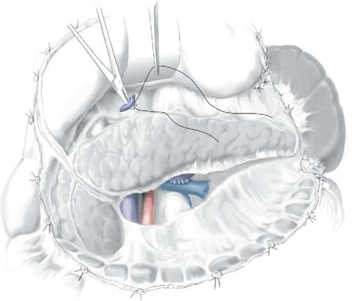
Exposure and dissection of the splenic vein |
|
The splenic vein is initially identified by a combination of palpation and vision. In an |
|
|
|
obese patient the vein may not be readily seen on the posterior surface of the pancreas, |
|
but it can always be felt. Initial dissection of the splenic vein is started where it is most |
|
easily seen or palpated. The goal is to mobilize all the overlying adventitia along the |
|
inferior and posterior surface of the splenic vein from the superior mesenteric vein |
|
to the splenic hilus. |
|
The inferior mesenteric vein enters the splenic vein in 50% of patients and the |
|
superior mesenteric vein in the other 50%. This is a useful landmark as it is always |
|
the first significant vein coming inferiorly as dissection proceeds from left to right. |
|
It should be ligated and interrupted. |
|
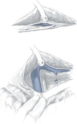
The other key plane in this phase of the procedure is the posterior plane at the splenic |
|
superior mesenteric venous junction, as illustrated in the Figure. This is a safe plane to |
|
open with a finger or the tip of the sucker. The value of opening this plane is if any |
|
bleeding is encountered as the anterior dissection of the splenic vein is started, it can |
|
always be controlled by finger compression of the vessels. |

658 |
SECTION 5 |
Portal Hypertension |
|
|
|
STEP 4 |
Dissection of pancreatic tributaries to the splenic vein |
|
|
|
|
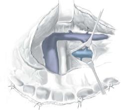
Pancreatic tributaries to the splenic vein always lie on the superior and anterior surface of the splenic vein. The trick in dissecting these small and fragile vessels is to open the plane on each side of these by spreading directly in the line of the small vessels. They can then be surrounded with a small right-angled clamp, and they should be ligated on the splenic vein side and clipped on the pancreatic side. This is the most delicate and difficult part of this procedure and requires considerable patience. It is facilitated by the step described in the Figure of fully mobilizing the posterior and inferior border of the splenic vein before commencing this dissection.
Sufficient splenic vein needs to be fully mobilized to allow for subsequent positioning of the vessel down to the left renal vein. The splenic vein should not be divided at this point.
Distal Splenorenal Shunt |
659 |
|
|
STEP 5 |
Exposure and dissection of the left renal vein |
|
The left renal vein crosses in front of the aorta and behind the superior mesenteric |
|
|
|
artery. Palpation of these landmarks aids identification of the renal vein at this point. |
|
The retroperitoneum is bluntly opened until the vein is identified and then dissection is |
|
carried out along the anterior surface of the left renal vein. Tissue should be ligated at |
|
this point as it contains numerous lymphatics that if left open may cause chylous ascites. |
|
The renal vein is mobilized over sufficient length to allow it to come up comfortably into |
|
a Satinsky clamp for subsequent anastomosis. The left adrenal vein should be ligated as |
|
it always acts as an inflow to the renal vein. The left gonadal vein should be left intact as |
|
it often acts as an outflow tract and helps accommodate increased renal vein flow after |
|
the shunt is open. |
660 |
SECTION 5 |
Portal Hypertension |
|
|
|
STEP 6 |
Preparation of the splenic vein |
|
|
|
|
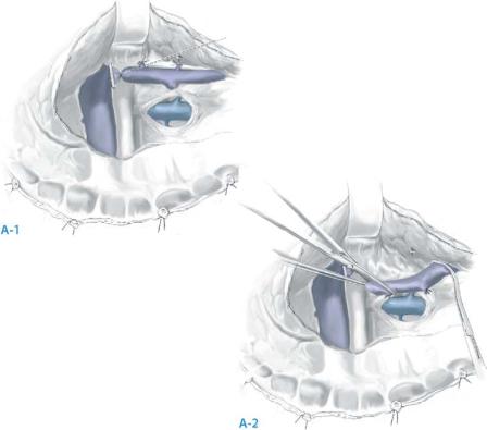
Once the left renal vein has been dissected and is ready for the anastomosis, the splenic vein is ligated flush at the superior mesenteric vein. Experience has shown that this is best achieved with a silk tie on the splenic vein stump and a large clip placed behind that. This is given the lowest instance of mesenteric venous thrombus at this site.
The splenic vein can then be moved downwards as shown in Fig. A-1, A-2, its relationship to the left renal vein confirmed, and the vein trimmed to an appropriate length for comfortable anastomosis. The goal is to have the splenic vein come down to the left renal vein without any kinking as the splenic vein emerges from the pancreas.
Distal Splenorenal Shunt |
661 |
|
|
STEP 7 |
Anastomosis: |
|
The anastomosis is made with a running suture to the posterior wall and interrupted |
|
|
|
sutures to the anterior wall. This is the preferred method to minimize the risk of a |
|
purse-string effect at this anastomosis. Suture material should be the surgeon’s pre- |
|
ference. We use a running 5-0 Ethibond stitch on the posterior wall and interrupted 5-0 |
|
silks to the anterior wall. This has proved very satisfactory with a low rate of thrombosis. |
|
This can be a difficult anastomosis because it is sewn in a deep hole, particularly in |
|
the obese patient. Minimizing the movement at the depth of this hole aids in the com- |
|
pletion of the anastomosis. The left renal vein clamp needs to be held forward by an |
|
assistant in the left upper quadrant and the splenic vein clamp may need to be held to |
|
keep the tension off the anastomosis. Careful positioning, with stability of these clamps, |
|
is key at this stage. |
662 |
SECTION 5 |
Portal Hypertension |
|
|
|
STEP 8 |
Completion of the anastomosis |
|
|
|
|
Clamps are removed on completion of the anastomosis and the pancreas allowed to come down toward the left renal vein. A small amount of bleeding is usually controlled with some Surgicel and light packing for several minutes. Checking the position of this anastomosis is important to make sure there is no kinking or twisting of the splenic vein.
Completion of devascularization. The left gastric vein is identified if possible either as it joins the splenic vein or as it joins the portal vein. If it can be clipped at this site, it should be. It is also identified at the superior margin of the pancreas as shown in this figure and completely interrupted at this site.
Completion of the procedure at this point has now created a low pressure decompression of the spleen, gastric fundus, and distal esophagus, while maintaining portal hypertension and portal flow in the superior mesenteric and portal venous system.

Distal Splenorenal Shunt |
663 |
|
|
Postoperative Tests
■Follow liver labs on alternate days and electrolytes daily for 1week.
■Diet: low sodium (2g/day) and low fat diet (30g/day) for 6weeks. The latter is necessary because of the disruption of lymphatics around the left renal vein that can lead to a chylous ascites.
■Radiologic shunt study: direct catheterization of the shunt at 5–7days to document patency and no significant pressure gradient.
Postoperative Complications
■Early:
–Liver decompensation
–Ascites
–Shunt thrombosis
–Managing these is primarily based on prevention. Careful patient selection will select good risk patients that should tolerate this operative procedure. Ascites is minimized by careful fluid management perioperatively and diet restrictions postoperatively. Shunt thrombosis is low risk (1–4%) and is minimized if the operative steps outlined above are followed.
■Late:
–Progressive liver disease. These patients should be kept in long-term follow-up, primarily for monitoring of their cirrhosis.
–Late shunt thrombosis is unusual, but any recurrence of bleeding requires documentation of shunt patency, usually by catheterization with pressure measurements.
Tricks of the Senior Surgeon
■Proper positioning of the patient helps later exposure.
■Exposure is critical – see Step 2.
■The posterior surface of the splenic vein is the safe dissection plane.
■Mobilize enough splenic vein for it to come down to the left renal vein easily.
■The left renal vein is best located by palpation as described in Step5.

Low-Diameter Mesocaval Shunt
Miguel A. Mercado, Hector Orozco
Introduction
The mesocaval shunt decompresses portal hypertension with an interposition graft between the superior mesenteric vein (SMV) and the inferior vena cava (IVC). Popularized by Drapanas in the 1970s, it has had several proponents since, and most highlight that it is a shunt performed remote from the hepatic hilus. Similar in physiology to a side-to-side portacaval shunt, a mesocaval shunt diverts all portal flow if ≥12mm diameter, while if 8–10mm diameter some prograde portal flow is maintained.
Indications and Contraindications
Indications |
■ |
Variceal bleeding refractory to endoscopic treatment |
|
■ |
Child’s A and B patients |
|
|
Advanced liver disease (Child’s C) |
Contraindications |
■ |
|
|
■ |
Mesenteric venous thrombosis |
Investigations
History: variceal bleeding
Absence of advanced liver disease Laboratory studies: Child’s A/B class
Vascular imaging: Ultrasound with Doppler
Angiography with venous phase imaging if SMV patency is questioned
Preparation
■Elective operation preferred
■Stabilize from acute bleed
–Correct coagulation
–Diurese ascites
–Improve nutrition status