
clavien_atlas_of_upper_gastrointestinal_and_hepato-pancreato-biliary_surgery2007-10-01_3540200045_springer
.pdf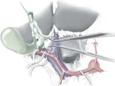
570 |
SECTION 4 |
Biliary Tract and Gallbladder |
|
|
|
|
Left Hepatic Resection |
|
|
|
|
STEP 5 |
Dividing the hepatic artery |
|
|
Once the arterial anatomy and possible arterial variation are clearly identified, the left |
|
|
||
|
and middle hepatic arteries (and cystic artery) or right hepatic artery are divided at the |
|
|
origin (see Figure B of STEP 4). The remaining left hepatic artery is skeletonized more |
|
|
distally to encircle the right anterior and posterior branches at the right extremity of the |
|
|
hilar plate or the middle and left hepatic artery at Rex’s recess. |
|
|
|
|
STEP 6 |
Transection of the left portal vein |
|
|
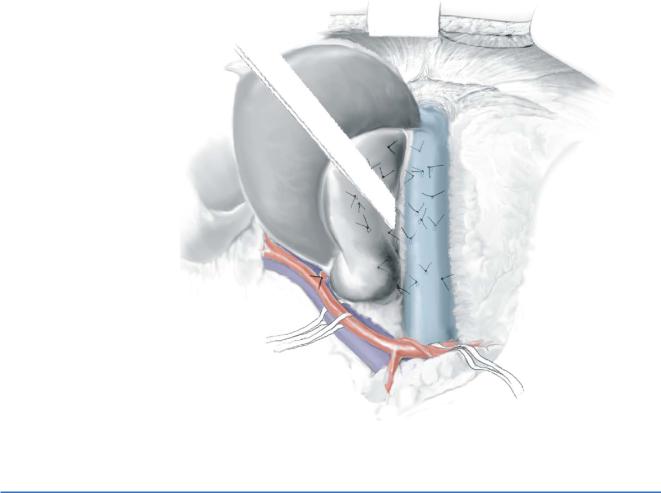
The left portal vein is divided and ligated distally to the bifurcation. An alternative is to |
|
|
||
|
use a small vascular clamp on the proximal side and oversew the venous stump with a |
|
|
running suture of 5-0 Prolene. |
|
Bile Duct Resection |
571 |
|
|
STEP 7 |
Mobilization of the caudate lobe with division of the short hepatic veins |
|
In case of left side hepatectomy, the short hepatic veins are divided in the same manner |
|
|
|
from the left caudal side to the right cranial side. Finally the distal end of the canal of |
|
Arantius is ligated and divided at the confluence of the left hepatic vein or the vena cava. |
STEP 8 |
Exposure and transection of the left hepatic vein |
|
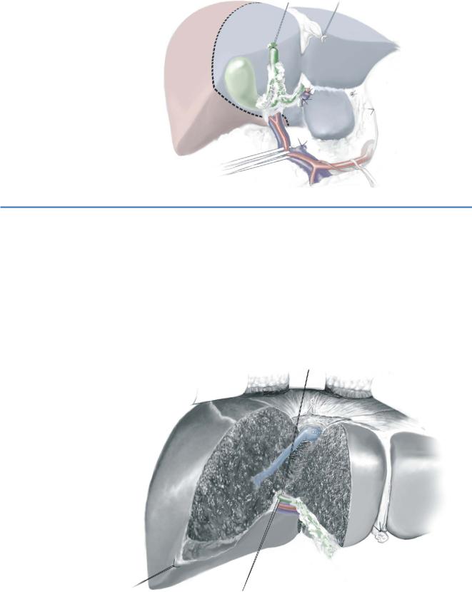
In case of a left hepatectomy, the left hepatic vein is not transected before liver |
|
|
|
dissection. A vessel loop is placed around the common trunk of the left and middle |
|
hepatic vein. In case of a left trisectionectomy, the common trunk of the left and |
|
middle hepatic veins is transected before the liver. |
572 |
SECTION 4 |
Biliary Tract and Gallbladder |
|
|
|
STEP 9 |
Demarcation and incision of the liver capsule |
|
|
|
|
In case of a left hepatectomy with caudate lobe resection, dorsal demarcation appears between the caudate process and segment 7 after complete devascularization of the caudate lobe.
STEP 10 |
Division of the draining veins of the pericaval segment |
|
In case of a total caudate lobe resection, draining veins of the pericaval segment |
|
|
|
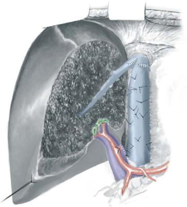
(segment 9) are identified behind the middle hepatic vein and carefully divided. Liver |
|
transection is continued until the left hepatic vein is reached, divided and closed at its |
|
confluence of the middle hepatic vein. The right intrahepatic bile duct is identified |
|
behind the middle hepatic vein, and the posterior wall is carefully detached from the |
|
right anterior branch of the hepatic artery, which runs in the connective tissues between |
|
the bile duct and the portal vein. Two stay sutures are placed caudally and cranially and |
|
the bile duct is incised from the caudal edge where the anterior branch or the bile duct |
|
of segment 5 is opened. |
Bile Duct Resection |
573 |
|
|
STEP 11 |
Intrahepatic bile duct resection |
|
Further extension of the incision opens the segmental or subsegmental bile ducts of |
|
|
|
segment 8 (B8, B8a, B8bc) and the right posterior duct. The surgical field after removal |
|
of the left lobe and caudate lobe is shown. |
574 |
SECTION 4 |
Biliary Tract and Gallbladder |
|
|
|
STEP 12 |
Extended lymph node dissection |
|
|
|
|
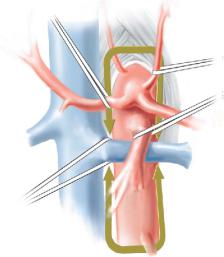
After removing the hemiliver and the caudate lobe, para-aortic node dissection is carried out from the level of the ligamentum crus to the origin of the inferior mesenteric artery. The lymph nodes behind the left renal vein are carefully dissected by taping the left renal vein and the right renal artery. Right celiac ganglionectomy is also performed during this procedure.
Bile Duct Resection |
575 |
|
|
STEP 13 |
Biliary reconstructions |
|
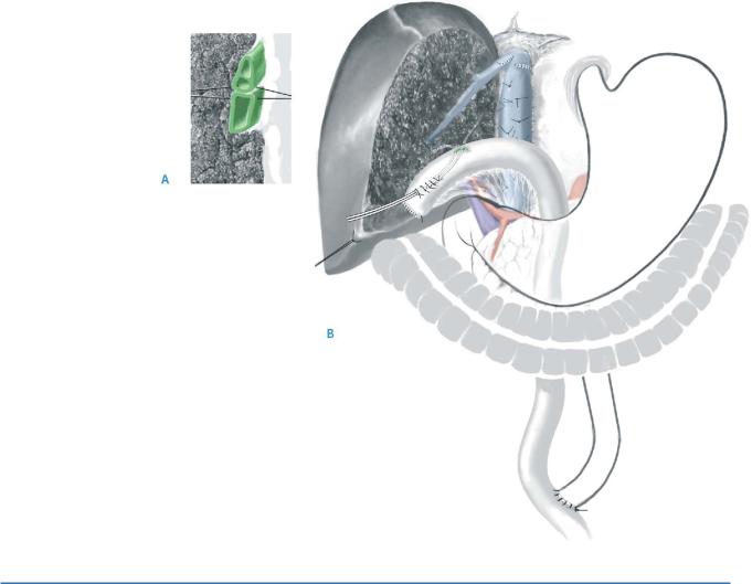
Before bilioenteric anastomosis, hepaticoplasty should be performed with 5-0 PDS |
|
|
|
sutures to minimize the number of anastomoses (A). |
|
A Roux-en-Y jejunal loop is lifted through the shortest route: the retrocolic and |
|
retrogastric route. A jejunostomy tube is also introduced from the proximal edge |
|
of the jejunal limb before hepaticojejunostomy (B). |
|
The posterior wall is first anastomosed with 4-0 PDS sutures and a biliary drainage |
|
tube is placed in each anastomosis. Finally the anterior wall is anastomosed. See chapter |
|
on biliary anastomosis. |
STEP 14 |
Drainage after reconstruction |
|
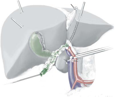
After completing hemostasis in the surgical field, closed drains are placed in the |
|
|
|
foramen of Winslow, the pericaval space along the aorta and along the cut surface |
|
of the liver. Biliary drainage catheters and a jejunostomy tube are fixed on the skin, |
|
and the abdomen is closed. Although some groups do not use biliary stents, externally |
|
drained bile is mixed with elemental diet and ingested through the jejunostomy tube |
|
from the second postoperative day. |
576 |
SECTION 4 |
Biliary Tract and Gallbladder |
|
|
|
|
Right Hepatic Resection |
|
|
|
|
STEP 5 |
Dividing the hepatic artery and portal vein |
|
|
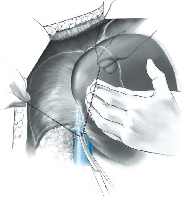
Once the arterial anatomy and possible arterial variations are clearly identified, the right |
|
|
||
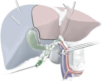
hepatic artery is divided at its origin. The remaining right hepatic artery is skeletonized more distally to encircle the right anterior and posterior branches at the right extremity of the hilar plate or the middle and left hepatic artery at Rex’s recess. The figure demonstrates a right predominant lesion and ligation of the right portal vein and segment 4 veins that is necessary. Also illustrated is the ligated right hepatic artery.
Bile Duct Resection |
577 |
|
|
STEP 6 |
Mobilization of the right hepatic lobe and caudate lobe with division of the short |
|
hepatic veins |
|
|
|
The right liver is mobilized and the entire short hepatic veins are divided between ties on |
|
the caval side and clips on the liver side from the right caudal side to the left cranial side. |
578 |
SECTION 4 |
Biliary Tract and Gallbladder |
|
|
|
STEP 7 |
Exposure and transection of the right hepatic vein |
|
|
A vessel loop is placed around the right hepatic vein, and vascular clamps are placed on |
|
|
||
|
the caval side and the liver side. The transection can be performed between the clamps. |
|
|
The caval side is secured by a running 4-0 Prolene suture and the other side with a 3-0 |
|
|
silk suture. An alternative technique is to transect the right hepatic vein with a vascular |
|
|
stapler (see the chapter on right hemihepatectomy). |
|
|
|
|
STEP 8 |
Demarcation and marking of the liver capsule |
|
|
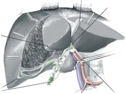
A stay suture is placed at the inferior margin of the ischemic side of the liver, and |
|
|
||
the liver capsule is incised with monopolar diathermy or bipolar scissors along the demarcation. At this point, central venous pressure (CVP) should be maintained below 3cm H2O.
In case of a right hepatectomy with caudate lobectomy, a transection line on the visceral surface of the liver is turned transversely from the Cantlie line about 1cm above the hilar plate to maintain a surgical margin and reach to the right edge
of Rex’s recess.
Bile Duct Resection |
579 |
|
|
STEP 9 |
Transection of the liver |
|
The liver dissection is started from the inferior margin under intermittent occlusion |
|
|
|
of the hepatic artery and the portal vein. The dissection is continued cranially |
|
and posteriorly, preserving the middle hepatic vein. |
