

clavien_atlas_of_upper_gastrointestinal_and_hepato-pancreato-biliary_surgery2007-10-01_3540200045_springer
.pdfOrthotopic Liver Transplantation |
477 |
|
|
Postoperative Tests
■Laboratory tests: liver tests, coagulation studies, kidney function, monitoring of levels of immunosuppressive drugs
■Doppler ultrasound: check for patency of the liver vessels
■Liver biopsy when rejection is clinically suspected
Postoperative Complications
■Early (<14days):
–Hepatic artery thrombosis or stenosis
–Primary non-function (death of patient without retransplantation)
–Initial poor function (definition center dependent)
–Acute rejection
–Massive ascites
–Budd-Chiari syndrome
–Portal vein thrombosis
–Biliary leakage
–Bleeding
■Intermediate/late (>14days):
–Acute or chronic rejection
–Abdominal infections
–Intra-abdominal abscess
–Infected ascites
–Systemic infections
–Viral [e.g., cytomegalovirus (CMV) or Epstein-Barr virus (EBV)]
–Fungal
–Bacterial
–Biliary complications
Intraand/or extrahepatic stenosis and/or sludge formation
–Vascular complications
–Hepatic artery thrombosis
–Portal vein thrombosis
–Venous outflow tract obstruction
–Recurrent disease
–Malignancies
–Post-transplant lymphoproliferative disorders
–Skin cancer

478 |
SECTION 3 |
Liver |
|
|
|
|
|
|
Management of the Most Common Complications |
|
|
|
■ |
Primary non-function (<5%): early retransplantation |
|
|
■ |
Rejection: increased immunosuppression |
|
|
■ |
Hepatic artery thrombosis: immediate surgical thrombectomy with or without intra- |
|
|
|
hepatic thrombolysis. If severe ischemic biliary strictures or hepatic necrosis develop, |
|
|
|
retransplantation should be considered |
|
|
■ |
Infectious complications should be treated as usual (e.g., surgical or percutaneous |
|
|
|
drainage and appropriate anti-infectious chemotherapy) |
|
|
■ |
Ascites/edema: Avoid fluid overload in the first week after transplantation, diuretics |
|
|
|
if necessary |
|
Tricks of the Senior Surgeon
■For both the classical approach and the cava-sparing technique, the central venous pressure should be kept as low as possible. Otherwise the dissection of the liver from the retrohepatic IVC and the dissection of the Spigelian veins is rendered more difficult, which leads to increased blood loss.
■A short period of hypotension is usually seen upon reperfusion of the graft. This can be due to bleeding or metabolic changes (i.e.,“post-reperfusion syndrome”). When venous bleeding occurs from the (retrohepatic) inferior vena cava or dorsal side of the liver, provide temporary packing for the retrohepatic space and wait for hemodynamic stabilization before attempting to place any sutures.
■In case of preexisting portal vein thrombosis, adapt donor and recipient operations to reduce ischemic time. Be assured, before starting the allograft implantation, of the method of portal revascularization. First make the arterial anastomosis and revascularize the liver via the artery. This will provide more time to remove clots and perform the anastomosis of the portal vein, without extending the cold or warm ischemia time.

Partial Cadaveric Liver Transplantation:
Donor Procedure and Implantation
Massimo Del Gaudio, Xavier Rogiers, Daniel Azoulay
In 1989 Pichlmayr et al. were the first to report a case of splitting a cadaveric liver for two recipients, an adult and a child. In the same year, Bismuth et al. performed the first transplantation of a single liver into two adult recipients with fulminant hepatitis. Today, donor livers are split for an adult and a pediatric recipient or, less frequently, for two adults.
Indications and Contraindications
General Donor Criteria |
■ |
Age <55years |
|
■ |
Weight >70kg |
|
■ |
Hemodynamic stability |
|
■ |
Normal liver function tests |
|
■ |
No macroscopic aspect of liver steatosis |
|
■ |
Graft-to-recipient body weight ratio >1% |
Donor Procedure: Ex-Situ Versus In-Situ Splitting
In general, the liver graft can be split either during the procurement procedure (i.e., in situ) or on the back-table after a conventional donor procedure (ex situ).
For ex-situ splitting of the liver, the whole organ is retrieved as described in the chapter “Technique of Multi-Organ Procurement.” Grafts are then prepared in the recipient transplant center. An alternative is the in-situ splitting technique, which is closely related to the techniques established for living related donor procurement.
Although the ex-situ split is the most widely used method to transplant two patients with one liver, extended cold ischemic time and some rewarming due to the longer backtable procedure as compared to conventional liver transplantation increase the risk of graft dysfunction in the recipient. While in-situ splitting potentially eliminates this problem, its application is limited due to a more time-consuming and technically more demanding explantation procedure.

480 SECTION 3 Liver
Back-Table Procedure for Ex-Situ Splitting
During back-table splitting, attention should be paid to keep the liver cold. After standard procurement of the liver graft, the presence of a portal bifurcation is checked by inserting a blunt metallic probe into the portal trunk. The anatomy of the hepatic artery and the bile duct is identified by dissection, probing or back-table X-ray with contrast medium.
The ultimate dissection of the portal vein, the hepatic artery, the biliary tree and the suprahepatic veins is performed on the back-table.
Partial Cadaveric Liver Transplantation: Donor Procedure and Implantation |
481 |
|
|
Procedures
|
In-Situ Split Liver Donor Procedure for an Adult and a Pediatric Recipient |
|
|
The goal of this procedure is to obtain the following grafts: |
|
|
■ |
Graft for adult recipient: Segments 4–8 |
|
■ |
Graft for pediatric recipient: Segments 2 and 3 |
|
|
|
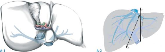
STEP 1 |
Mobilization and transection of the left lateral segments |
|
|
Segments 2 and 3 are mobilized and prepared in an identical manner as for a living |
|
|
||
|
donor procedure. The left hepatic artery and the left portal vein are isolated. Particular |
|
|
attention is paid to preserve the arterial branch to segment 4 whenever possible. The left |
|
hepatic vein is identified and controlled by placing a vessel loop around it to allow vessel-loop guided parenchymal transection (see the chapter “Hanging Maneuver for Right Hepatectomy”).
Optionally a cholangiography can be performed as shown in the chapters on living donor procedures.
Parenchymal transection is performed along the falciform ligament (resection line 1 in A-2). The hepatic veins are separated, the left hepatic vein remaining with the left graft, whereas the right and the middle hepatic veins remain with the right graft in continuity with the inferior vena cava.
Once the division of the parenchyma reaches the hilar plate it is cut straight with a scalpel slightly toward the left side, thus cutting the left hepatic duct blindly. This avoids unnecessary dissection of the bile duct which would compromise the biliary arterial blood supply.
Segment 1 is partially resected, with ligation of portal branches originating from the posterior aspect of the portal bifurcation and the hepatic veins draining into the inferior vena cava.
This will help the implantation of the right graft on the recipient’s inferior vena cava. The resection encompasses the left part of segment 1 and extends to the right side of
the IVC. Although the resection of the left part of segment 1 is not mandatory, it is recommended as it permits better exposure of the left hepatic vein during the implantation procedure.
In case of a too large right graft or when the perfusion of segment 4 is not optimal, segment 4 can be resected after implantation (resection line 2 in A-2).
482 |
SECTION 3 |
Liver |
|
|
|
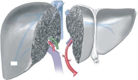
STEP 2 |
Procurement of the grafts |
|
|
|
|
Now the graft is prepared for procurement. Flushing of the organs is performed as in a standard multiorgan procurement. The left or right portal vein is cut at its origin and the orifice is closed with a 7-0 monofilament running suture. If the portal vein is to be cut at the origin of the left branch, the branches from the first centimeters of the left branch of the portal vein to segment 1 are secured between ligatures and divided in order to gain length and to allow for coaxial anastomosis to the recipient’s portal vein. In a standard situation, the artery is divided at the origin of the right hepatic artery, leaving the celiac trunk with the left graft. The left hepatic vein is divided and the left graft is removed and stored. Then the right graft is removed.
Partial Cadaveric Liver Transplantation: Donor Procedure and Implantation |
483 |
|
|
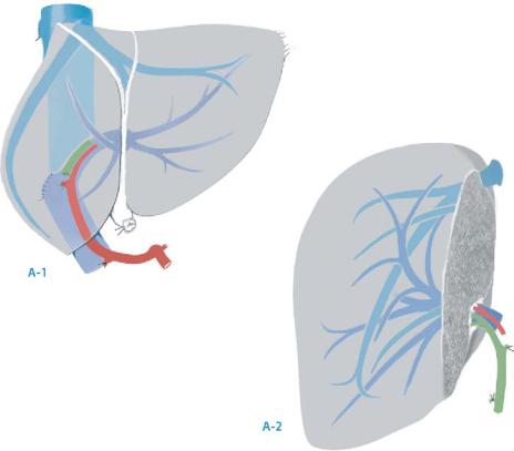
In-Situ Splitting for Two Adult Recipients
The goal of this procedure is to obtain the following grafts:
■Right graft: Segments 5–8
■Left graft: Segments 2–4
The hepatic dissection starts with complete mobilization of the right liver including isolation of the right hepatic vein, which is prepared for vessel loop guided parenchymal transection. Short accessory hepatic veins draining the right liver are preserved if they are larger than 5mm as they need to be anastomosed during implantation of the right graft.
The portal vein and the hepatic artery are prepared as described in the section on adult and pediatric split liver procedure. In a standard situation, the celiac axis remains with the left graft. Regarding portal bifurcation, the portal vein trunk is kept in continuity with the left graft. In case of portal trifurcation, the portal trunk is kept with the right graft. The plane of transection is to the right of the middle hepatic vein, so the whole of segment 4 is included in the left graft. This procedure yields two grafts as shown in A-1, A-2. In contrast to the split liver procedure for an adult and a pediatric recipient, in this situation the cava remains with the left graft.
484 |
SECTION 3 |
Liver |
|
|
|
|
Implantation |
|
|
Implantation of the Right Graft When the Liver is Split for an Adult |
|
|
and a Pediatric Recipient |
|
|
|
|
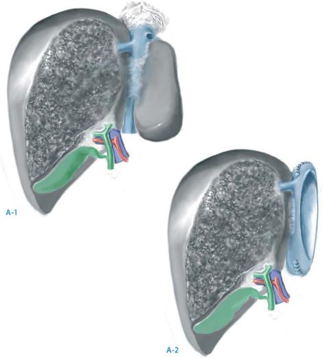
STEP 1 |
Preparation of the graft |
|
|
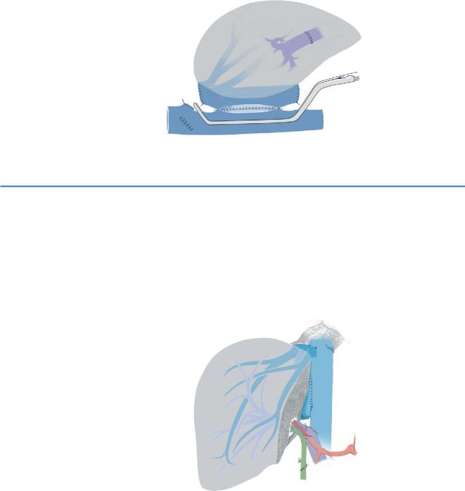
As in this situation the cava remains with the right graft, side-to-side cavocavostomy is |
|
|
||
possible and yields optimal allograft outflow. The preparation of the graft consists of resection of the upper and lower inferior vena cava cuffs at a level beneath the first major hepatic veins draining the residual part of Sg1. A 6-cm-long cavotomy at the right posterior side of the inferior vena cava encompasses the orifices of the major hepatic veins (A-1, A-2).
Partial Cadaveric Liver Transplantation: Donor Procedure and Implantation |
485 |
|
|
|
|
STEP 2 |
Side-to-side cavocavostomy |
|
|
|
|
A side-to-side cavocavostomy using partial clamping of the recipient inferior vena cava is performed (see also chapter “Orthotopic Liver Transplantation”). Resection of the retrocaval and left part of Sg1 during back-table procedure improves exposure and thereby easy anastomosis between the two caval veins. The same technique can be applied for the left graft in a procedure for two adult recipients, as in this situation the cava remains with the left liver.
STEP 3 |
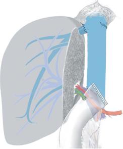
Portal vein, hepatic artery and bile duct anastomoses |
|
An end-to-end portal vein anastomosis is performed between the right branch of the |
|
|
|
portal vein of the graft and the common trunk of portal vein of the recipient. If long |
|
enough, the right hepatic artery is anastomosed to the hepatic artery of the recipient at |
|
the level of the bifurcation with the gastroduodenal artery. Otherwise it is anastomosed |
|
to the right or common hepatic artery. Finally, the right hepatic duct is connected to the |
|
common bile duct of the recipient applying the same technique as in orthotopic liver |
|
transplantation. |
486 |
SECTION 3 |
Liver |
|
|
|
Implantation of the Right Graft After Splitting for Two Adult Grafts or After Right Living Donor Procurement
Implantation of the left graft when the liver is split for two adult recipients is described in the chapter on left living donor transplantation.
In the situation of living donor procurement or split liver procedure for two adult recipients, the cava remains in the donor or with the left graft, respectively. Therefore, the venous outflow is reconstructed by anastomosing the donor’s hepatic veins to the recipient’s vena cava. The hepatectomy in the recipient is performed as for an orthotopic liver transplantation with preservation of the inferior vena cava. The orifices of the middle and left hepatic veins are oversewn or stapled; the right hepatic vein is directly anastomosed to the stump of the right hepatic vein or to a wider orifice in the recipient’s vena cava. Any inferior hepatic veins more than 5mm in diameter are also anastomosed directly to the inferior vena cava. A significant hepatic vein from segment 5 or 8 needs to be drained. This can be achieved by constructing a jump graft by means of a saphenous vein or, depending on the anatomic situation, by creating a common orifice with the right hepatic vein.
Anastomoses of the portal vein and the hepatic artery are performed as for the split graft for adult and pediatric recipients and the biliary continuity can be restored by biliodigestive anastomosis as shown in the Figure or by the direct connection between right hepatic duct of the right graft and common bile duct of the recipient.