

clavien_atlas_of_upper_gastrointestinal_and_hepato-pancreato-biliary_surgery2007-10-01_3540200045_springer
.pdf298 |
SECTION 2 |
Esophagus, Stomach and Duodenum |
|
|
|
STEP 3 |
Retrogastric preparation and band placement |
|
|
|
|
The retrogastric passage from right to left crus is created by blunt dissection using an articulating “finger” dissector which exits at the angle of His. However, the lesser sac should not be entered during this maneuver. If the lesser sac is entered, retrogastric passage is too far distal along the lesser curvature of the stomach.
The Silicone band is then pulled through the retrogastric tunnel from the left to right side of the stomach, encircling the proximal stomach, anterior vagus nerve, and upper part of the lesser omentum.
The Silastic band is locked around the inflated calibrating catheter and is now inflated with 15ml saline, which is positioned proximal to the band, thereby determining the size of the proximal pouch. The calibration catheter is deflated but left in place.
Operation for Morbid Obesity |
299 |
|
|
STEP 4 |
Band fixation |
|
The band is fixed in place along the anterior stomach by placing three to five interrupted |
|
|
|
seromuscular sutures of non-absorbable material, approximating the gastric wall proxi- |
|
mal and distal to the band. The left gastric wall distal to the band is fixed to the left crus |
|
of the diaphragm. |
|
The procedure is finished by implantation of the reservoir subcutaneously just below |
|
the xiphoid, allowing easy access to the port. |
300 |
SECTION 2 |
Esophagus, Stomach and Duodenum |
|
|
|
|
|
|
Standard Postoperative Investigations |
|
|
|
■ |
Postoperative surveillance in an intermediate care unit if the patient has sleep apnea |
|
|
|
or a history of severe cardiac disease |
|
|
■ |
Oral liquid diet after routine Gastrografin swallow radiography on the first or second |
|
|
|
postoperative day if the operative procedure is done laparoscopically |
|
|
■ |
Clinical and metabolic follow-up at 2 and 6weeks and at 3, 6, 9, and 12months and |
|
|
|
once yearly thereafter |
|
|
■ |
Multivitamin administration routinely, parenteral vitamin B12 after RYGB or BPD, |
|
|
|
and iron supplementation according to the blood tests |
|
Postoperative Complications
General
■Intra-abdominal bleeding
■Wound infection
■Abdominal wound dehiscence
■Deep vein thrombosis/pulmonary embolism
■Adhesive small bowel obstruction
Procedure-Specific
■RYGB:
–Anastomotic leakage
–Internal hernia
–Stenosis at gastrojejunostomy
–Stomal ulcer/stomal bleeding
–Afferent (pancreatobiliary) limb obstruction
■LAGB:
–Pouch dilatation or band slippage
–Reservoir infection
–Band migration into the stomach (late)
–Band leakage (late)
–Reservoir/band (balloon) dysfunction
■VBG:
–Stricture at the stoma
–Gastroesophageal reflux
–Staple line rupture
–Band erosion into stoma
–Maladaptive eating disorder

Operation for Morbid Obesity |
301 |
|
|
Tricks of the Senior Surgeon
■Creation of pneumoperitoneum with the Veress needle technique avoids a large incision for the first trocar with a subsequent annoying gas leak. The Veress needle should be placed just below (<1cm) the left costal margin; others prefer the Opti-View trocar.
■Generally a high placement of trocars is recommended, especially the upper two 10/12-mm trocars below the xiphoid and left costal arch.
■The circular EEA staplers for the laparoscopic approach are wrapped with a plastic cover to protect the abdominal wall incision from contact with the
contaminated outside of the cartridge of the stapler after intraluminal insertion and firing.
■Conservative weight reduction with dietary measures of 5–10kg before surgery is believed by some to facilitate technical performance by “shrinking liver size,” especially in superobese male patients.

Pancreas-Sparing Duodenectomy
Claus F. Eisenberger, Jakob R. Izbicki, Michael G. Sarr
Introduction
Pancreas-sparing duodenectomy (PSD) is reserved for premalignant lesions of the duodenum and the papilla of Vater, when local excision is not appropriate due to the size or multiplicity of the lesions. PSD involves complete or near complete resection of the duodenum with total preservation of the pancreas. Although the duodenum and the pancreas share the same blood supply, the duodenum may be resected without compromising the viability of the pancreas, but reinsertion of the bile and pancreatic duct into a “neoduodenum” is necessary.
Indications and Contraindications
Indications |
■ |
Multiple premalignant lesions of the duodenal mucosa and of the papilla of Vater |
|
|
(e.g., familial adenomatous polyposis syndrome) |
|
■ |
Localized benign or premalignant tumors of the duodenum |
|
|
Malignant disease |
Contraindications |
■ |
|
|
■ |
Previous surgical procedures of the duodenum, the stomach, or the pancreatic head |
Preoperative Investigations
■Gastroduodenoscopy with biopsy
■Endoscopic ultrasonography
■Endoscopic retrograde cholangiopancreatography (ERCP) [alternatively magnetic resonance cholangiopancreatography (MRCP)] for duct anatomy and morphology
304 |
SECTION 2 |
Esophagus, Stomach and Duodenum |
|
|
|
|
Procedure |
|
|
|
|
STEP 1 |
Exposure of the duodenum |
|
|
The abdomen is explored via a transverse or midline upper celiotomy. The gallbladder is |
|
|
||
|
resected, allowing later passage of a probe to localize the ampulla. The hepatic flexure of |
|
|
the colon is mobilized inferiorly and the lesser sac is opened widely. An extensive Kocher |
|
|
maneuver well to the left of the midline is performed, thus exposing the first through |
|
|
third portions of the duodenum. |
|
|
|
|
STEP 2 |
Preparation and resection of the duodenum |
|
|
|
|
The ligament of Treitz is incised. The proximal jejunum is then transected with a GIA stapler, after which the mesentery to the proximal jejunum and fourth portion of the duodenum is transected and ligated close to the bowel wall. The freed proximal jejunum is transposed behind the mesenteric root to the right upper abdomen.
The third and fourth portions of the duodenum are detached from the pancreas by meticulous dissection with ligation of these small and fragile mesenteric vessels. This dissection preparation is performed proximally up to the level of the papilla.
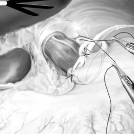
Precise localization of the papilla is important to facilitate dissection of the periampullary region. This is accomplished by passing a bile duct probe into the duodenum either via the cystic duct remnant (after cholecystectomy) or a choledochotomy. Retrograde cannulation of the bile duct with a probe via a lateral duodenotomy will also identify the papilla. In the region of the papilla of Vater, the duodenum is dissected carefully from the pancreas, thus exposing the estuaries of the common bile duct and the main pancreatic duct (A).
Next, the extraduodenal bile and pancreatic ducts are transected close to the duodenum. This step is shown in B. If a long common channel (pancreatic and bile ducts) is present and the duodenal disease does not extend past the ampullary region, this common channel can be transected, leaving only one “ductal structure” for reimplantation into the neoduodenum. The pancreatic and bile ducts are intubated separately with two catheters (C).
In the duodenum proximal to the papilla, the pancreas is densely adherent to the duodenum. One can dissect and develop a narrow subserosal plane outside the muscularis propria of the medial wall of the duodenum up to the distal part of the first part of the duodenum where a “duodenal mesentery” becomes present. Alternatively, dissection can also be initiated at the proximal duodenum and continued distally to the papilla. Small vessels are ligated. A careful search for the separate minor pancreatic duct should be made; if found and identified, we recommend suture ligation of this duct. Often, the duct is not identified and thus all vessels and “fibrous” connections to the second portion of the duodenum should be ligated.
The duodenum is transected either 1–2cm distal to the pylorus if this area is diseasefree or directly distal to pylorus. The resected specimen should be sent for pathologic examination with intraoperative frozen section to exclude the presence of invasive malignancy.
Pancreas-Sparing Duodenectomy |
305 |
|
|
STEP 2 (continued) |
Preparation and resection of the duodenum |
|
|
A
|
|
C |
|
B |
|||
|
|
306 |
SECTION 2 |
Esophagus, Stomach and Duodenum |
|
|
|
STEP 3 |
Reconstruction |
|
|
|
|
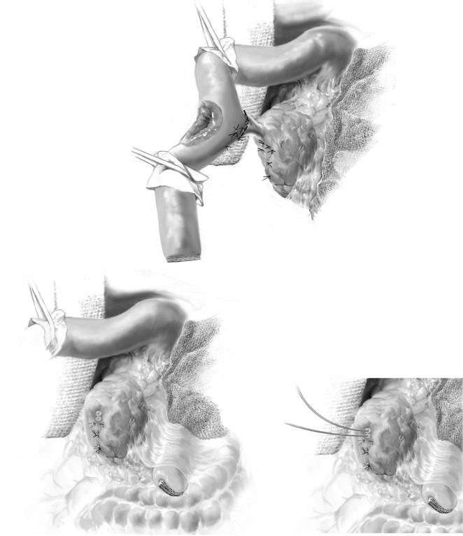
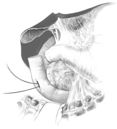
The proximal jejunum which will now become the “neoduodenum” is passed either behind the superior mesenteric vessels or retrorolically through the mesocolon.
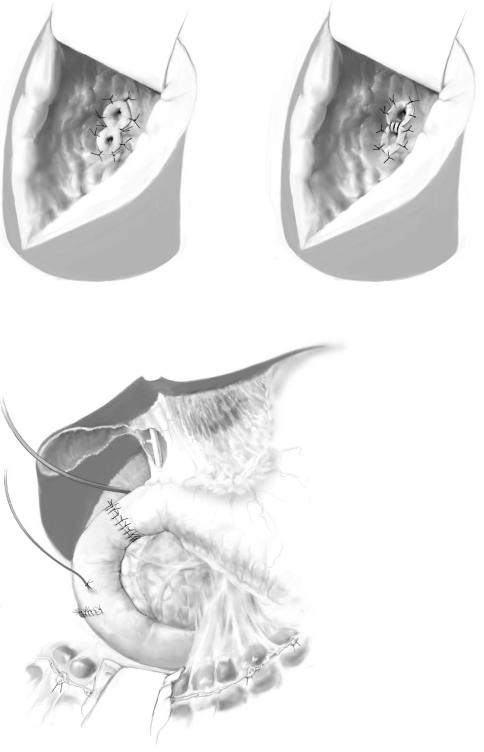
The bile and pancreatic duct anastomoses are performed at an equivalent distance from the pylorus as the native papilla. The ducts are implanted into the neoduodenum via a 2-cm enterotomy opposite to the proposed site of reimplantation. The anastomoses are done with interrupted transmural fine (6-0 or 7-0) monofilament resorbable sutures (A). Several techniques for ductal reimplantation should be available in the surgeon’s armamentarium. If a common channel can be preserved, one anastomosis will suffice. In contrast, if the disease involves the distal ducts and/or ampulla, usually the ductal transection leaves two ducts connected only by the interductal septum. In this situation, a single anastomosis can be fashioned by carefully including both ducts into the anastomosis (B-1). If the ductal transection leaves two individual ducts without preservation of the interductal septum (an unusual situation), the best approach is to sew together the adjoining walls of the ducts using 6- or 7-0 absorbable suture material and then reimplant the joined ductal structures as one anastomosis (B-2).
The pancreatic stent is passed through the neoduodenum via a hollow needle, thus creating a long submucosal tunnel, and is passed percutaneously through the abdominal wall. The access enterotomy is closed transversely, and the pancreatic stent is left in situ, to be removed 4–6weeks after surgery.
Gastrointestinal continuity is reestablished by end-to-end anastomosis of the neoduodenum with either the proximal duodenum or pylorus with a single layer of interrupted sutures. A t-tube is inserted in the common bile duct. Soft drains are placed behind the neoduodenum. The complete reconstruction is shown in C.
A
Pancreas-Sparing Duodenectomy |
307 |
|
|
STEP 3 (continued) |
Reconstruction |
|
|
B-1 |
|
B-2 |
C

308 |
SECTION 2 |
Esophagus, Stomach and Duodenum |
|
|
|
|
|
|
Standard Postoperative Investigations |
|
|
|
■ |
T-tube and pancreatic duct drain are left in-situ for 6weeks postoperatively |
|
|
■ |
T-tube cholangiography is performed prior to removal |
|
Postoperative Complications
■Pancreatic duct drain or t-tube dislocation
■Pancreatic and/or biliary fistula
■Anastomotic dehiscence
■Pancreatitis
■Cholangitis
■Anastomotic strictures
Tricks of the Senior Surgeon
■Fixation of the pancreatic duct drain by a resorbable stay suture at the “neo-papilla.”
■Tension-free anastomoses are mandatory; sometimes it is necessary to use an excluded jejunal interponate.