

clavien_atlas_of_upper_gastrointestinal_and_hepato-pancreato-biliary_surgery2007-10-01_3540200045_springer
.pdf214 |
SECTION 2 |
Esophagus, Stomach and Duodenum |
|
|
|
STEP 4 |
Transection of duodenum and resection of gastrohepatic ligament |
|
|
|
|
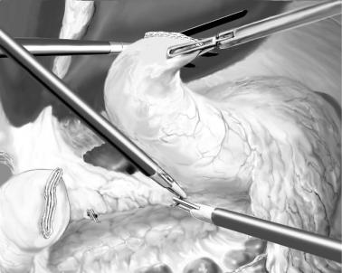
After detachment of the greater omentum, the right gastroepiploic vessels are identified and secured with clips at the level of the duodenum. Mayo’s vein will locate the exact position of the pylorus. Identification of the pylorus can be facilitated by gentle palpation with a clamp in the postpyloric area. Care should be taken not to damage
the pancreatic parenchyma as this will result in pancreatitis. Sharp dissection at the posterior side of the postpyloric part of the duodenum creates space to introduce a 45-mm stapling device. A vessel loop can be used to facilitate safe insertion of the stapler. Prior to the closure of the stapler, care should be taken that the vessel loop and vascular clips are not included in the line of stapling (A).
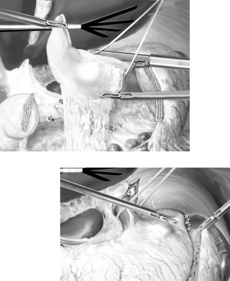
The assistant retracts the liver to allow exposure of the smaller omentum. The gastrohepatic ligament is opened at the level of the hepatoduodenal ligament. The right gastric artery is transected using Ultracision (B). The assistant retracts the liver to allow exposure of the liver hilum. Following the common, proper, and left hepatic artery, the smaller omentum is freed, securing lymph nodes of the pyloric group up to the right pericardial group. This en bloc lymphadenectomy is part of a level D2 resection and
is optional. A replaced or aberrant left hepatic artery, originating from the left gastric artery, can be safely dealt with, using clips if necessary. Alternatively this lymphadenectomy can be done after transection of the stomach.
Laparoscopic Gastrectomy |
215 |
|
|
STEP 5 |
Securing of left gastric vessels |
|
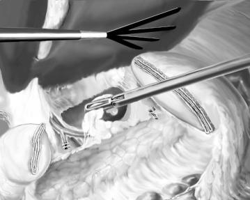
The posterior aspect of the stomach is freed from the anterior surface of the pancreas |
|
|
|
by sharp dissection of adhesions. At this stage a vessel loop can be used to allow easier |
|
manipulation of the stomach. Cranial to the pancreas the splenic artery is identified in |
|
most cases. More cranially, the left gastric vessels are identified and transected with |
|
clips or a vascular stapler. Optional D2 lymphadenectomy of the stomach implies |
|
truncal lymphadenectomy at this stage. |
216 |
SECTION 2 |
Esophagus, Stomach and Duodenum |
|
|
|
STEP 6 |
Transection of the stomach |
|
|
|
|
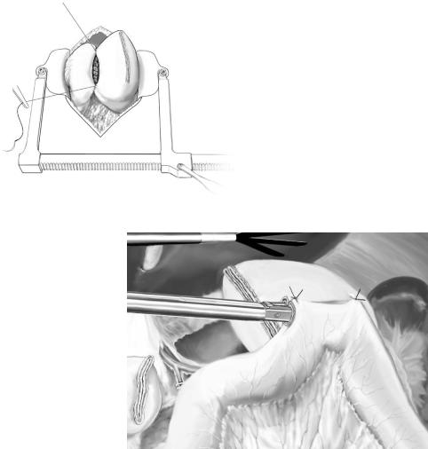
The transection line of the stomach is performed 5cm orally to the tumor (A). If the tumor cannot be identified adequately on the serosal side of the stomach, intraoperative gastroscopy is mandatory to determine the exact line of transection. Location of the tumor high in the body of the stomach may require opening of the gastrosplenic ligament and securing of short gastric vessels with Ultracision (B).
D2 lymphadenectomy requires resection of lymph nodes of the gastrohepatic ligament and between the hepatic artery. If the nodal clearance has not been performed en bloc, it is feasible to do it at this stage (C).
After transection of the stomach, the specimen is placed in a retrieval bag for safe extraction. Extraction is done through a mini-laparotomy. This laparotomy can be conducted at a cosmetically preferred site (e.g., Pfannenstiehl). Alternatively a midline mini-laparotomy is performed in the upper abdominal region. In the latter option the anastomosis can be done in an open fashion.
A
B
Laparoscopic Gastrectomy |
217 |
|
|
STEP 6 (continued) |
Transsection of the stomach |
|
|
C
218 |
SECTION 2 |
Esophagus, Stomach and Duodenum |
|
|
|
STEP 7 |
Anastomosis |
|
|
|
|
Open anastomosis:
Through a small midline laparotomy a standard Billroth II or Roux-en-Y reconstruction can be performed (A).
Laparoscopic anastomosis (Billroth II):
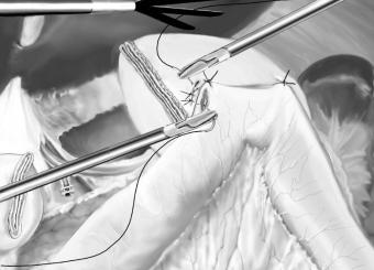
To perform a laparoscopic side-to-side gastrojejunostomy, ligament of Treitz and the proximal jejunum are identified by lifting the transverse colon and tilting the table into a Trendelenburg position (head down). A loop of proximal jejunum is brought up in an antecolic or retrocolic fashion. This loop of jejunum is sutured to the anterior aspect of the stomach remnant with two resorbable, seromuscular stay sutures, approximately 2cm apart. A stab incision in both the stomach and the jejunum is made with diathermia. Care should be taken that the incision in the stomach is made through all gastric wall layers. The stab incisions are enlarged, and the endostapler is introduced with one blade in the stomach and the other in the jejunum. Subsequently the stapler is fired one or two times, dependent on the size of the cartridges (60 or 45mm). In case a 60-mm stapler is used, a 15-mm trocar should be introduced (B).
A
B
Laparoscopic Gastrectomy |
219 |
|
|
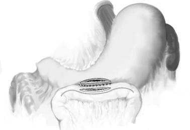
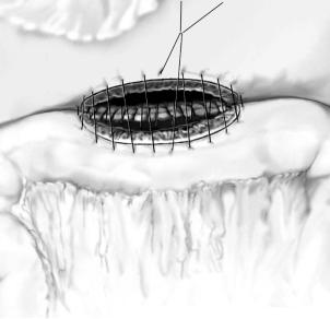
STEP 8 |
Closure of stab incisions |
|
The incision that remains in the stomach and the jejunum after firing the endostapler is |
|
|
|
closed using a single layer resorbable, polyfilament suture. Closure with an endostapler |
|
should not be attempted as the anastomosis is easily compromised because it is difficult |
|
to ensure inclusion of all tissue of the stomach and jejunum on both sides of the stab |
|
incisions in the staple line without narrowing the anastomosis. |

220 SECTION 2 Esophagus, Stomach and Duodenum
Postoperative Investigations
See chapter “Total Gastrectomy with Conventional Lymphadenectomy.”
Postoperative Complications
■Short term:
–Anastomotic insufficiency (including duodenal stump insufficiency)
–Acute pancreatitis
–Chylous ascites (particularly after R2 resection)
■Long term (all indications):
–Biliary gastritis (particularly after Billroth II reconstruction)
–Jejunal peptic ulcer disease
■Long term (in case of malignancy):
–Local recurrence (duodenal stump or resection line of stomach)
–Distant metastases
Tricks of the Senior Surgeon
■Instead of a vessel loop, a heavy resorbable suture can be used to force the stomach or duodenum into the endostapler. Even if this suture is included in the staple line, this will not compromise your anastomosis.
■In lean patients it is often possible to remove one of the 10to 12-mm trocars and directly introduce the 60-mm stapler or the retrieval bag, instead of using a 15-mm trocar.
■Suturing is best done with the scope in the middle and two needle holders on either side of the scope with a 60–90° angle between the two needle holders.

Gastroenterostomy
John Tsiaoussis, Gregory G. Tsiotos
Introduction
In this chapter both techniques for gastroenterostomy, the open and laparoscopic approaches, are described in patients with gastric outlet obstruction syndrome.
Indications and Contraindications
Indications |
■ |
Palliation of gastric outlet obstruction caused by advanced gastric, duodenal, |
|
|
or periampullary tumor |
|
■ |
Gastric drainage following vagotomy when pyloroplasty is not feasible |
|
|
Severe hypoalbuminemia |
Contraindications |
■ |
|
|
■ |
Evidence of diffuse metastatic spread, indicating extremely low life expectancy |
|
■ |
Prohibitive comorbidity |
Preoperative Investigation/Preparation for the Procedure
History: |
Persistent vomiting |
Laboratory tests: |
Electrolytes, albumin, coagulation parameters |
Radiology: |
Upper GI contrast study |
CT scan: |
Assessment of primary disease/condition |
Endoscopy: |
Assessment of gastric outlet obstruction, periampullary biopsy |
|
for tissue diagnosis |
A large-bore nasogastric tube is placed the day prior to the operation for gastric decompression and irrigation.
The patient’s water and electrolyte balance are corrected preoperatively.
222 |
SECTION 2 |
Esophagus, Stomach and Duodenum |
|
|
|
|
Procedure |
|
|
Open, Hand-Sewing Technique |
|
|
Access |
|
|
Midline incision from xiphoid process to the umbilicus |
|
|
|
|
STEP 1 |
Preparation of the jejunal loop using the retrocolic route |
|
|
The gastroepiploic vessels are dissected, clamped, divided and ligated starting about |
|
|
||
|
5cm proximal to the pylorus and moving 6–7cm proximally along the greater curvature |
|
|
of the stomach, so this is completely dissected free from the omentum. |
|
The jejunal loop can be brought either anterior to the transverse colon (antecolic),
or through a window in the transverse mesocolon (retrocolic).Although a retrocolic gastrojejunostomy has been considered more prone to obstruction because of its closer proximity to an ever enlarging unresectable periampullary tumor, this has never proved true, especially since patient survival in this context rarely exceeds 6months. On the other hand, the retrocolic route allows more proximal placement of the jejunal stoma and smoother angles between afferent, efferent loops and stomach in both the coronal and the sagittal planes.
The window (wide enough to allow comfortable sliding of both afferent and efferent jejunal loops) is made in an avascular plane of the mesocolon left to the middle colic vessels. The ligament of Treitz is identified by lifting up the transverse colon, and the jejunal loop is brought up through the mesocolic window in apposition to the greater curvature (now free from omental vessels). The length of the afferent jejunal limb should not exceed 20cm.
The gastrojejunostomy can be placed either on the anterior (easier and thus preferable) or the posterior gastric wall; the latter has not proved superior in terms of gastric emptying. Then 3-0 silk traction seromuscular sutures are placed, taking into account that the incision in the jejunum will not be made exactly at the antimesenteric border, but at a level closer to its mesentery on the stomal side. This provides for more comfortable lining of the completed anastomosis without any undue angles in the transverse plane. At 5-mm intervals 3-0 silk interrupted seromuscular Lembert sutures are placed and tied to create the posterior outer suture line. Two incisions along the gastric and the jejunal apposite segments are then made. Although the gastric incision should be about 4cm, the jejunal incision should be a bit shorter, since it always tends to dilate and ends up being realistically longer than initially planned or thought to be.
Gastroenterostomy |
223 |
|
|
STEP 2 |
Technique of anastomosis |
|
The posterior full-thickness inner anastomotic layer is made by two 3-0 PDS running |
|
|
|
sutures in an over-and-over fashion starting at the middle of the posterior layer and |
|
moving in opposite directions towards each corner of the anastomosis, where the two |
|
corner traction seromuscular stitches are still present. Then, the two 3-0 PDS running |
|
suture lines continue into the anterior wall of the gastrojejunostomy (again full-thick- |
|
ness) using the Connell technique in order to invert all gastric and jejunal mucosa, |
|
which might otherwise protrude out through the anastomosis. Moving from the two |
|
corners towards the middle, the sutures meet and are tied together. The anastomosis is |
|
completed by placing the anterior seromuscular layer with interrupted 3-0 silk Lembert |
|
sutures starting at the corner away from the surgeon and moving towards the surgeon, |
|
so that there are no sutures tangling in the middle of the operative field. These outer |
|
sutures should be first all placed and then tied; “tying as we go” will lead to packing of |
|
the serosa towards the inner suture line and thus placement of each successive suture at |
|
an ever increasing distance away from the inner suture line, which may then lead to |
|
entrapment of a lot of seromuscular tissue within the suture lines and protrusion of this |
|
soft tissue mass towards the anastomosis itself with its potential obliteration. |
|
After completion, the anastomosis is brought below the mesocolic window, and the |
|
gastric wall (not the jejunal) is tacked circumferentially on the mesocolon with inter- |
|
rupted 3-0 Vicryl sutures. A drain tube does not need to be placed. |