

clavien_atlas_of_upper_gastrointestinal_and_hepato-pancreato-biliary_surgery2007-10-01_3540200045_springer
.pdfAbdominothoracic En Bloc Esophagectomy with High Intrathoracic Anastomosis |
91 |
|
|
STEP 3 (continued)
|
|
B |
A |
||
|
|
|
C
D

92 |
SECTION 2 |
Esophagus, Stomach and Duodenum |
|
|
|
|
See the transhiatal approach for standard postoperative investigations and complicatons. |
|
Tricks of the Senior Surgeon
■An additional length of the gastric tube is achieved by mobilization of the duodenum (Kocher maneuver). In this way, the end of the gastric tube designated for the anastomosis is located closer to the gastroepiploic pedicle. The improved vascular supply reduces the risk of anastomotic leakage.
■In contrast to cervical esophagogastric anastomosis, in any case of suspected intrathoracic leakage, emergency endoscopy should be done. Even if an insufficiency cannot be definitely visualized, the indication for stenting should be established generously as long as clinical signs suggest a leakage to prevent catastrophic mediastinitis.

Limited Resection of the Gastroesophageal Junction with Isoperistaltic Jejunal Interposition
Asad Kutup, Emre F. Yekebas, Jakob R. Izbicki
Introduction
Limited en bloc resection of the gastroesophageal junction includes complete removal of the esophageal segment with metaplastic mucosa, the lower esophageal sphincter and a part of the lesser gastric curvature and formation of a neofundus. Since even early adenocarcinomas of the distal esophagus (T1b) seed lymph node metastases in up to 20% of patients, removal of the lymph nodes of the lesser curvature, the hepatic and splenic arteries, the celiac trunk, the para-aortal region, and the inferior mediastinum is an essential part of the operation.
In patients with early tumors, staged as uT1a or b on preoperative endosonography or severe dysplasia in the distal esophagus (Barrett’s esophagus), a limited resection of the proximal stomach, cardia and distal esophagus with interposition of a pedicled isoperistaltic jejunal segment offers excellent functional and oncological results.
Indications and Contraindications
Indications |
■ |
Severe dysplasia in the distal esophagus (Barrett’s esophagus) |
|
■ |
Distal adenocarcinoma of the esophagus (stage T1a and b) (UICC 2005) |
|
■ |
For palliative reasons (stenotic tumor with severe dysphagia or profuse |
|
|
hemorrhage in selective patients) |
|
|
Esophageal carcinoma staged T2 and more |
Contraindications |
■ |
|
|
■ |
Long Barrett’s segment above the carina |
Preoperative Investigations/Preparation for the Procedure
■Esophagogastroscopy with extensive biopsies
■Endosonography of the esophagus
■Computed tomography of the chest and abdomen
■Abdominal ultrasound
■Pulmonary function test
■Orthograde cleansing of the intestines
Positioning
■ Supine position with hyperlordosis
94 |
SECTION 2 |
Esophagus, Stomach and Duodenum |
|
|
|
|
|
|
Procedure |
|
|
|
Access |
|
|
|
■ |
Upper transverse incision with median T-shaped |
|
|
■ |
Insertion of Rochard retractor to elevate costal margin |
|
|
|
||
STEP 1 |
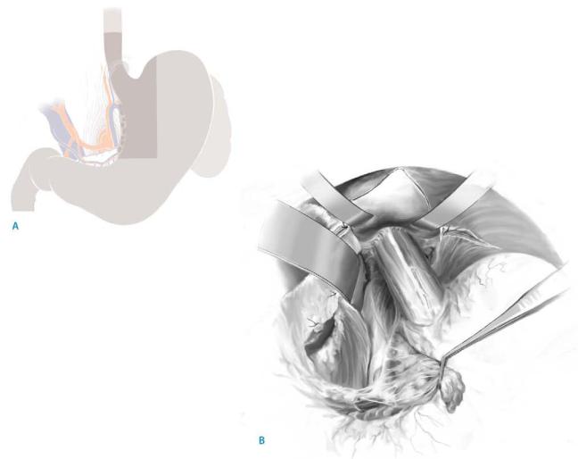
Exposure of the inferior posterior mediastinum; diaphanoscopy |
||
|
The left liver lobe is completely mobilized and the lesser omentum is incised just medial |
||
|
|||
|
to the anterior and posterior gastric vagal branches. A longitudinal median diaphragmal |
||
|
incision enables exposure of the inferior posterior mediastinum. The distal esophagus is |
||
|
then mobilized including the paraesophageal tissue. The vagal nerves are divided. |
||
|
|
Intraoperative esophagoscopy identifies the cranial limit of the Barrett’s segment by |
|
|
diaphanoscopy. This also marks the proximal limit of resection. |
||
|
|
A lymphadenectomy around the splenic and hepatic artery is performed, the left |
|
|
gastric vein is divided, and the left gastric artery is divided at the celiac trunk. Then the |
||
|
celiac trunk and the para-aortic region above the celiac trunk are cleared from |
||
|
lymphatic tissue (A, B). |
|
|

Limited Resection of the Gastroesophageal Junction with Isoperistaltic Jejunal Interposition |
95 |
|
|
|
|
STEP 2 |
Transection of the esophagus |
|
|
|
|
Approximately 1cm proximal to the cranial limit of the Barrett’s segment, a pursestring clamp is placed and the esophagus is divided.
Removal of the cardia and lesser curvature is performed by placing multiple linear staplers down to the border between antrum and body. Thus, a neofundus is formed.
In case an advanced tumor stage is encountered, possible extension of the operation including transhiatal esophagectomy or esophagogastrectomy should be performed.

96 |
SECTION 2 |
Esophagus, Stomach and Duodenum |
|
|
|
STEP 3 |
Transposition of the proximal jejunum segment; esophagojejunostomy |
|
|
|
|
A 15to 20-cm-long segment of the proximal jejunum is isolated and is transposed with its mesenteric root to the diaphragmatic region through the mesocolon and behind the stomach. Care has to be taken while dissecting the vascular pedicle of this jejunal interposition to provide adequate length. It is imperative to form an isoperistaltic jejunal interposition which should be pulled up retrogastric and retrocolic. The proximal anastomosis is then performed by a circular stapling device as a terminolateral esophagojejunostomy. The stapler is introduced into the end of the jejunal interponate (A-1).
After firing of the anastomosis, the blind end of the loop is then resected and closed by a linear stapler and then oversewn (A-2).
A-1
A-2

Limited Resection of the Gastroesophageal Junction with Isoperistaltic Jejunal Interposition |
97 |
|
|
|
|
STEP 4 |
Jejunogastrostomy |
|
|
|
|
Close to the base of the neofundus the gastric stapler line is removed over a distance of 3–4cm and a terminolateral or laterolateral jejunogastrostomy is performed.
The remaining gastric suture line is oversewn. A terminoterminal jejunojejunostomy reconstructs the enteric passage. Drainage of the mediastinum is warranted by two soft drains from the abdomen. Finally an anterior and/or posterior hiatal repair
is performed.

98 SECTION 2 Esophagus, Stomach and Duodenum
Standard Postoperative Investigations
■ Daily check the drains for insufficiency of the intrathoracic esophagojejunostomy
Postoperative Complications
■Insufficiency of the esophagojejunostomy or the intra-abdominal anastomosis
■Mediastinitis
■Pancreatitis
■Peritonitis
■Pleural empyema
■Necrosis of the transposed jejunal segment
■Reflux
■Delayed gastric emptying
Tricks of the Senior Surgeon
■Intraoperative endoscopy in any case of long-segment irregularities or evidence of multicentric lesions.
■Insertion of the stapler device through the oral end of the pedicled jejunal segment.

Three-Field Lymphadenectomy for Esophageal Cancer
Masamichi Baba, Shoji Natsugoe, Takashi Aikou
Introduction
Lymphatic drainage from the upper two-thirds of the thoracic esophagus occurs mainly towards the neck and upper mediastinum, although there is also some drainage to the nodes along the left gastric artery. In 1981, the first reported study of three-field lymphadenectomy in Japan noted that 10 of 36 patients with esophagectomy had skip metastases to the neck or abdominal lymph nodes in the absence of associated intrathoracic spread. In this chapter, we focus on the lymph node dissection of the upper mediastinal and cervical regions.
Indications and Contraindications
Indications |
■ |
Tumors of the supracarinal esophagus (>T1m stage) |
|
|
Superficial carcinoma (T1m stage) |
Contraindications |
■ |
|
|
■ |
Severe comorbidity (heart disease, pulmonary and/or liver dysfunction) |
|
■ |
No evidence of cervical lymph node metastases preoperatively in high risk patients |
|
|
(relative) |
|
■ |
Infracarinal tumors (relative) |
Preoperative Investigation/Preparation for the Procedure
■See transhiatal approach
■US+CT scan of the neck
■In locally advanced tumors: primary radiochemotherapy and surgery is done secondarily
Procedure
Access
■Anterior-lateral thoracotomy through the right 5th ICS
■Supine position and T-shaped incision in the neck

100 |
SECTION 2 |
Esophagus, Stomach and Duodenum |
|
|
|
STEP 1 |
Exposure and lymphadenectomy of the right upper mediastinum |
|
|
|
|
The arch of the azygous vein is resected, and the right bronchial artery is ligated and secured at its root to evaluate the tumor for its resectability. This procedure provides a good exposure of the upper and middle mediastinum. The brachiocephalic and right subclavian arteries are exposed in order to remove the right recurrent nerve nodes and right paratracheal nodes followed by carefully ligating the branches of the inferior thyroid artery (arrow indicates the direction of lymphadenectomy).