

Психология
.pdfAn advantage of the cadaver approach is that the brains can be fully studied, but an obvious disadvantage is that the brains are no longer active. In other cases, however, we can study living brains. The brains of living human beings may be damaged, for instance, as a result of strokes, falls, automobile accidents, gunshots, or tumors. These damages are called lesions. In rare occasions, brain lesions may be created intentionally through surgery, such as that designed to remove brain tumors or (as in split-brain patients) to reduce the effects of epilepsy. Psychologists also sometimes intentionally create lesions in animals to study the effects on their behavior. In so doing, they hope to be able to draw inferences about the likely functions of human brains from the effects of the lesions in animals.
Lesions allow the scientist to observe any loss of brain function that may occur. For instance, when an individual suffers a stroke, a blood clot deprives part of the brain of oxygen, killing the neurons in the area and rendering that area unable to process information. In some cases, the result of the stroke is a specific lack of ability. For instance, if the stroke influences the occipital lobe, then vision may suffer, and if the stroke influences the areas associated with language or speech, these functions will suffer. In fact, our earliest understanding of the specific areas involved in speech and language were gained by studying patients who had experienced strokes.
It is now known that a good part of our moral reasoning abilities are located in the frontal lobe, and at least some of this understanding comes from lesion studies. For instance, consider the well-known case of Phineas Gage, a 25-year-old railroad worker who, as a result of an explosion, had an iron rod driven into his cheek and out through the top of his skull, causing major damage to his frontal lobe (Macmillan, 2000). [2] Although remarkably Gage was able to return to work after the wounds healed, he no longer seemed to be the same person to those who knew him. The amiable, soft-spoken Gage had become irritable, rude, irresponsible, and dishonest. Although there are questions about the interpretation of this case study (Kotowicz, 2007),[3] it did provide early evidence that the frontal lobe is involved in emotion and morality (Damasio et al., 2005). [4]
More recent and more controlled research has also used patients with lesions to investigate the source of moral reasoning. Michael Koenigs and his colleagues (Koenigs et al., 2007) [5] asked
Attributed to Charles Stangor |
Saylor.org |
Saylor URL: http://www.saylor.org/books/ |
141 |
groups of normal persons, individuals with lesions in the frontal lobes, and individuals with lesions in other places in the brain to respond to scenarios that involved doing harm to a person, even though the harm ultimately saved the lives of other people (Miller, 2008). [6]
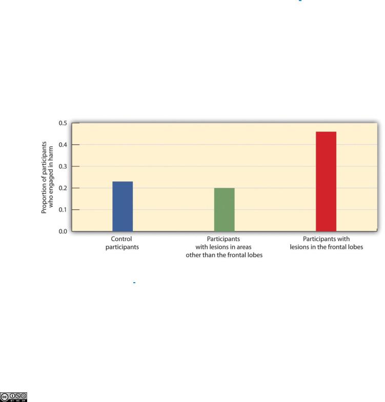
In one of the scenarios the participants were asked if they would be willing to kill one person in order to prevent five other people from being killed. As you can see in Figure 3.14 "The Frontal Lobe and Moral Judgment", they found that the individuals with lesions in the frontal lobe were significantly more likely to agree to do the harm than were individuals from the two other groups.
Figure 3.14 The Frontal Lobe and Moral Judgment
Koenigs and his colleagues (2007) [7] found that the frontal lobe is important in moral judgment. Persons with lesions in the frontal lobe were more likely to be willing to harm one person in order to save the lives of five others than were control participants or those with lesions in other parts of the brain.
Recording Electrical Activity in the Brain
In addition to lesion approaches, it is also possible to learn about the brain by studying the electrical activity created by the firing of its neurons. One approach, primarily used with animals,
Attributed to Charles Stangor |
Saylor.org |
Saylor URL: http://www.saylor.org/books/ |
142 |

is to place detectors in the brain to study the responses of specific neurons. Research using these techniques has found, for instance, that there are specific neurons, known as feature detectors, in the visual cortex that detect movement, lines and edges, and even faces (Kanwisher, 2000). [8]
A less invasive approach, and one that can be used on living humans, is electroencephalography (EEG). The EEG is a technique that records the electrical activity produced by the brain’s neurons through the use of electrodes that are placed around the research participant’s head. An EEG can show if a person is asleep, awake, or anesthetized because the brain wave patterns are known to differ during each state. EEGs can also track the waves that are produced when a person is reading, writing, and speaking, and are useful for understanding brain abnormalities, such as epilepsy. A particular advantage of EEG is that the participant can move around while the recordings are being taken, which is useful when measuring brain activity in children who often have difficulty keeping still. Furthermore, by following electrical impulses across the surface of the brain, researchers can observe changes over very fast time periods.
Peeking Inside the Brain: Neuroimaging
Although the EEG can provide information about the general patterns of electrical activity within the brain, and although the EEG allows the researcher to see these changes quickly as they occur in real time, the electrodes must be placed on the surface of the skull and each electrode measures brain waves from large areas of the brain. As a result, EEGs do not provide a very clear picture of the structure of the brain.
But techniques exist to provide more specific brain images.
Functional magnetic resonance imaging (fMRI) is a type of brain scan that uses a magnetic field to create images of brain activity in each brain area. The patient lies on a bed within a large cylindrical structure containing a very strong magnet. Neurons that are firing use more oxygen, and the need for oxygen increases blood flow to the area. The fMRI detects the amount of blood flow in each brain region, and thus is an indicator of neural activity.
Attributed to Charles Stangor |
Saylor.org |
Saylor URL: http://www.saylor.org/books/ |
143 |

Very clear and detailed pictures of brain structures (see, e.g., Figure 3.16 "fMRI Image") can be produced via fMRI. Often, the images take the form of cross-sectional “slices‖ that are obtained as the magnetic field is passed across the brain. The images of these slices are taken repeatedly and are superimposed on images of the brain structure itself to show how activity changes in different brain structures over time. When the research participant is asked to engage in tasks while in the scanner (e.g., by playing a game with another person), the images can show which parts of the brain are associated with which types of tasks. Another advantage of the fMRI is that is it noninvasive. The research participant simply enters the machine and the scans begin.
Although the scanners themselves are expensive, the advantages of fMRIs are substantial, and they are now available in many university and hospital settings. fMRI is now the most commonly used method of learning about brain structure.
There is still one more approach that is being more frequently implemented to understand brain function, and although it is new, it may turn out to be the most useful of
all. Transcranial magnetic stimulation (TMS) is a procedure in which magnetic pulses are applied to the brain of living persons with the goal of temporarily and safely deactivating a small brain region. In TMS studies the research participant is first scanned in an fMRI machine to determine the exact location of the brain area to be tested. Then the electrical stimulation is provided to the brain before or while the participant is working on a cognitive task, and the effects of the stimulation on performance are assessed. If the participant‘s ability to perform the task is influenced by the presence of the stimulation, then the researchers can conclude that this particular area of the brain is important to carrying out the task.
The primary advantage of TMS is that it allows the researcher to draw causal conclusions about the influence of brain structures on thoughts, feelings, and behaviors. When the TMS pulses are applied, the brain region becomes less active, and this deactivation is expected to influence the research participant’s responses. Current research has used TMS to study the brain areas responsible for emotion and cognition and their roles in how people perceive intention and
Attributed to Charles Stangor |
Saylor.org |
Saylor URL: http://www.saylor.org/books/ |
144 |

approach moral reasoning (Kalbe et al., 2010; Van den Eynde et al., 2010; Young, Camprodon, Hauser, Pascual-Leone, & Saxe, 2010). [9] TMS is also used as a treatment for a variety of psychological conditions, including migraine, Parkinson’s disease, and major depressive disorder.
Research Focus: Cyberostracism
Neuroimaging techniques have important implications for understanding our behavior, including our responses to those around us. Naomi Eisenberger and her colleagues (2003) [10] tested the hypothesis that people who were excluded by others would report emotional distress and that images of their brains would show that they experienced pain in the same part of the brain where physical pain is normally experienced. In the experiment, 13 participants were each placed into an fMRI brain-imaging machine. The participants were told that they would be playing a computer ―Cyberball‖ game with two other players who were also in fMRI machines (the two opponents did not actually exist, and their responses were controlled by the computer).
Each of the participants was measured under three different conditions. In the first part of the experiment, the participants were told that as a result of technical difficulties, the link to the other two scanners could not yet be made, and thus at first they could not engage in, but only watch, the game play. This allowed the researchers to take a baseline fMRI reading. Then, during a second inclusion scan, the participants played the game, supposedly with the two other players. During this time, the other players threw the ball to the participants. In the third, exclusion, scan, however, the participants initially received seven throws from the other two players but were then excluded from the game because the two players stopped throwing the ball to the participants for the remainder of the scan (45 throws). The results of the analyses showed that activity in two areas of the frontal lobe was significantly greater during the exclusion scan than during the inclusion scan. Because these brain regions are known from prior research to be active for individuals who are experiencing physical pain, the authors concluded that these results show that the physiological brain responses associated with being socially excluded by others are similar to brain responses experienced upon physical injury.
Further research (Chen, Williams, Fitness, & Newton, 2008; Wesselmann, Bagg, & Williams, 2009) [11] has documented that people react to being excluded in a variety of situations with a variety of emotions and behaviors. People who feel that they are excluded, or even those who observe other people being excluded, not only experience
Attributed to Charles Stangor |
Saylor.org |
Saylor URL: http://www.saylor.org/books/ |
145 |

pain, but feel worse about themselves and their relationships with people more generally, and they may work harder
to try to restore their connections with others.
K EY TA KEA WAY S
Studying the brains of cadavers can lead to discoveries about brain structure, but these studies are limited due to the fact that the brain is no longer active.
Lesion studies are informative about the effects of lesions on different brain regions.
Electrophysiological recording may be used in animals to directly measure brain activity.
Measures of electrical activity in the brain, such as electroencephalography (EEG), are used to assess brain-wave patterns and activity.
Functional magnetic resonance imaging (fMRI) measures blood flow in the brain during different activities, providing information about the activity of neurons and thus the functions of brain regions.
Transcranial magnetic stimulation (TMS) is used to temporarily and safely deactivate a small brain region, with the goal
of testing the causal effects of the deactivation on behavior.
EXE RCIS E AND CRI TICA L THINKING
1.Consider the different ways that psychologists study the brain, and think of a psychological characteristic or behavior that could be studied using each of the different techniques.
[1]Diamond, M. C. (1999). Why Einstein’s brain? New Horizons for Learning. Retrieved
from http://www.newhorizons.org/neuro/diamond_einstein.htm
[2]Macmillan, M. (2000). An odd kind of fame: Stories of Phineas Gage. Cambridge, MA: MIT Press.
[3]Kotowicz, Z. (2007). The strange case of Phineas Gage. History of the Human Sciences, 20(1), 115–131.
[4]Damasio, H., Grabowski, T., Frank, R., Galaburda, A. M., Damasio, A. R., Cacioppo, J. T., & Berntson, G. G. (2005). The return of Phineas Gage: Clues about the brain from the skull of a famous patient. In Social neuroscience: Key readings (pp. 21–28). New York, NY: Psychology Press.
[5]Koenigs, M., Young, L., Adolphs, R., Tranel, D., Cushman, F., Hauser, M., & Damasio, A. (2007). Damage to the prefontal cortex increases utilitarian moral judgments. Nature, 446(7138), 908–911.
[6]Miller, G. (2008). The roots of morality. Science, 320, 734–737.
Attributed to Charles Stangor |
Saylor.org |
Saylor URL: http://www.saylor.org/books/ |
146 |

[7]Koenigs, M., Young, L., Adolphs, R., Tranel, D., Cushman, F., Hauser, M., & Damasio, A. (2007). Damage to the prefontal cortex increases utilitarian moral judgments. Nature, 446(7138), 908–911.
[8]Kanwisher, N. (2000). Domain specificity in face perception. Nature Neuroscience, 3(8), 759–763.
[9]Kalbe, E., Schlegel, M., Sack, A. T., Nowak, D. A., Dafotakis, M., Bangard, C.,…Kessler, J. (2010). Dissociating cognitive from affective theory of mind: A TMS study. Cortex: A Journal Devoted to the Study of the Nervous System and Behavior, 46(6), 769–
780; Van den Eynde, F., Claudino, A. M., Mogg, A., Horrell, L., Stahl, D.,…Schmidt, U. (2010). Repetitive transcranial magnetic stimulation reduces cue-induced food craving in bulimic disorders. Biological Psychiatry, 67(8), 793–795; Young, L., Camprodon, J. A., Hauser, M., Pascual-Leone, A., & Saxe, R. (2010). Disruption of the right temporoparietal junction with transcranial magnetic stimulation reduces the role of beliefs in moral judgments. PNAS Proceedings of the National Academy of Sciences of the United States of America, 107(15), 6753–6758.
[10]Eisenberger, N. I., Lieberman, M. D., & Williams, K. D. (2003). Does rejection hurt? An fMRI study of social exclusion. Science, 302(5643), 290–292.
[11]Chen, Z., Williams, K. D., Fitness, J., & Newton, N. C. (2008). When hurt will not heal: Exploring the capacity to relive social and physical pain. Psychological Science, 19(8), 789–795; Wesselmann, E. D., Bagg, D., & Williams, K. D. (2009). “I feel your
pain”: The effects of observing ostracism on the ostracism detection system. Journal of Experimental Social Psychology, 45(6),
1308–1311.
Attributed to Charles Stangor |
Saylor.org |
Saylor URL: http://www.saylor.org/books/ |
147 |

3.4 Putting It All Together: The Nervous System and the Endocrine System
LE ARNING OB JECT I VE S
1.Summarize the primary functions of the CNS and of the subsystems of the PNS.
2.Explain how the electrical components of the nervous system and the chemical components of the endocrine system work together to influence behavior.
Now that we have considered how individual neurons operate and the roles of the different brain areas, it is time to ask how the body manages to “put it all together.‖ How do the complex activities in the various parts of the brain, the simple all-or-nothing firings of billions of interconnected neurons, and the various chemical systems within the body, work together to allow the body to respond to the social environment and engage in everyday behaviors? In this section we will see that the complexities of human behavior are accomplished through the joint actions of electrical and chemical processes in the nervous system and the endocrine system.
Electrical Control of Behavior: The Nervous System
The nervous system (see Figure 3.17 "The Functional Divisions of the Nervous System"), the electrical information highway of the body, is made up ofnerves—bundles of interconnected neurons that fire in synchrony to carry messages. The central nervous system (CNS), made up of the brain and spinal cord, is the major controller of the body’s functions, charged with interpreting sensory information and responding to it with its own directives. The CNS interprets information coming in from the senses, formulates an appropriate reaction, and sends responses to the appropriate system to respond accordingly. Everything that we see, hear, smell, touch, and taste is conveyed to us from our sensory organs as neural impulses, and each of the commands that the brain sends to the body, both consciously and unconsciously, travels through this system as well.
Attributed to Charles Stangor |
Saylor.org |
Saylor URL: http://www.saylor.org/books/ |
148 |
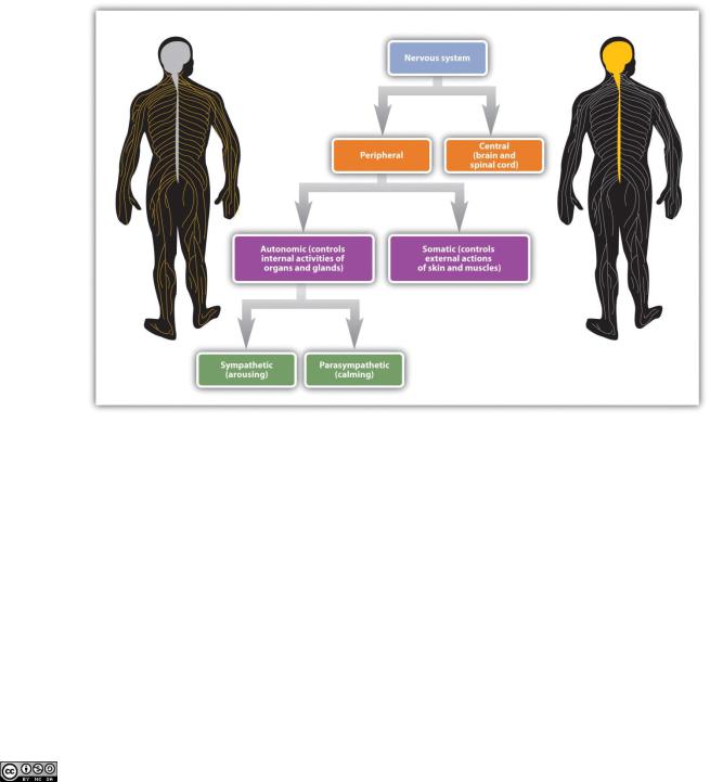
Figure 3.17 The Functional Divisions of the Nervous System
Nerves are differentiated according to their function. A sensory (or afferent) neuron carries information from the sensory receptors, whereas a motor (or efferent) neuron transmits information to the muscles and glands. An interneuron, which is by far the most common type of neuron, is located primarily within the CNS and is responsible for communicating among the neurons. Interneurons allow the brain to combine the multiple sources of available information to create a coherent picture of the sensory information being conveyed.
The spinal cord is the long, thin, tubular bundle of nerves and supporting cells that extends down from the brain. It is the central throughway of information for the body. Within the spinal cord, ascending tracts of sensory neurons relay sensory information from the sense organs to the brain
Attributed to Charles Stangor |
Saylor.org |
Saylor URL: http://www.saylor.org/books/ |
149 |
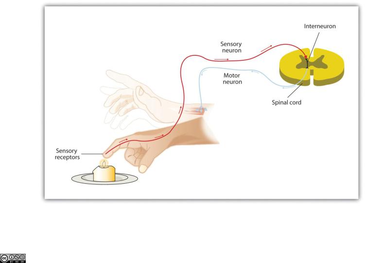
while descending tracts of motor neurons relay motor commands back to the body. When a quicker-than-usual response is required, the spinal cord can do its own processing, bypassing the brain altogether. A reflex is an involuntary and nearly instantaneous movement in response to a stimulus. Reflexes are triggered when sensory information is powerful enough to reach a given threshold and the interneurons in the spinal cord act to send a message back through the motor neurons without relaying the information to the brain (see Figure 3.18 "The Reflex"). When you touch a hot stove and immediately pull your hand back, or when you fumble your cell phone and instinctively reach to catch it before it falls, reflexes in your spinal cord order the appropriate responses before your brain even knows what is happening.
Figure 3.18 The Reflex
Attributed to Charles Stangor |
Saylor.org |
Saylor URL: http://www.saylor.org/books/ |
150 |