Garrett R.H., Grisham C.M. - Biochemistry (1999)(2nd ed.)(en)
.pdf12.3 ● Denaturation and Renaturation of DNA |
371 |
Sar = Sarcosine = H3C |
N |
CH2 |
COOH (N-Methylglycine) |
||
|
H |
|
|
|
|
|
|
|
CH3 |
|
|
Meval = Mevalonic acid = HOCH2 |
CH2 C |
CH2 |
COOH |
||
|
|
|
OH |
|
|
B-DNA before |
|
|
Intercalating agents |
B-DNA after |
|
intercalation |
|
|
|
|
intercalation |
|
|
H2N |
|
|
NH2 |
|
|
|
+ |
Br– |
|
|
|
|
N |
||
|
|
|
|
CH2CH3 |
|
|
|
|
Ethidium bromide |
|
|
|
|
|
or |
|
|
|
|
|
+ |
|
|
+ |
(CH3)2N |
N |
|
N(CH3)2 |
|
H |
|
||||
|
|
|
Acridine orange |
|
|
|
|
|
or |
|
|
|
|
Sarcosine |
|
Sarcosine |
|
|
Pro |
L-Meval |
Pro |
L-Meval |
|
D-Val |
|
O |
D-Val |
O |
|
|
|
Thr |
O |
|
Thr |
|
|
|
|
|
|
|
|
C |
|
C |
|
|
|
O |
N |
NH2 |
|
O O
CH3 CH3
Actinomycin D
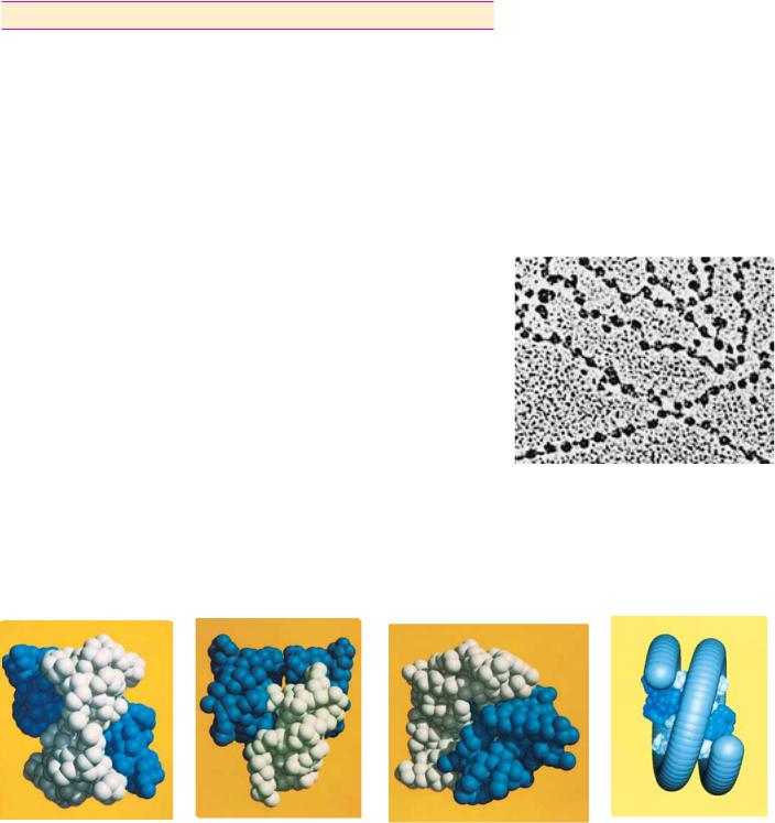
FIGURE 12.16 ● The structures of ethidium bromide, acridine orange, and actinomycin
D, three intercalating agents, and their effects on DNA structure.
12.3 ● Denaturation and Renaturation of DNA
Thermal Denaturation and Hyperchromic Shift
When duplex DNA molecules are subjected to conditions of pH, temperature, or ionic strength that disrupt hydrogen bonds, the strands are no longer held together. That is, the double helix is denatured and the strands separate as individual random coils. If temperature is the denaturing agent, the double helix is said to melt. The course of this dissociation can be followed spectrophotometrically because the relative absorbance of the DNA solution at 260 nm increases as much as 40% as the bases unstack. This absorbance increase, or hyperchromic shift, is due to the fact that the aromatic bases in DNA interact via their electron clouds when stacked together in the double helix. Because the UV absorbance of the bases is a consequence of electron transitions, and because the potential for these transitions is diminished when the bases stack, the bases in duplex DNA absorb less 260-nm radiation than expected for their numbers. Unstacking alleviates this suppression of UV absorbance. The rise in absorbance coincides with strand separation, and the midpoint of the absorbance increase is termed the melting temperature, Tm

12.3 ● Denaturation and Renaturation of DNA |
373 |
Native DNA |
Denatured DNA |
Renatured DNA |
||||||||||||||
|
|
|
|
|
|
|
|
|
|
|
|
|
|
|
|
|
|
|
|
|
|
|
|
|
|
|
|
|
|
|
|
|
|
|
|
|
|
|
|
|
|
|
|
|
|
|
|
|
|
|
|
|
|
|
|
|
|
|
|
|
|
|
|
|
|
|
|
|
|
|
|
|
|
|
|
|
|
|
|
|
|
|
|
|
|
|
|
|
|
|
|
|
|
|
|
|
|
|
|
|
|
|
|
|
|
|
|
|
|
|
|
|
|
|
|
|
|
|
|
|
|
|
|
|
|
|
|
|
|
|
|
|
|
|
|
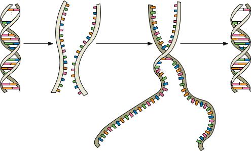
Heat |
Nucleation |
Zippering |
(second-order) |
(first-order) |
|
|
Slow |
Fast |
FIGURE 12.19 ● Steps in the thermal denaturation and renaturation of DNA. The nucleation phase of the reaction is a second-order process depending on sequence alignment of the two strands. This process takes place slowly because it takes time for complementary sequences to encounter one another in solution and then align themselves in register. Once the sequences are aligned, the strands zipper up quickly.
Renaturation Rate and DNA Sequence Complexity—c0t Curves
The renaturation rate of DNA is an excellent indicator of the sequence complexity of DNA. For example, bacteriophage T4 DNA contains about 2 105 nucleotide pairs, whereas Escherichia coli DNA possesses 4.64 106. E. coli DNA is considerably more complex in that it encodes more information. Expressed another way, for any given amount of DNA (in grams), the sequences represented in an E. coli sample are more heterogeneous, that is, more dissimilar from one another, than those in an equal weight of phage T4 DNA. Therefore, it will take the E. coli DNA strands longer to find their complementary partners and reanneal. This situation can be analyzed quantitatively.
If c is the concentration of single-stranded DNA at time t, then the secondorder rate equation for two complementary strands coming together is given by the rate of decrease in c:
dc/dt k2c2
where k2 is the second-order rate constant. Starting with a concentration, c0, of completely denatured DNA at t 0, the amount of single-stranded DNA remaining at some time t is
c/c0 1/(1 k2c0t)
where the units of c are mol of nucleotide per L and t is in seconds. The time for half of the DNA to renature (when c/c0 0.5) is defined as t t1/2. Then,
0.5 1/(1 k2c0t1/2) and thus 1 k2c0t1/2 2
yielding
c0t1/2 1/k2
A graph of the fraction of single-stranded DNA reannealed (c/c0) as a function of c0t on a semilogarithmic plot is referred to as a c0t (pronounced “cot”) curve (Figure 12.20). The rate of reassociation can be followed spectrophotometrically by the UV absorbance decrease as duplex DNA is formed. Note that
 supercoil
 B
B 
 A
A 
 B
B 


378 Chapter 12 ● Structure of Nucleic Acids
|
|
|
|
|
|
|
|
|
|
|
|
|
|
|
|
|
|
|
|
|
|
|
|
|
|
|
|
|
|
|
|
|
|
|
|
|
T T G |
|||||||
|
|
|
|
|
|
|
|
|
|
|
|
|
|
|
|
|
|
|
|
|
|
|
|
|
|
|
|
|
|
|
|
|
|
|
|
A |
|
T |
||||||
|
|
|
|
|
|
|
|
|
|
|
|
|
|
|
|
|
|
|
|
|
|
|
|
|
|
|
|
|
|
|
|
|
|
|
|
T |
|
C |
||||||
|
|
|
|
|
|
|
|
|
|
|
|
|
|
|
|
|
|
|
|
|
|
|
|
|
|
|
|
|
|
|
|
|
|
|
|
|
C |
|
|
G |
|
|
|
|
|
|
|
|
|
|
|
|
|
|
|
|
|
|
|
|
|
|
|
|
|
|
|
|
|
|
|
|
|
|
|
|
|
|
|
|
|
|
|
|
|
|
|
||
|
|
|
|
|
|
|
|
|
|
|
|
|
|
|
|
|
|
|
|
|
|
|
|
|
|
|
|
|
|
|
|
|
|
|
|
|
C |
|
|
G |
|
|
|
|
|
|
|
|
|
|
|
|
|
|
|
|
|
|
|
|
|
|
|
|
|
|
|
|
|
|
|
|
|
|
|
|
|
|
|
|
|
|
|
|
|
|
|
||
|
|
|
|
|
|
|
|
|
|
|
|
|
|
|
|
|
|
|
|
|
|
|
|
|
|
|
|
|
|
|
|
|
|
|
|
|
T |
|
|
A |
|
|
|
|
|
|
|
|
|
|
|
|
|
|
|
|
|
|
|
|
|
|
|
|
|
|
|
|
|
|
|
|
|
|
|
|
|
|
|
|
|
|
|
|
|
|
|
||
|
|
|
|
|
|
|
|
|
|
|
|
|
|
|
|
|
|
|
|
|
|
|
|
|
|
|
|
|
|
|
|
|
|
|
|
|
G |
|
|
C |
|
|
|
|
|
|
|
|
|
|
|
|
|
|
|
|
|
|
|
|
|
|
|
|
|
|
|
|
|
|
|
|
|
|
|
|
|
|
|
|
|
|
|
|
|
|
|
||
|
|
|
|
|
|
|
|
|
|
|
|
|
|
|
|
|
|
|
|
|
|
|
|
|
|
|
|
|
|
|
|
|
|
|
|
|
C |
|
|
G |
|
|
|
|
|
|
|
|
|
|
|
|
|
|
|
|
|
|
|
|
|
|
|
|
|
|
|
|
|
|
|
|
|
|
|
|
|
|
|
|
|
|
|
|
|
|
|
||
|
|
|
|
|
|
|
|
|
|
|
|
|
|
|
|
|
|
|
|
|
|
|
|
|
|
|
|
|
|
|
|
|
|
|
|
|
A |
|
|
T |
|
|
|
|
|
|
|
|
|
|
|
|
|
|
|
|
|
|
|
|
|
|
|
|
|
|
|
|
|
|
|
|
|
|
|
|
|
|
|
|
|
|
|
|
|
|
|
||
|
|
|
|
|
|
|
|
|
|
|
|
|
|
|
|
|
|
|
|
|
|
|
|
|
|
|
|
|
|
|
|
|
|
|
|
|
A |
|
|
T |
|
|
|
|
|
|
|
|
|
|
|
|
|
|
|
|
|
|
|
|
|
|
|
|
|
|
|
|
|
|
|
|
|
|
|
|
|
|
|
|
|
|
|
|
|
|
|
||
|
|
|
|
|
|
|
|
|
|
|
|
|
|
|
|
|
|
|
|
|
|
|
|
|
|
|
|
|
|
|
|
|
|
|
|
|
G |
|
|
C |
|
|
|
|
|
|
|
|
|
|
|
|
|
|
|
|
|
|
|
|
|
|
|
|
|
|
|
|
|
|
|
|
|
|
|
|
|
|
|
|
|
|
|
|
|
|
|
||
. . .C A T |
G A A C G T C C |
T A T T G T C |
G G A C G T T C |
T G A . . . |
|
. . .C A T |
|
|
|
|
T G A . . . |
|||||||||||||||||||||||||||||||||
|
|
|
|
|
|
|
|
|
|
|
|
|
|
|
|
|
|
|
|
|
|
|
|
|
|
|
|
|
|
|
|
|
|
|
|
|
|
|
|
|
|
|
|
|
. . .G T A |
C T T G C A G G |
A T A A C A G |
C C T G C A A G |
A C T . . . |
|
. . .G T A |
|
|
A C T . . . |
|||||||||||||||||||||||||||||||||||
|
|
|
|
|
|
|
|
|
|
|
|
|
|
|
|
|
|
|
|
|
|
|
|
|
|
|
|
|
|
|
|
|
|
|
|
|
C |
|
|
G |
|
|
|
|
|
|
|
|
|
|
|
|
|
|
|
|
|
|
|
|
|
|
|
|
|
|
|
|
|
|
|
|
|
|
|
|
|
|
|
|
|
|
|
|
|
|
|
||
|
|
|
|
|
|
|
|
|
|
|
|
|
|
|
|
|
|
|
|
|
|
|
|
|
|
|
|
|
|
|
|
|
|
|
|
|
T |
|
|
A |
|
|
|
|
|
|
|
|
|
|
|
|
|
|
|
|
|
|
|
|
|
|
|
|
|
|
|
|
|
|
|
|
|
|
|
|
|
|
|
|
|
|
|
|
|
|
|
||
|
|
|
|
|
|
|
|
|
|
|
|
|
|
|
|
|
|
|
|
|
|
|
|
|
|
|
|
|
|
|
|
|
|
|
|
|
T |
|
|
A |
|
|
|
|
|
|
|
|
|
|
|
|
|
|
|
|
|
|
|
|
|
|
|
|
|
|
|
|
|
|
|
|
|
|
|
|
|
|
|
|
|
|
|
|
|
|
|
||
|
|
|
|
|
|
|
|
|
|
|
|
|
|
|
|
|
|
|
|
|
|
|
|
|
|
|
|
|
|
|
|
|
|
|
|
|
G |
|
|
C |
|
|
|
|
|
|
|
|
|
|
|
|
|
|
|
|
|
|
|
|
|
|
|
|
|
|
|
|
|
|
|
|
|
|
|
|
|
|
|
|
|
|
|
|
|
|
|
||
|
|
|
|
|
|
|
|
|
|
|
|
|
|
|
|
|
|
|
|
|
|
|
|
|
|
|
|
|
|
|
|
|
|
|
|
|
C |
|
|
G |
|
|
|
|
|
|
|
|
|
|
|
|
|
|
|
|
|
|
|
|
|
|
|
|
|
|
|
|
|
|
|
|
|
|
|
|
|
|
|
|
|
|
|
|
|
|
|
||
|
|
|
|
|
|
|
|
|
|
|
|
|
|
|
|
|
|
|
|
|
|
|
|
|
|
|
|
|
|
|
|
|
|
|
|
|
A |
|
|
T |
|
|
|
|
|
|
|
|
|
|
|
|
|
|
|
|
|
|
|
|
|
|
|
|
|
|
|
|
|
|
|
|
|
|
|
|
|
|
|
|
|
|
|
|
|
|
|
||
|
|
|
|
|
|
|
|
|
|
|
|
|
|
|
|
|
|
|
|
|
|
|
|
|
|
|
|
|
|
|
|
|
|
|
|
|
G |
|
|
C |
|
|
|
|
|
|
|
|
|
|
|
|
|
|
|
|
|
|
|
|
|
|
|
|
|
|
|
|
|
|
|
|
|
|
|
|
|
|
|
|
|
|
|
|
|
|
|
||
|
|
|
|
|
|
|
|
|
|
|
|
|
|
|
|
|
|
|
|
|
|
|
|
|
|
|
|
|
|
|
|
|
|
|
|
|
G |
|
|
C |
|
|
|
|
|
|
|
|
|
|
|
|
|
|
|
|
|
|
|
|
|
|
|
|
|
|
|
|
|
|
|
|
|
|
|
|
|
|
|
|
|
|
|
|
|
|
|
||
|
|
|
|
|
|
|
|
|
|
|
|
|
|
|
|
|
|
|
|
|
|
|
|
|
|
|
|
|
|
|
|
|
|
|
|
A |
|
G |
||||||
|
|
|
|
|
|
|
|
|
|
|
|
|
|
|
|
|
|
|
|
|
|
|
|
|
|
|
|
|
|
|
|
|
|
|
|
T |
|
A |
||||||
|
|
|
|
|
|
|
|
|
|
|
|
|
|
|
|
|
|
|
|
|
|
|
|
|
|
|
|
|
|
|
|
|
|
|
|
|
A A C |
|||||||
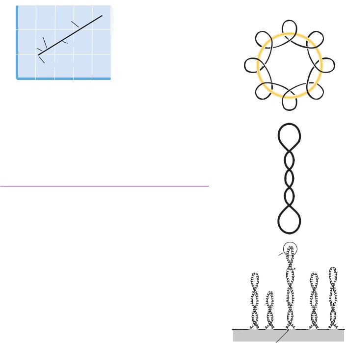
● The formation of a cruciform structure from a palindromic sequence within DNA. The self-complementary inverted repeats can rearrange to form hydrogenbonded cruciform loops.
formation of DNA is found in protein : DNA interactions that are the basis of phenomena as diverse as chromosome structure (see Figure 12.31) and gene expression.
Cruciforms
Palindromes are words, phrases, or sentences that are the same when read backward or forward, such as “radar,” “sex at noon taxes,” “Madam, I’m Adam,” and “a man, a plan, a canal, Panama.” DNA sequences that are inverted repeats, or palindromes, have the potential to form a tertiary structure known as a cruciform (literally meaning “cross-shaped”) if the normal interstrand base pairing is replaced by intrastrand pairing (Figure 12.27). In effect, each DNA strand folds back on itself in a hairpin structure to align the palindrome in base-pair- ing register. Such cruciforms are never as stable as normal DNA duplexes because an unpaired segment must exist in the loop region. However, negative supercoiling causes a localized disruption of hydrogen bonding between base pairs in DNA and may promote formation of cruciform loops. Cruciform structures have a twofold rotational symmetry about their centers and potentially create distinctive recognition sites for specific DNA-binding proteins.
12.5 ● Chromosome Structure
A typical human cell is 20 m in diameter. Its genetic material consists of 23 pairs of dsDNA molecules in the form of chromosomes, the average length of which is 3 109 bp/23 or 1.3 108 nucleotide pairs. At 0.34 nm/bp in B-DNA, this represents a DNA molecule 5 cm long. Together, these 46 dsDNA molecules amount to more than 2 m of DNA that must be packaged into a nucleus perhaps 5 m in diameter! Clearly, the DNA must be condensed by a factor of more than 105. This remarkable task is accomplished by neatly wrapping the DNA around protein spools called nucleosomes and then packing the nucleosomes to form a helical filament that is arranged in loops associated with the nuclear matrix, a skeleton or scaffold of proteins providing a structural framework within the nucleus.