

Dewick P.M. Medicinal natural products VCH-Wiley, Weinheim, 2002 / booktext@id88013689placeboie
.pdfMACROLIDES AND POLYETHERS |
105 |
(Continued )
up to 100 times more potent than cyclosporin A, but produces similar side-effects including neurotoxicity and nephrotoxicity.
Rapamycin (sirolimus) (Figure 3.69) is produced by cultures of Streptomyces hygroscopicus and is also being investigated as an immunosuppressant drug. Although tacrolimus and rapamycin possess a common structural unit, and both inhibit T-cell activation, they appear to achieve this by somewhat different mechanisms. The first-formed rapamycin–receptor protein binds not to calcineurin, but to a different protein. Rapamycin suppresses lymphocyte production. Rapamycin also possesses pronounced antifungal activity, but is not active against bacteria.
O |
HS |
|
|
HS |
||
|
|
|
|
O |
||
SCoA |
|
|
|
|
N CO2H |
|
H2N CO2H |
|
|
||||
|
|
|||||
|
L-Cys |
|
|
H |
||
|
|
|
|
|
||
|
O |
R |
||||
|
12 |
|
|
|||
S |
|
|
|
|
||
|
|
|
|
|
|
|
N |
|
|
|
|
|
OH |
|
|
|
|
|
||
|
|
|
|
|
|
|
|
O |
4 |
|
|||
|
|
|
|
|||
|
|
|
|
|
|
|
|
O |
|
OH O |
|||
R = Me, epothilone B
R = H, epothilone A
S |
|
|
|
|
CO2H |
CO2H |
||
|
|
|
|
|
||||
N |
|
|
SCoA |
+ 3 x |
|
SCoA + 5 x |
|
SCoA |
|
|
|
||||||
|
|
|
|
|
|
|
|
|
|
|
|
|
|
|
|
|
|
|
|
O |
|
O |
O |
|||
S
N

 Me
Me 
 OH
OH
OH
SEnz
O OH O
Figure 3.70
of the substituent at C-12, which is hydrogen in epothilone A but a methyl group in epothilone B. Genetic evidence shows that the polyketide synthase enzyme can accept either malonyl-CoA or methylmalonyl-CoA extender units for this position. Thus, epothilone B is constructed from three malonate and five methylmalonate extender units as shown in Figure 3.70, whilst epothiolone A requires four units of each type. The epothilones display marked antitumour properties with a mode of action paralleling that of the highly successful anticancer drug taxol (see page 205). However, the epothilones have a much higher potency (2000–5000 times) and are active against cell lines which are resistant to taxol and other drugs. There appears to be considerable potential for developing the epothilones or analogues into valuable anticancer drugs.
A further group of macrolides in which nonadjacent positions on an aromatic ring are bridged by the long aliphatic chain is termed ansa
macrolides . These are actually lactams rather than lactones, and the nitrogen atom originates from 3-amino-5-hydroxybenzoic acid, which acts as the starter unit for chain extension with malonyl-CoA or methylmalonyl-CoA. 3-Amino- 5-hydroxybenzoic acid (Figure 3.71) is a simple phenolic acid derivative produced by an unusual variant of the shikimate pathway (see Chapter 4), in which aminoDAHP is formed in the initial step, and then the pathway continues with amino analogues. This proceeds through to aminodehydroshikimic acid which yields 3-amino- 5-hydroxybenzoic acid on dehydration. In the biosynthesis of rifamycin B (Figure 3.71) in
Amycolatopsis mediterranei , this starter unit, plus two malonyl-CoA and eight methylmalonylCoA extenders, are employed to fabricate proansamycin X as the first product released from the enzyme. The enzyme-bound intermediate shown in Figure 3.71 is not strictly correct, in that the naphthoquinone ring system is now
106 |
|
|
|
|
|
|
|
|
|
|
|
|
|
|
|
|
|
|
|
|
THE ACETATE PATHWAY |
|
|
|
|
|
|
|
|
|
|
|
|
|
|
|
||||||||||||
PEP |
CO2H |
|
|
|
|
|
|
|
|
|
|
CO2H |
|
|
|
|
|
|
|
|
CO2H |
|
|
|
|
|
|
|
|
|
|
|
|
|
|
|
||||||||||||
PO |
|
|
NH3 |
|
|
|
|
|
|
|
|
|
|
|
|
|
|
|
|
CO2H |
|
|
|
|
|
|
||||||||||||||||||||||
|
|
|
|
|
|
|
|
|
|
|
|
|
PO O |
|
|
|
|
|
|
|
|
|
|
|
|
|
|
|
– H2O |
|
|
|
|
|
|
|
||||||||||||
PO |
|
|
|
|
|
|
|
|
|
|
|
|
|
|
|
|
|
|
|
|
|
|
|
|
|
|
|
|
|
|
|
|
|
|
|
|
|
|
|
|
|
|
|
|
|
|
|
|
|
|
|
|
|
|
|
|
|
|
|
|
HO |
|
|
NH2 |
|
|
|
|
O |
|
|
NH2 |
|
|
|
|
|
|
|
|
NH2 |
|
|
|
|
|
|
||||||||||
HO |
|
O |
|
|
|
|
HO |
|
|
|
|
|
|
|||||||||||||||||||||||||||||||||||
|
|
|
|
|
|
|
|
|
|
|
OH |
|
|
|
|
|
|
|
|
OH |
|
|
|
|
|
|
|
|||||||||||||||||||||
|
OH |
|
|
|
|
|
|
|
|
|
|
|
|
|
|
|
|
|
|
|
3-amino-5-hydroxy- |
|
|
|
|
|
||||||||||||||||||||||
|
|
|
|
|
|
|
|
|
|
aminoDAHP |
|
|
|
|
aminodehydroshikimic |
|
|
|
|
|
|
|||||||||||||||||||||||||||
D-erythrose 4-P |
|
|
|
|
|
|
|
|
|
|
|
|
|
|
|
|
|
|
|
|||||||||||||||||||||||||||||
|
|
|
|
|
|
|
|
|
|
|
|
|
|
|
|
|
|
|
|
|
acid |
|
|
|
|
benzoic acid |
|
|
|
|
|
|
||||||||||||||||
|
|
|
|
|
|
|
|
|
|
|
|
|
|
|
|
|
|
|
|
|
|
|
|
|
|
|
|
|
|
|
|
|
|
|
|
|
|
|||||||||||
|
|
|
|
|
|
|
|
|
|
|
|
|
|
|
|
|
|
|
|
|
|
|
|
|
|
|
|
|
HSCoA |
|
|
|
|
|
|
|
|
|
|
|
|
|
|
|
||||
HO |
|
|
|
|
|
|
|
|
|
|
|
|
|
|
|
|
|
|
|
|
|
|
|
O |
|
SCoA |
|
|
|
|
|
|
|
|
|
|
|
|
|
|
|
|||||||
HO |
|
OH |
OH |
|
|
|
|
|
|
|
|
|
|
|
|
|
|
|
|
|
|
|
+ 2 x |
CO2H |
+ 8 x |
CO2H |
|
|
||||||||||||||||||||
|
|
|
|
|
|
|
|
|
|
|
|
|
|
|
|
|
|
|||||||||||||||||||||||||||||||
|
|
|
|
|
|
|
|
|
|
|
|
|
|
|
|
|
|
|
|
|
|
|
|
|
|
|
|
|
|
|
|
|
SCoA |
|
|
|
SCoA |
|||||||||||
|
O |
|
EnzS |
|
|
|
|
|
|
|
|
|
|
|
|
|
|
|
|
|
|
|
|
|
|
|
||||||||||||||||||||||
|
|
|
|
|
|
|
|
|
|
|
|
|
|
|
|
|
|
|
|
|
|
|||||||||||||||||||||||||||
|
|
|
|
|
|
|
|
|
|
|
|
|
|
|
HO |
|
|
NH2 |
|
|
|
|
|
|
|
|
|
|
|
|
|
|
|
|||||||||||||||
|
|
|
|
|
|
|
|
NH2 |
|
|
|
|
|
|
|
|
|
|
|
|
|
|
|
O |
|
|
O |
|
|
|||||||||||||||||||
|
|
|
|
|
|
|
|
|
|
O |
|
|
|
|
|
|
|
|
|
|
|
|
|
|
|
|
|
|
|
|
|
|
|
|
|
|
|
|
|
|
|
|
||||||
O |
|
|
|
|
|
|
|
|
|
|
|
|
|
|
|
|
|
|
|
|
|
|
|
|
|
|
|
|
|
|
|
|
|
|
|
|
|
|
|
|
|
|
|
|
|
|
|
|
|
|
|
OH |
|
|
|
|
|
|
|
|
|
|
|
|
|
|
|
|
|
|
|
|
|
|
|
|
|
|
|
|
|
|
|
|
|
|
|
|
|
|
|
|
|
|
|||
HO |
|
O |
|
|
|
|
|
|
|
|
|
|
|
|
|
HO |
|
|
|
|
|
|
|
|
|
|
|
|
|
|
|
O |
|
|
|
|
|
|
||||||||||
|
|
|
|
|
|
|
|
|
|
|
|
|
|
|
|
|
|
|
|
|
|
|
|
|
|
|
|
|
|
|
|
|
|
|
||||||||||||||
|
|
|
|
|
|
|
|
|
|
|
|
|
|
|
|
|
|
|
|
|
|
|
|
|
|
|
|
|
|
|
|
|
|
|
|
|
|
|
|
|||||||||
|
|
|
|
|
|
|
|
|
|
|
|
|
|
|
|
|
|
|
|
|
|
|
|
|
|
|
|
|
|
|
|
|
|
|
|
|
|
|
|
|||||||||
|
|
|
|
|
|
|
|
|
|
|
|
|
|
|
|
|
|
|
|
|
|
|
|
|
|
|
|
|
|
|
|
|
|
|
|
|
|
|
|
|||||||||
|
|
OH |
OH |
|
|
|
|
|
|
|
|
|
|
|
|
|
|
|
OH |
OH |
|
|
|
|
|
|
|
|
O |
|
OH |
|
OH |
|
|
|||||||||||||
HO |
|
|
|
|
|
HO |
|
|
|
|
|
|
|
|
|
|
|
|
MeO |
|
|
|
||||||||||||||||||||||||||
|
|
|
|
|
|
|
|
|
|
|
|
|
|
|
|
|
|
|
||||||||||||||||||||||||||||||
H OH O |
|
|
|
|
|
|
|
|
|
|
|
|
|
|
|
|
|
|
O |
|
|
|
|
|
|
|
|
|
|
|
|
|
|
|
|
|||||||||||||
|
|
|
|
|
|
|
|
|
|
|
|
|
|
|
|
|
|
OH |
|
|
|
|
|
|
|
|
|
|
|
|
|
|
|
|
|
|||||||||||||
H |
|
H |
|
|
|
|
|
|
|
|
|
H |
|
|
|
|
|
|
|
|
|
|
|
OH |
OH |
H |
||||||||||||||||||||||
|
|
|
|
|
|
|
|
|
|
|
|
|
|
|
|
|
||||||||||||||||||||||||||||||||
|
|
|
|
|
|
|
N |
|
|
|
|
|
|
|
|
|
|
|
|
|
|
N |
|
|
|
|
|
|
|
|
|
|
|
|
|
|
|
N |
||||||||||
|
|
|
|
|
|
|
|
|
|
|
|
|
|
|
|
|
|
|
|
|
|
|
|
|
|
|
|
|
|
|
|
|
|
|
|
|
|
|
|
|
|
|
|
|
|
|
||
HO |
|
|
|
|
O |
HO |
|
|
HO |
|
|
O |
|
|
|
|
|
|
|
|
O |
|
|
|
|
O |
||||||||||||||||||||||
|
|
|
|
|
|
|
|
|
|
|
|
|
|
|
|
|
|
|
|
|
|
|
|
|
|
|
|
|
|
|
|
|
|
|
|
|
||||||||||||
O |
|
|
O |
|
|
|
|
|
|
|
|
|
|
carbon |
|
|
|
|
|
O |
O |
|
|
|
|
|
|
|
|
|
|
|
|
|
O |
|
O |
CO2H |
||||||||||
|
O |
|
|
|
|
|
|
|
|
|
|
|
|
|
|
|
|
|
|
|
|
|
|
|
|
|
|
|
|
|
|
|
O |
|
|
|
|
|
||||||||||
|
|
|
|
|
|
|
|
|
|
|
|
|
|
|
|
|
lost |
|
|
|
|
|
|
|
|
|
|
|
|
|
|
|
|
|
|
|
|
|
|
|
|
|
|
|
|
|||
|
proansamycin X |
|
|
|
|
rifamycin W |
|
|
|
|
|
|
|
|
|
rifamycin B |
|
|
||||||||||||||||||||||||||||||
|
|
|
|
|
|
|
|
|
|
|
|
|
|
|
|
|
|
|
||||||||||||||||||||||||||||||
Figure 3.71
known to be constructed during, not after, chain assembly. Rifamycin W and then the antibiotic rifamycin B are the result of further modifications including cleavage of the double bond, loss of one carbon, then formation of the ketal. Maytansine (Figure 3.72) is a plant-derived ansa macrolide from Maytenus serrata (Celastraceae), though other esters of the parent alcohol, maytansinol, are produced by species of the fungus Nocardia. Maytansine has been extensively investigated for its potential antitumour activity.
The macrolide systems described above are produced by formation of an intramolecular ester or amide linkage, utilizing appropriate functionalities in the growing polyketide chain. Macrolide formation does not always occur, and similar acetate–propionate precursors might also be expected to yield molecules which are essentially linear in nature. Good examples of such molecules
|
|
O |
Me |
|||
|
|
|
|
|
|
|
|
|
|
|
N |
||
|
Cl Me |
O O |
||||
|
|
|
|
|||
|
|
|
|
|||
|
|
O |
||||
MeO |
N |
O |
|
|||
|
|
|
|
|
|
|
|
|
|
|
|
O |
|
|
|
|
|
|
||
|
|
|
|
|
||
N O
MeO OHH
maytansine
Figure 3.72
are lasalocid A (Figure 3.74) from Streptomyces lasaliensis and monensin A (Figure 3.75) from
Streptomyces cinnamonensis, representatives of a large group of compounds called polyether antibiotics. These, and other examples, are of value in veterinary medicine, being effective in preventing

MACROLIDES AND POLYETHERS |
107 |
Ansa Macrolides
Ansamycins are a class of macrocyclic compounds in which non-adjacent positions on an aromatic ring system are spanned by the long aliphatic bridge (Latin: ansa = handle). The aromatic portion may be a substituted naphthalene or naphthaquinone, or alternatively a substituted benzene ring. The macrocycle in the ansamycins is closed by an amide rather than an ester linkage, i.e. ansamycins are lactams. The only ansamycins currently used therapeutically are semi-synthetic naphthalene-based macrocycles produced from rifamycin B.
The rifamycins are ansamycin antibiotics produced by cultures of Amycolatopsis mediterranei (formerly Nocardia mediterranei or Streptomyces mediterranei). The crude antibiotic mixture was found to contain five closely related substances rifamycins A–E, but if the organism was cultured in the presence of sodium diethyl barbiturate (barbitone or barbital), the product was almost entirely rifamycin B (Figure 3.71). Rifamycin B has essentially no antibacterial activity, but on standing in aqueous solution in the presence of air, it is readily transformed by oxidation and intramolecular nucleophilic addition into rifamycin O, which
|
|
OH |
OH |
H |
|
||||
|
|
|
|
|
|
|
|||
|
|
|
|
|
|
N |
|
||
|
|
|
|
|
|
|
|
|
|
|
|
|
|
|
|
|
O |
|
|
|
|
O |
|
|
|
|
|||
|
|
|
|
|
|
|
|
||
|
O |
O |
O |
CO2H |
|
||||
|
|
|
|
|
|
|
|
||
|
|
rifamycin B |
|
|
|
|
|||
|
O |
|
|
|
|
|
|
|
|
|
|
|
|
|
|
|
|
|
|
O |
OH |
OH |
|
|
|
|
|||
MeO |
|
|
|
|
|||||
|
|
|
|
|
|
|
|
||
|
|
OH |
OH |
H |
|
||||
|
|
|
|||||||
|
|
|
|
|
|
N |
|
||
|
|
|
|
|
|
|
|||
|
|
|
|
|
|
|
|
|
|
|
|
O |
|
|
|
O |
|
||
|
|
|
|
|
|||||
|
|
|
|
|
|
|
|
||
|
O |
|
|
OH |
N |
|
|||
|
|
O |
|
|
|
N |
|
||
|
|
|
|
|
|
|
|
||
rifampicin
|
|
|
|
|
|
|
OH |
|
O |
|
H |
|
|
|
|
|
|
|
|
|
OH |
O |
|
H |
||||||||||
|
|
O2 |
|
|
|
|
|
|
|
|
|
|
|
|
N |
|
|
|
|
|
|
|
|
|
|
|
|
|
|
N |
||||
|
|
|
|
|
|
|
|
|
|
|
|
|
|
|
|
|
|
|
|
|
|
|
|
|
||||||||||
|
HONO |
|
|
|
|
|
|
|
|
|
|
|
|
|
|
|
|
|
|
|
|
|
|
|
|
|
|
|
|
|
|
|
||
|
|
|
|
|
|
|
|
|
|
|
|
|
|
|
|
|
|
|
|
|
|
|
|
|
|
|
|
|
|
|
||||
|
|
or |
|
|
|
|
|
|
|
|
|
|
|
|
|
|
O |
|
|
|
|
|
|
|
|
|
|
|
|
|
|
|
O |
|
|
|
|
|
|
|
|
|
|
|
|
|
|
|
|
|
|
|
|
|
|
|
|
|
|
|
|
||||||||
|
|
|
|
|
O |
|
|
|
|
|
|
OH |
|
|
|
|
|
|
|
|
O |
|
|
|
|
O |
||||||||
|
|
|
|
|
|
|
|
|
|
|
|
|
|
|
|
|
|
|
|
|
||||||||||||||
electrochemical |
|
|
|
|
|
|
|
|
|
|
|
|
|
|
|
|
O |
|
|
|||||||||||||||
|
O |
|
|
|
|
O |
|
|
|
|
|
|
|
|
|
|
|
|
O |
|
|
|||||||||||||
oxidation |
|
|
|
O |
|
|
|
O |
|
|
|
|
|
|
|
|
O |
|
|
|
|
|
||||||||||||
|
|
|
|
|
|
|
|
|
|
|
|
|
|
|
|
|
|
|
|
|
|
|
|
|
|
|
|
|
||||||
|
|
|
|
|
|
|
|
|
|
|
|
|
|
|
|
|
|
|
|
|
|
|
|
|
|
|
|
|
O |
|||||
|
|
|
|
|
|
|
|
nucleophilic attack on |
|
|
|
|
|
|
|
|||||||||||||||||||
|
|
|
|
|
|
|
|
|
|
rifamycin O |
||||||||||||||||||||||||
|
|
|
|
|
|
|
|
to quinone system |
|
|
|
|
|
|
|
|
|
|||||||||||||||||
|
|
|
|
|
|
|
|
|
|
|
|
|
|
|
hydrolysis of |
|
|
|
H |
|||||||||||||||
|
|
|
|
|
|
|
|
|
|
|
|
|
|
|
|
|
|
|
|
|
|
|
|
|
|
|
|
|
||||||
|
|
|
|
|
|
|
|
|
|
|
|
|
|
|
|
|
|
|
|
|
|
|
|
|
|
ketal |
|
|
|
|
|
|||
H2N |
|
N |
N |
|
Me |
OH |
OH H |
|
|
|
|
|
|
|
OH |
O |
|
H |
||||||||||||||||
|
|
|
|
|
|
|
|
|
|
|
|
|||||||||||||||||||||||
|
|
|
|
|
|
|
|
|
|
|
|
|
|
|
|
|||||||||||||||||||
|
|
|
|
|
|
|
|
|
|
|
|
|
||||||||||||||||||||||
|
|
HCHO |
|
|
|
|
|
|
|
|
|
|
|
N |
reduction |
|
|
|
|
|
|
|
N |
|||||||||||
|
|
|
|
|
|
|
|
|
|
|
|
|
|
|
|
|
|
|
||||||||||||||||
|
|
|
|
|
|
|
|
|
|
|
|
|
|
|
|
|
|
|
|
|
|
|
|
|
|
|||||||||
|
|
|
|
|
|
|
|
|
|
|
|
|
|
|
|
|
|
|
|
|
|
|
|
|||||||||||
|
|
|
|
|
|
|
O |
|
|
|
|
|
|
O |
|
|
|
|
|
|
|
|
O |
|
|
|
|
|
|
O |
||||
|
Mannich |
|
|
|
|
|
|
|
|
|
|
|
|
|
|
|
|
|
|
|
|
|
|
|
||||||||||
|
|
|
|
|
|
|
|
|
|
ascorbic |
|
|
|
|
|
|
|
|||||||||||||||||
|
|
|
|
|
|
|
|
|
|
|
|
|
|
|
|
|
|
|
|
|
||||||||||||||
|
|
reaction |
|
|
O |
|
O |
OH |
|
|
|
|
|
|
acid |
|
O |
O |
O |
|
|
|
||||||||||||
|
|
|
|
|
|
|
|
|
|
|
|
|
|
|
|
|
|
|
|
|
|
|
|
|
|
|
|
|
|
|
||||
|
|
|
|
|
|
|
rifamycin SV |
|
|
|
|
|
|
|
|
|
rifamycin S |
|||||||||||||||||
N |
|
|
|
|
|
|
|
|
|
|
|
|
|
|
|
|
|
|
|
|
|
|
conjugate |
|
|
|
|
|
NH3 |
|||||
|
|
|
|
|
|
|
|
|
|
|
|
|
|
|
|
|
|
|
|
|
|
|
|
|
|
|
||||||||
|
|
|
|
|
|
|
|
|
|
|
|
|
|
|
|
|
|
|
|
|
|
addition on to |
|
|
|
|||||||||
Me |
|
O |
|
|
|
|
|
|
|
|
|
|
|
|
|
|
|
|
|
|
|
|
|
|
|
|
||||||||
|
|
|
|
|
|
|
|
|
|
|
|
|
|
|
|
|
|
|
|
|
|
|
|
|
|
quinone |
|
|
|
|
|
|
|
|
|
|
|
|
|
|
|
|
|
|
|
|
|
|
|
|
|
|
|
|
|
|
|
|
|
|
|
|
|
|
|
|
|
||
|
|
|
O |
|
|
OH |
OH |
|
|
|
|
|
|
|
|
|
|
|
|
|
|
|
|
|
|
|
|
|||||||
|
|
|
MeO |
|
|
|
|
|
|
|
|
|
|
|
|
|
OH |
OH |
|
H |
||||||||||||||
|
|
|
|
|
|
|
|
|
|
|
|
|
|
|
|
|
|
|
|
|
|
|
|
|
||||||||||
|
|
|
|
|
|
|
OH O |
|
|
|
|
|
|
|
|
|
|
|
|
|
|
|
|
|
|
|
|
|
|
|
||||
|
|
|
|
|
|
|
|
|
H |
|
|
|
|
|
|
|
|
|
|
|
|
|
|
|
|
|
|
N |
||||||
|
|
|
|
|
|
|
|
|
|
|
|
N |
|
|
|
|
|
|
|
|
|
|
|
|
|
|
|
|
|
|
|
|
||
|
|
|
|
|
|
|
|
|
|
|
|
|
|
|
|
|
|
|
|
|
|
|
|
|
|
|
|
|
|
|
|
|||
|
|
|
|
|
|
|
|
|
|
|
|
|
|
|
|
|
|
|
|
|
|
|
|
|
|
|
|
|
|
|
|
|
O |
|
|
|
|
|
|
|
O |
|
|
|
|
|
NH |
O |
|
|
|
|
|
|
|
|
O |
|
|
OH |
|
NH2 |
|||||||
|
|
|
|
|
|
|
|
|
|
|
|
|
|
|
|
|
|
|
|
|
||||||||||||||
|
|
|
|
|
|
|
|
|
|
|
|
|
|
|
|
|
|
|
|
|
|
|
O |
|
|
|
|
|
||||||
|
|
|
|
|
O |
|
|
N |
|
|
|
|
|
|
|
|
|
|
|
|
|
O |
|
|
|
|
|
|||||||
|
|
|
|
|
|
|
O |
|
|
|
|
|
|
|
|
|
|
|
|
|
|
|
|
|
|
3-amino-rifamycin SV |
||||||||
 N rifabutin

Figure 3.73
(Continues)

108 |
THE ACETATE PATHWAY |
(Continued )
under acidic conditions then hydrolyses and gives rifamycin S, a highly active antibacterial agent (Figure 3.73). Chemical reduction of rifamycin S using ascorbic acid (vitamin C) converts the quinone into a quinol and provides a further antibacterial, rifamycin SV. Rifamycins O, S, and SV can all be obtained by fermentation using appropriate strains of A. mediterranei. Rifamycin SV is actually the immediate biosynthetic precursor of rifamycin B under normal conditions, so this conversion can be genetically blocked and lead to accumulation of rifamycin SV. Several other rifamycin analogues have also been characterized. Rifamycin O is usually produced by chemical or electrochemical oxidation of rifamycin B, and converted into rifamycin SV as in Figure 3.73.
The most useful rifamycin employed clinically is rifampicin (Figure 3.73), a semisynthetic derivative produced from rifamycin SV via a Mannich reaction (see page 18) using formaldehyde and N-amino-N -methylpiperazine. Rifampicin has a wide antibacterial spectrum, with high activity towards Gram-positive bacteria and a lower activity towards Gram-negative organisms. Its most valuable activity is towards Mycobacterium tuberculosis and rifampicin is a key agent in the treatment of tuberculosis, usually in combination with at least one other drug to reduce the chances for development of resistant bacterial strains. It is also useful in control of meningococcal meningitis and leprosy. Rifampicin’s antibacterial activity arises from inhibition of RNA synthesis by binding to DNA-dependent RNA polymerase. RNA polymerase from mammalian cells does not contain the peptide sequence to which rifampicin binds, so RNA synthesis is not affected. In contrast to the natural rifamycins which tend to have poor absorption properties, rifampicin is absorbed satisfactorily after oral administration, and is also relatively free of toxic side-effects. The most serious side-effect is disturbance of liver function. A trivial, but to the patient potentially worrying, side-effect is discoloration of body fluids, including urine, saliva, sweat, and tears, to a red–orange colour, a consequence of the naphthalene/naphthoquinone chromophore in the rifamycins. Rifamycin, the sodium salt of rifamycin SV (Figure 3.73), has also been used clinically in the treatment of Gram-positive infections, and particularly against tuberculosis. Rifabutin (Figure 3.73) is a newly introduced derivative, synthesized via 3-amino-rifamycin SV, which also has good activity against the Mycobacterium avium complex frequently encountered in patients with AIDS.
CO2H
HO
|
O |
OH |
|
||
OH O |
O |
|
|
|
|
lasalocid A
Figure 3.74
and controlling coccidiae and also having the ability to improve the efficiency of food conversion in ruminants. The polyether antibiotics are characterized by the presence of a number of tetrahydrofuran and/or tetrahydropyran rings along the basic chain. The polyether acts as an ionophore, increasing influx of sodium ions into the parasite, causing a resultant and fatal
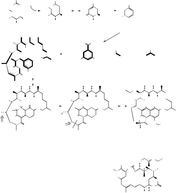
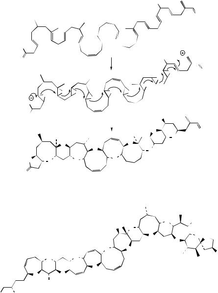
increase in osmotic pressure. Current thinking is that these ring systems arise via a cascade cyclization mechanism, probably involving epoxide intermediates. Thus, in the biosynthesis of monensin A (Figure 3.75), chain assembly from acetate, malonate, methylmalonate, and ethylmalonate precursors could produce the triene shown. If the triepoxide is then formed, a concerted stereospecific cyclization sequence initiated by a hydroxyl and involving carbonyls and epoxides could proceed as indicated.
Even more remarkable polyether structures are found in some toxins produced by marine dinoflagellates, which are in turn taken up by shellfish and pass on their toxicity to the shellfish. Okadaic acid (Figure 3.76) and related polyether structures from Dinophysis species are responsible for

|
|
|
|
|
|
|
|
|
MACROLIDES AND POLYETHERS |
|
|
|
|
|
109 |
||||||||
|
|
|
|
CO2H |
|
|
|
CO2H |
|
|
CO2H |
|
|
|
|
|
|
|
|||||
|
SCoA + 4 x |
|
|
|
SCoA + 7 x |
|
SCoA |
+ |
|
|
SCoA |
|
|
|
|
|
|
||||||
|
|
|
|
|
|
|
|
|
|
|
|
|
|
|
|
|
|
|
|
|
|
|
|
O |
|
|
|
O |
|
|
|
O |
|
|
O |
|
|
|
|
|
|
|
|||||
|
|
|
|
|
|
|
|
|
|
|
|
epoxidations |
|
|
|
|
|
|
|
||||
|
HO |
|
|
|
|
|
|
|
|
|
|
|
|
|
HO |
|
|
|
|
|
|
|
|
|
|
|
|
|
|
|
|
|
|
|
|
|
|
|
|
|
|
|
|
|
|
|
|
|
|
|
|
|
|
|
|
|
|
|
|
|
|
|
|
|
|
|
|
|
|
|
|
|
|
O |
|
|
|
|
|
|
|
|
|
|
|
|
|
O |
|
O |
O |
O |
|
||
|
|
|
|
|
|
|
|
|
|
|
|
|
|
|
|
|
|||||||
|
|
OH |
|
|
|
|
|
|
|
|
|
|
|
|
|
OH |
|
|
|||||
|
HO |
|
|
|
|
|
|
|
O |
|
|
HO |
|
|
|
|
O |
|
|||||
|
|
|
|
|
|
|
|
|
|
|
|
|
|
|
|
|
|
||||||
EnzS |
|
|
|
|
|
|
|
|
|
|
|
EnzS |
|
|
|
|
concerted |
|
|
||||
|
|
|
|
|
|
|
|
|
|
|
|
|
|
|
|
|
|
|
|
|
|
|
|
O |
|
|
|
|
|
|
|
|
|
|
|
|
O |
|
|
|
|
cyclization |
|
||||
|
|
|
|
|
|
|
|
|
|
|
|
|
|
|
|
sequence |
|
|
|||||
|
|
|
|
|
|
|
|
|
|
|
|
|
|
|
|
|
|
||||||
|
|
|
|
|
|
|
|
|
|
|
|
|
|
|
|
|
|
|
|
|
|
|
|
|
|
|
|
|
|
|
|
|
|
|
|
|
|
|
HO |
|
|
|
|
|
|
|
|
|
|
|
|
|
|
|
|
|
|
|
|
|
|
|
|
|
O |
O |
O |
O |
O |
|
|
|
|
|
|
|
|
|
|
|
|
|
|
|
|
|
MeO |
|
|
|
|
|
|||
|
|
|
|
|
|
|
|
|
|
|
|
|
|
|
|
|
|
|
|
|
|||
|
|
|
|
|
|
|
|
|
|
|
|
|
|
|
|
|
|
|
|
|
|
||
|
|
|
|
|
|
|
|
|
|
|
|
|
|
|
|
|
|
|
|
|
|
HO |
OH |
|
|
|
|
|
|
|
|
|
|
|
|
|
|
HO2C |
|
|
monensin A |
|
|
||||
|
|
|
|
|
|
|
|
|
|
|
|
Figure 3.75 |
|
|
|
|
|
|
|
||||
|
|
|
|
|
|
|
|
|
|
|
|
|
|
|
OH |
|
|
|
|
|
|
|
|
|
|
HO2C |
|
|
|
|
O |
|
|
|
|
O |
|
|
|
|
|
|
|
||||
|
|
|
|
|
|
OH |
|
|
O |
|
|
O |
|
|
|
|
O |
|
|
||||
|
|
|
|
|
|
|
|
OH |
|
|
|
|
O |
|
|
O |
|
|
|
||||
|
|
|
|
|
|
|
|
|
|
|
|
|
|
|
|
|
|
|
|||||
OH
okadaic acid
Figure 3.76
diarrhoeic shellfish poisoning in mussels, causing severe diarrhoea in consumers of contaminated shellfish in many parts of the world. Brevetoxin A (Figure 3.77) is an example of the toxins associated with ‘red tide’ blooms of dinoflagellates, which affect fishing and also tourism especially in Florida and the Gulf of Mexico. The red tide toxins are derived from Gymnodimium breve and are the causative agents of neurotoxic shellfish poisoning, leading to neurological disorders as well as gastrointestinal troubles. The toxins are known to bind to sodium channels, keeping them in an open state. Fatalities among marine life, e.g. fish, dolphins, whales, and in humans, are associated with these toxins synthesized by organisms at the base of the marine food chain. These compounds are postulated to be produced from a polyunsaturated fatty acid by epoxidation of the double bonds, and then a
concerted sequence of epoxide ring openings leads to the extended polyether structure (Figure 3.77). The carbon skeleton does not conform to a simple polyketide chain, and biosynthetic studies have shown that fragments from the citric acid cycle and a four-carbon starter unit from mevalonate are also involved, and that some of the methyls originate from methionine. Ciguatoxin (Figure 3.78) is one of the most complex examples of a polyether structure found in nature. This is found in the moray eel (Gymnothorax javanicus) and in a variety of coral reef fish, such as red snapper (Lutjanus bohar). Ciguatoxin is remarkably toxic even at microgram levels, causing widespread food poisoning (ciguatera) in tropical and subtropical regions, characterized by vomiting, diarrhoea, and neurological problems. Most sufferers slowly recover, and few cases are fatal, due principally to the very low
110 |
THE ACETATE PATHWAY |
HO
O
EnzS
O |
|
|
|
|
|
HO |
H |
|
|
epoxidations |
|
|
|
|
|
|
|
|
|
|
|
|
|
|
|
|
|
|
|
O |
O |
|
|
|
|
|
|
O |
|
|
|
|
|
|
|
O |
|
|
O |
O |
O |
O |
O |
|
|
|
|
O |
|
|
|
|
|
O |
|
|
|
|
|
|
|
|
|
|
|
|
|
|
|
O |
|
concerted cyclization |
|
|
HO |
|
|
|
|
|
|
|
|
||
|
|
sequence |
|
|
O |
O |
|
|
|
|
|
||||
|
|
|
|
|
|
||
|
|
|
|
|
|
O |
|
|
|
O |
O |
|
|
O |
|
|
|
|
|
|
O |
|
|
|
|
|
|
|
|
|
|
O |
|
O |
O |
|
O |
|
|
|
|
|
|
|
|
||
O
brevetoxin-A
Figure 3.77
|
|
|
|
HO |
O |
OH |
|
|
|
|
O |
|
|
|
|
|
|
|
|
|
|
|
|
|
|
O |
O O |
|
|
|
|
|
|
O |
|
|
|
|
O |
O |
OH |
|
|
|
O |
O |
|
|
|
|
|
|
|
||
|
|
|
|
O |
|
|
|
|
|
O |
O |
|
|
|
|
|
OH |
ciguatoxin |
|
|
|
|
|
|
|
||
HO |
OH |
|
|
|||
Figure 3.78
levels of toxin actually present in the fish. A dinoflagellate Gambierdiscus toxicus is ultimately responsible for polyether production, synthesizing a less toxic analogue, which is passed through the food chain and eventually modified into the very toxic ciguatoxin by the fish.
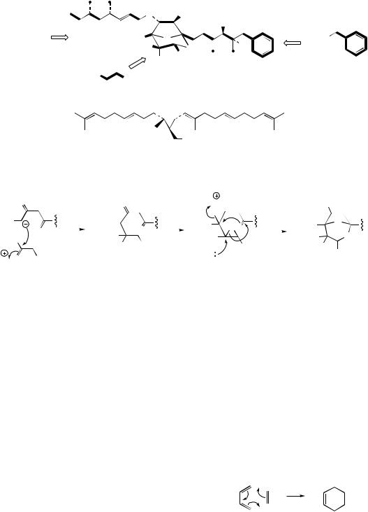
The zaragozic acids (squalestatins) are not macrolides, but they are primarily acetate derived, and the central ring system is suggested to be
formed by an epoxide-initiated process resembling the polyether derivatives just described. Thus, zaragozic acid A (Figure 3.79) is known to be constructed from two acetate-derived chains and a C4 unit such as the Krebs cycle intermediate oxaloacetate (see Figure 2.1). One chain has a benzoyl-CoA starter (from the shikimate pathway, see page 141), and both contain two methioninederived side-chain substituents (Figure 3.79). The
|
|
|
|
CYCLIZATION THROUGH DIELS–ALDER REACTIONS |
111 |
||||||||||||||||||
|
|
|
|
|
|
|
|
|
O |
|
|
OH |
|
|
|
|
|
|
|
|
O |
||
|
|
|
|
|
|
|
|
|
|
|
|
|
|
OAc |
|
|
|
|
|
||||
|
|
|
|
|
|
|
|
|
|
|
|
|
|
|
|
|
|
|
|
|
|
|
|
H3C |
|
COSCoA |
|
|
|
|
O |
|
|
|
|
|
|
|
|
|
|
|
|
|
|
||
|
|
|
|
|
HO2C |
O |
|
|
|
|
|
|
|
CoAS |
|
||||||||
• |
Met |
|
|
|
|
|
HO2C |
|
|
O |
|
|
|
H |
|
|
|
|
|
|
|
||
|
|
|
|
|
|
|
|
|
|
|
CO2H |
|
|
|
|
|
|
|
|
|
|
|
|
|
|
|
|
|
|
|
|
|
|
OH |
|
|
|
|
|
|
|
|
|
|
|
||
|
|
|
|
|
|
HO2C |
|
|
|
|
zaragozic acid A |
|
|
|
|
|
|
|
|
|
|||
|
|
|
|
|
|
|
CO2H |
|
|
(squalestatin S1) |
|
|
|
|
|
|
|
|
|
||||
|
|
|
|
|
|
|
succinic acid |
|
|
|
|
|
|
|
|
|
|
|
|||||
|
|
|
|
|
|
|
|
|
|
|
|
|
|
|
|
|
|
|
|
|
|||
|
|
|
|
|
|
|
|
|
|
|
|
OPP |
|
|
|
|
|
|
|
|
|
|
|
|
|
|
|
|
|
|
|
|
presqualene PP |
|
|
|
|
|
|
|
|
|
|
|
|||
|
|
|
|
|
|
|
|
|
|
Figure 3.79 |
|
|
|
|
|
|
|
|
|
||||
|
|
|
|
aldol |
|
|
|
formation of |
H |
|
|
|
concerted |
|
|
|
|||||||
|
|
O |
|
|
O |
|
|
|
|
nucleophilic |
HO |
||||||||||||
|
|
|
|
|
epoxide functions |
|
|
|
|||||||||||||||
|
|
|
reaction |
|
|
|
|
|
|
||||||||||||||
|
|
|
|
|
|
|
|
|
|
O |
|
|
|
reactions |
|
|
|
||||||
|
|
|
|
|
|
|
|
|
|
|
|
|
|
|
|
|
|
|
|||||
HO2C |
O |
|
|
|
HO2C |
O |
|
|
HO2C |
O O |
|
|
HO2C |
|
O |
||||||||
HO2C |
|
|
|
HO2C |
|
|
|
HO2C |
|
|
HO2C |
|
O |
||||||||||
|
|
|
|
|
|
|
|
|
|||||||||||||||
|
|
|
|
|
CO2H |
|
|
|
|
|
|
||||||||||||
|
|
|
|
|
|
|
HO |
|
|
|
|
CO2H |
|
|
HO |
||||||||
H |
|
O |
CO2H |
|
|
|
|
|
|
|
|
|
H2O |
|
|
|
|
|
|
|
CO2H |
||
|
|
|
|
|
|
|
|
|
|
|
|
|
|
|
|
|
|
|
|
|
|||
oxaloacetic acid
Figure 3.80
heterocyclic ring system can be envisaged as arising via nucleophilic attack on to oxaloacetic acid, formation of a diepoxide, then a concerted sequence of reactions as indicated (Figure 3.80). The zaragozic acids are produced by a number of fungi, including Sporomiella intermedia and
Leptodontium elatius, and are attracting considerable interest since they are capable of reducing blood cholesterol levels in animals by acting as potent inhibitors of the enzyme squalene synthase (see page 212). This is achieved by mimicking the steroid precursor presqualene PP (Figure 3.79) and irreversibly inactivating the enzyme. They thus have considerable medical potential for reducing the incidence of coronary-related deaths (compare the statins, below).
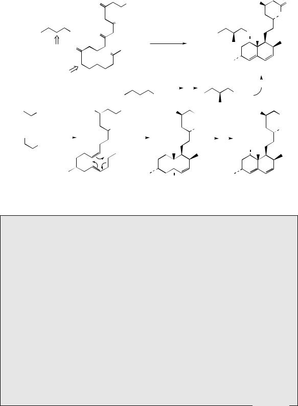
the acetate pathway, but experimental evidence supports cyclization processes different from the aldol and Claisen reactions seen in the biosynthesis of aromatic compounds. They can, however, be rationalized in terms of an enzymic Diels–Alder reaction, represented as the electrocyclic sequence shown in Figure 3.81. Thus, lovastatin can be formulated as arising from two polyketide chains with C-methylation as outlined in Figure 3.82, with relatively few of the oxygen functions being retained in the final product. Accordingly, it is possible that lovastatin is formed by cyclization of the trienoic acid (Figure 3.82), which is likely to arise by a variant of the macrolide
CYCLIZATION THROUGH DIELS–ALDER REACTIONS
diene dienophile
Diels−Alder reaction
A number of cyclic structures, typically containing |
|
cyclohexane rings, are known to be formed via |
Figure 3.81 |
112
+ 8 x
THE ACETATE PATHWAY
|
|
|
|
|
|
O |
|
|
|
|
SEnz |
|
|
|
|
|
|
|
|
|
HO |
O |
|||||||
|
|
|
|
|
|
|
|
|
|
|
|
|
|
|
|
|
|
|
|
|
|
|
|
|
|
|
|
|
O |
|
|
|
|
|
|
|
|
|
|
|
|
|
|
|
|
|
|
|
|
|
|
|
|
|
|
|
|
|
|
|
|
O |
|
|
|
|
|
|
O |
|
|
|
|
|
|
|
|
O |
|||||||||||
|
|
|
|
|
|
|
|
O |
|
|
|
|
|
|
|
|
|
|
|
|
|
|
|||||||
|
|
|
SEnz |
|
O |
|
|
|
|
|
|
|
|
|
|
|
|
|
|
|
|
|
O |
|
|||||
|
|
|
|
|
|
|
|
|
|
|
|
|
|
|
|
|
|
|
|
|
|
|
|||||||
|
|
|
|
|
|
|
|
|
|
|
|
|
|
|
|
|
|
|
|
|
|
|
|||||||
|
|
|
|
|
|
|
|
|
|
|
|
|
|
|
|
|
|
|
|
|
|
|
|
|
|
H |
|
||
|
|
|
O |
|
|
|
|
|
|
|
|
|
|
|
|
|
|
|
|
|
|
|
|
|
|
|
|
|
|
|
Me |
|
|
|
|
O |
|
|
|
|
|
|
|
|
|
|
|
|
|
|
|
|
|
|
|
|
|||
|
|
|
|
|
|
|
|
|
|
|
|
|
|
|
|
|
|
|
|
|
|
|
|
||||||
|
|
|
|
|
|
|
|
|
|
|
|
|
|
|
|
|
|
|
|
|
|
|
|
|
|
|
|
|
|
|
|
|
|
|
|
O |
|
|
|
|
|
|
|
|
|
|
|
|
|
|
|
|
|
|
lovastatin |
|
|||
|
Me |
|
|
|
|
|
|
|
|
|
|
|
|
|
|
|
|
|
|
|
|
|
|
||||||
|
|
|
|
|
|
|
|
|
|
|
|
|
|
|
|
|
|
|
|
|
|
|
|
|
|
||||
O |
O |
|
|
O O |
SAM |
|
O |
|
|
|
|||||||||||||||||||
|
|
|
|
|
|
|
|
|
|
|
|||||||||||||||||||
|
|
|
|
|
|
|
|
|
|||||||||||||||||||||
|
|
|
|
|
|
|
|
|
|
|
|
|
|
|
|
|
|
|
|
|
|
|
|
|
SEnz |
|
|
|
|
|
|
|
|
|
|
|
|
|
|
|
|
|
|
|
|
|
SEnz |
|
|
|
|
|
|||||||
|
|
|
|
|
|
|
|
|
|
|
|
|
|
|
|
|
|
|
|
|
|
|
|
||||||
|
|
|
|
HO |
|
|
|
|
SEnz |
HO |
|
|
|
|
|
|
|
||||||||||||
|
SCoA |
|
|
|
|
|
|
|
HO |
|
|||||||||||||||||||
|
|
|
|
|
|
|
|
|
|
|
|
|
|
|
|
|
|
|
|
|
CO2H |
|
|
|
|
|
|
CO2H |
|
|
|
|
|
|
|
|
|
|
|
O |
|
|
|
|
|
|
|
|
|
|
|
|
|
|
|
|
|
|
|
O |
|
|
|
|
|
|
|
|
|
|
|
|
|
|
OH |
|
|
|
|
|
|
OH |
|||||||
|
|
|
|
|
OH |
Diels–Alder |
|
|
|
|
|
|
|
|
|
|
|||||||||||||
|
|
|
|
|
|
|
|
|
|
|
|
|
|
|
|
|
|
|
|
||||||||||
CO2H |
|
|
|
|
|
|
|
|
|
|
|
|
|
|
|
|
|
|
|
|
|
|
|
||||||
|
|
|
|
|
|
|
|
|
|
|
|
|
|
|
|
|
|
|
|
|
|
|
|
|
|
|
|||
|
SCoA |
|
|
|
|
|
|
|
|
|
|
|
|
H |
|
|
|
HO |
|
||||||||||
|
|
|
|
|
|
|
|
|
|
|
|
|
|
|
|
|
|
|
|
|
|||||||||
|
|
|
|
|
|
|
|
|
|
|
|
|
|
|
|
|
|
|
|
H |
|
||||||||
|
O |
|
|
|
|
|
|
|
|
|
|
|
|
|
|
|
|
|
|
|
|
|
|
|
|
|
|||
|
|
|
|
|
|
|
|
|
|
|
|
|
|
|
|
|
|
|
|
|
|
|
|
|
|||||
+ SAM |
|
|
|
|
|
|
|
|
|
|
|
|
H |
|
|
|
|
|
|
|
|||||||||
|
|
|
|
|
|
|
|
|
|
|
|
|
|
|
|
|
|
|
|||||||||||
|
|
|
|
|
|
|
|
|
|
|
|
|
|
|
|
|
|
|
|
|
|
|
|
||||||
|
|
|
|
|
|
|
|
|
|
|
|
|
|
|
|
dihydromonacolin L |
|
|
|
|
|
|
|
||||||
Figure 3.82
Mevastatin and other Statins
Mevastatin (formerly compactin) (Figure 3.83) is produced by cultures of Penicillium citrinum and P. brevicompactum, and was shown to be a reversible competitive inhibitor of HMGCoA reductase, dramatically lowering sterol biosynthesis in mammalian cell cultures and animals, and reducing total and low density lipoprotein cholesterol levels (see page 236). Mevastatin in its ring-opened form (Figure 3.84) mimics the half-reduced substrate mevaldate hemithioacetal during the two-stage reduction of HMG-CoA to mevalonate (see page 170), and the affinity of this agent towards HMG-CoA reductase is 10 000-fold more than the normal substrate. High blood cholesterol levels contribute to the incidence of coronary heart disease (see page 236), so mevastatin, or analogues, are of potential value in treating high risk coronary patients, and some agents are already in use. Although lowering of cholesterol levels reduces the risk of heart attacks, there is evidence that the beneficial effects of statins may extend beyond simply cholesterol reduction.
Lovastatin (formerly called mevinolin or monacolin K) (Figure 3.83) is produced by Monascus ruber and Aspergillus terreus and is slightly more active than mevastatin, but has been superseded by more active agents. Simvastatin is obtained from lovastatin by ester hydrolysis and then re-esterification, and is two to three times as potent as lovastatin. Pravastatin is prepared from mevastatin by microbiological hydroxylation using Streptomyces carbophilus and is consequently more hydrophilic than the other drugs, with an activity similar to lovastatin. Lovastatin and simvastatin are both lactones, and are inactive until metabolized
(Continues)

CYCLIZATION THROUGH DIELS–ALDER REACTIONS |
113 |
(Continued )
|
HO |
O |
O |
O |
|
|
||
|
O |
|
|
|
|
|
H |
|
R 
 R = H, mevastatin R = Me, lovastatin
(mevinolin; monacolin K)
HO
CO2H

 OH
OH
F
MeO 
 N
N
cerivastatin
HO
CO2H
OH
O
H
O
H
mevastatin (opened lactone form)
|
|
HO |
|
|
|
O |
|
|
HO |
||||
|
|
|
|
|
|
|
|
|
|
CO2H |
|||
|
O |
|
|
O |
|
|
|
OH |
|||||
|
|
|
|
|
|
O |
|||||||
|
|
O |
|
|
|
|
|
|
O |
||||
|
|
|
|
|
|
|
|
||||||
|
|
H |
|
|
|
|
|
|
H |
||||
|
|
|
|
|
|
|
HO |
|
|
|
|
|
|
|
|
simvastatin |
|
|
pravastatin |
||||||||
|
|
|
|
|
|
|
|
|
HO |
||||
|
|
HO |
|
|
|
|
|
|
|
CO2H |
|||
|
|
|
|
CO2H |
|
|
|
OH |
|||||
|
|
|
|
|
|
|
|
||||||
|
|
|
|
|
OH |
F |
|
|
|
|
|
|
|
|
|
|
|
|
|
|
|
|
|
|
|
|
|
F |
|
|
|
|
|
|
|
N |
|||||
|
|
|
|
|
|
|
|||||||
|
|
|
|
|
N |
|
|
|
|||||
|
|
|
|
|
|
|
|||||||
|
|
|
|
|
|
|
|
H |
|||||
|
|
|
|
|
|
|
|
||||||
|
|
|
|
|
|
|
|
|
|
||||
|
|
|
|
|
|
|
|
|
|
N |
|||
|
|
|
|
|
|
|
|
|
|||||
|
|
|
|
|
|
|
|
|
|
O |
|
|
|
|
|
|
|
|
|
|
|
|
|
||||
|
|
fluvastatin |
|
|
atorvastatin |
||||||||
|
|
Figure 3.83 |
|
|
|
|
|
|
|
||||
HO |
|
|
|
HO |
CO2H |
|
|
HO |
|||||
|
|
CO2H |
NADPH |
|
|
|
|
CO2H |
|||||
|
|
O |
|
|
|
|
OH |
|
|
|
|
O |
|
|
|
|
|
|
|
|
H |
|
|
mevaldic acid |
|||
CoAS |
|
|
|
|
CoAS |
|
|
||||||
|
|
|
|
|
|
|
|
|
|
||||
HMG-CoA |
|
|
|
|
mevaldic acid |
|
|
|
|
NADPH |
|||
|
|
|
|
|
|
|
hemithioacetal |
|
|
|
|
|
|
|
|
|
|
|
|
|
|
|
HO |
||||
|
|
|
|
|
|
|
|
|
|
||||
|
|
|
|
|
|
|
|
|
|
|
|
CO2H |
|
|
|
|
|
|
|
|
|
|
|
|
|
OH |
|
|
|
|
|
|
|
|
|
|
|
mevalonic acid |
|||
Figure 3.84
in the liver to the open-ring hydroxy acids typified by pravastatin. Other agents currently in use are synthetic, though they feature the same dihydroxycarboxylic acid side-chain as in pravastatin. Atorvastatin, cerivastatin, and fluvastatin have all been introduced recently.
biosynthetic processes, though C-methylation must occur during chain assembly whilst activating carbonyl groups are available. The Diels–Alder reaction can then account for formation of the decalin system and further reactions will allow the other functional groups in lovastatin to be
produced. The ester side-chain is derived as a separate unit from two acetates with a methyl from methionine, again with C-methylation preceding reduction processes. Lovastatin was isolated from cultures of Aspergillus terreus and was found to be a potent inhibitor of hydroxymethylglutaryl-CoA
114 |
THE ACETATE PATHWAY |
(HMG-CoA) reductase, a rate-limiting enzyme in the mevalonate pathway (see page 169). Analogues of lovastatin (statins ) (Figure 3.83) find drug use as HMG-CoA reductase inhibitors, thus lowering blood cholesterol levels in patients.
GENETIC MANIPULATION OF THE
ACETATE PATHWAY
With only a few exceptions, the transformations in any particular biosynthetic pathway are catalysed by enzymes. These proteins facilitate the chemical modification of substrates by virtue of binding properties conferred by a particular combination of functional groups in the constituent amino acids. As a result, enzymes tend to demonstrate quite remarkable specificity towards their substrates, and usually catalyse only a single transformation. This specificity means enzymes do not accept alternative substrates, or, if they do, they convert a limited range of structurally similar substrates and usually much less efficiently. Any particular organism thus synthesizes a range of secondary metabolites dictated largely by its enzyme complement and the supply of substrate molecules. Occasionally, where enzymes do possess broader substrate specificities, it is possible to manipulate an organism’s secondary metabolite pattern by supplying an alternative, but acceptable, substrate. A good example of this approach is in the directed biosynthesis of modified penicillins by the use of phenylacetic acid analogues in cultures of Penicillium chrysogenum (see page 437), but its scope is generally very limited. It has also been possible, particularly with microorganisms, to select natural mutants, or to generate mutants artificially, where the new strain synthesizes modified or substantially different products. For example, mutant strains of Streptomyces aureofaciens synthesize tetracycline or demeclocycline rather than chlortetracycline (see page 90). Such mutants are usually deficient in a single enzyme and are thus unable to carry out a single transformation, but the broader specificity of later enzymes in the sequence means subsequent modifications may still occur. However, as exemplified throughout this book, the vast bulk of modified natural products of medicinal importance are currently obtained by chemical synthesis or semi-synthesis.
Rapid advances in genetic engineering have now opened up tremendous scope for manipulating the processes of biosynthesis by providing an organism with, or depriving it of, specific enzymes. The genes encoding a particular protein (see page 407) can now be identified, synthesized, and inserted into a suitable organism for expression; to avoid complications with the normal biosynthetic machinery, this is usually different from the source organism. Specific genes can be damaged or deleted to prevent a particular enzyme being expressed. Genes from different organisms can be combined and expressed together so that an organism synthesizes abnormal combinations of enzyme activities, allowing production of modified products. Although the general approaches for genetic manipulation are essentially the same for all types of organism and/or natural product, it has proved possible to make best progress using the simpler organisms, especially bacteria, and in particular there have been some substantial achievements in the area of acetatederived structures. Accordingly, some results from this group of compounds are used to exemplify how genetic manipulation may provide an extra dimension in the search for new medicinal agents. However, it is important that an organism is not viewed merely as a sackful of freely diffusible and always available enzymes; biosynthetic pathways are under sophisticated controls in which there may be restricted availability or localization of enzymes and/or substrates (see the different localizations of the mevalonate and deoxyxylulose phosphate pathways to terpenoids in plants, page 172). Enzymes involved in the biosynthesis of many important secondary metabolites are often grouped together as enzyme complexes, or may form part of a multifunctional protein.
A detailed study of amino acid sequences and mechanistic similarities in various polyketide synthase (PKS) enzymes has led to two main types being distinguished. Type I enzymes consist of one or more large multifunctional proteins that possess a distinct active site for every enzyme-catalysed step. On the other hand, Type II enzymes are multienzyme complexes that carry out a single set of repeating activities. Like fatty acid synthases, PKSs catalyse the condensation of coenzyme A esters of simple carboxylic acids. However, the variability at each step in