

Dewick P.M. Medicinal natural products VCH-Wiley, Weinheim, 2002 / booktext@id88013689placeboie
.pdf
|
|
UNSATURATED FATTY ACIDS |
45 |
|
CO SR |
R = SCoA in animals/fungi |
|
|
|
R = ACP in plants |
|
stearic |
|
|
|
18:0 |
desaturation towards |
|
|
|
|
||
|
methyl terminus |
|
|
|
|
|
|
plants |
|
|
|
|
|
|
|
plants |
||
|
|
|
|
|
|
|
|
|
|
|
fungi |
|||
|
|
|
CO SR |
fungi |
|
|
|
|
|
|
|
|||
|
|
|
|
|
|
|
|
|
CO SR |
|||||
|
|
|
|
|
|
|
|
|
|
|
|
|
|
|
oleic |
|
|
|
|
|
linoleic |
|
|
|
|||||
18:1 (9c) |
|
|
|
|
|
18:2 (9c,12c) |
||||||||
animals |
|
desaturation |
|
|
|
|
|
|
|
|
|
|
|
|
|
|
|
|
|
|
|
|
|
|
|
|
|||
|
towards carboxyl |
|
|
|
|
|
|
animals |
||||||
|
|
|
terminus |
|
|
|
|
|
|
|
|
|
|
|
|
|
|
CO SR |
|
|
|
|
|
|
|
|
CO SR |
||
|
|
|
|
|
|
|
|
|
|
|
||||
|
|
|
|
|
|
|
|
|
|
|
|
|
||
18:2 (6c,9c) |
|
|
|
γ-linolenic |
|
|
|
|||||||
|
|
|
|
|
|
|
18:3 (6c,9c,12c) |
|||||||
|
|
|
chain extension by Claisen |
+ C2 (malonate) |
||||||||||
|
|
|
||||||||||||
|
|
|
reaction with malonate; chain |
|||||||||||
|
|
|
|
|
|
|||||||||
|
|
|
length increased by two |
|
|
|
||||||||
|
|
|
carbons |
|
|
|
|
|
|
|
|
|
|
|
|
|
|
prostaglandins |
|
|
|
|
|
|
|
|
CO SR |
||
|
|
|
|
|
|
|
|
|
|
|
|
|
|
|
|
|
|
|
|
|
dihomo-γ-linolenic |
||||||||
|
|
|
1-series |
|
|
|
||||||||
|
|
|
|
|
|
20:3 (8c,11c,14c) |
||||||||
|
|
|
|
|
|
|
||||||||
|
|
|
|
|
|
|
|
|
|
|
|
CO SR |
||
|
|
|
|
|
|
|
|
|
|
|
|
|||
|
|
|
|
|
|
|
|
|
|
|
|
|||
|
|
|
|
|
|
|
|
|
|
|
|
|||
CO SR
α-linolenic
18:3 (9c,12c,15c)
animals
CO SR
stearidonic
18:4 (6c,9c,12c,15c)
+ C2 (malonate)
CO SR
eicosatetraenoic
20:4 (8c,11c,14c,17c)
CO SR
prostaglandins |
|
|
|
arachidonic |
|
|
|
|
eicosapentaenoic (EPA) |
||||||||
2-series |
|
|
|
|
|
|
|
||||||||||
|
|
|
20:4 (5c,8c,11c,14c) |
|
20:5 (5c,8c,11c,14c,17c) |
||||||||||||
|
|
|
|
|
|
||||||||||||
Note: the names given |
|
|
|
|
|
|
prostaglandins |
|
|
|
|
+ C2 (malonate) |
|||||
|
|
|
|
|
|
|
|
|
|
||||||||
are for the appropriate |
|
|
|
|
|
|
3-series |
|
|
|
|
||||||
|
|
|
|
|
|
|
|
|
|
|
|
|
|||||
fatty acid; the structures |
|
|
|
|
|
|
|
|
|
|
|
|
|
|
|
|
|
shown are actually the |
|
|
|
|
|
|
|
|
|
|
|
|
|
|
|
|
CO SR |
thioesters involved in |
|
|
|
|
|
|
CO SR |
|
|
|
|
|
|
||||
|
|
|
|
|
|
|
|
||||||||||
|
|
|
|
|
|
|
|
|
|
|
|
|
|||||
the conversions |
|
|
|
|
|
|
|
|
|
|
|
|
|
|
|
|
|
|
|
|
|
|
|
|
|
|
|
|
|
|
|
|
|
||
|
|
|
|
|
|
|
|
|
|
|
|
|
|
|
|
|
|
|
|
docosahexaenoic (DHA) |
|
|
|
docosapentaenoic (DPA) |
|||||||||||
|
|
22:6 (4c,7c,10c,13c,16c,19c) |
|
|
|
22:5 (7c,10c,13c,16c,19c) |
|||||||||||
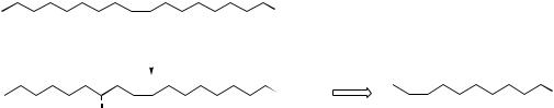
Figure 3.7
∆6,9-octadecadienoate rather than linoleate. However, animals need linoleate for the biosynthesis of dihomo-γ-linolenate (∆8,11,14-eicosatrienoate) and arachidonate (∆5,8,11,14-eicosatetraenoate), C20 polyunsaturated fatty acid precursors of
prostaglandins in the ‘one’ and ‘two’ series respectively (see page 52). Accordingly, linoleic acid must be obtained from plant material in the diet, and it is desaturated towards the carboxyl to yield γ-linolenate, which is then used as the

46 |
THE ACETATE PATHWAY |
substrate for further chain extension, adding a C2 unit from malonate, and producing dihomo- γ-linolenate. Arachidonate derives from this by additional desaturation, again towards the carboxyl
end of the chain (Figure 3.7). α-Linolenate is similarly a precursor on the way to ∆5,8,11,14,17-
eicosapentaenoate (EPA), required for the synthesis of prostaglandins of the ‘three’ series, and it is also obtained from the diet. A similar chain extension process using further molecules of malonate is encountered in the sequence from α- linolenate in animal systems (Figure 3.7). Chain extension/dehydrogenations lead to formation of eicosapentaenoate (EPA) with further elaborations producing docosapentaenoate (DPA) and then docosahexaenoate (DHA). DHA is a component of lipids in sperm, the retina, and the brain. It is thought to be important for brain development, and deficiency is associated with abnormalities in brain function. Linoleate and α-linolenate are referred to as ‘essential fatty acids’ (EFAs) since they and their metabolites are required in the diet for normal good health. Some food sources such as the
oils present in fish are rich in the later metabolites derived from α-linolenic acid, e.g. EPA and DHA, and are also beneficial to health. Since these fatty acids all have a double bond three carbons from the methyl end of the chain, they are grouped together under the term ω-3 fatty acids (omega- 3 fatty acids). Regular consumption of fish oils is claimed to reduce the risk of heart attacks and atherosclerosis.
Although most plant-derived oils contain high amounts of unsaturated fatty acid glycerides, including those of linoleic and α-linolenic acids, the conversion of linoleate into γ-linolenate can be blocked or inhibited in certain conditions in humans. This restricts synthesis of prostaglandins. In such cases, the use of food supplements, e.g. evening primrose oil from Oenothera biennis (Onagraceae), which are rich in γ-linolenic esters, can be valuable and help in the disorder. Many plants in the Boraginaceae, e.g. borage (Borago afficinalis), also accumulate significant amounts of γ-linolenic acid glycerides, as does evening primrose, indicating their unusual ability
Evening Primrose Oil
Evening primrose oil is extracted from the seeds of selected strains of the evening primrose (Oenothera biennis; Onagraceae), a biennial plant native to North America, which is now widely cultivated in temperate countries. The seeds contain about 24% fixed oil, which has a high content of glycerides of the unsaturated fatty acids linoleic acid (65–80%) and γ-linolenic acid (gamolenic acid) (7–14%). Because of this high γ-linolenic acid content, evening primrose oil is widely used as a dietary supplement, providing additional quantities of this essential fatty acid, which is a precursor in the biosynthesis of prostaglandins, which regulate many bodily functions (see page 54). Genetic and a number of other factors may inhibit the desaturation of linoleic acid into γ-linolenic acid. Ageing, diabetes, excessive alcohol intake, catecholamines, and zinc deficiency have all been linked to inhibition of the desaturase enzyme. The conversion may also be inhibited if there is a high proportion of fatty acids in the diet, which compete for the desaturase enzyme, including saturated and trans-unsaturated fatty acids. The latter group may be formed during the partial hydrogenation of polyunsaturated fatty acids which is commonly practised during food oil processing to produce semi-solid fats. Evening primrose oil appears to be valuable in the treatment of premenstrual tension, multiple sclerosis, breast pain (mastalgia), and perhaps also in eczema. There is potential for further applications, e.g. in diabetes, alcoholism, and cardiovascular disease. In evening primrose, γ-linolenic acid is usually present in the form of a dilinoleoylmono-γ-linolenylglycerol. This triglyceride is also being explored as a drug material for the treatment of diabetes-related neuropathy and retinopathy. γ-Linolenic acid is also found in the fixed oil of other plants, e.g. blackcurrant, comfrey, and borage, and in human milk. Borage oil (starflower oil) from the seeds of Borago officinalis (Boraginaceae) is used in the same way as evening primrose oil. It contains higher concentrations of γ-linolenic acid (23–26%), but rather less linoleic acid.
|
|
|
|
ACETYLENIC FATTY ACIDS |
47 |
|
|
|
|
|
CO2H |
|
|
|
|
|
|
|
|
|
|
oleic acid |
|
|
|||
|
|
|
|
O2 / NADPH |
|
|
|
|
|
|
|
|
|
|
|
|
|
pyrolysis |
|
CO2H |
OH |
|
|
|
CO2H |
|
|
|
|
|
|
|||
|
ricinoleic acid |
|
undecenoic acid |
|||
|
|
|
|
|||
Figure 3.8
to desaturate linoleic esters towards the carboxyl terminus, rather than towards the methyl terminus as is more common in plants. Arachidonic acid itself has not been found in higher plants, but does occur in some algae, mosses, and ferns.
Ricinoleic acid (Figure 3.8) is the major fatty acid found in castor oil from seeds of the castor oil plant (Ricinus communis; Euphorbiaceae),
and |
is the 12-hydroxy derivative of oleic acid. |
It is |
formed by direct hydroxylation of oleic |
acid (usually esterified as part of a phospholipid) by the action of an O2- and NADPH-dependent mixed function oxidase, but this is not of the cytochrome P-450 type. Castor oil has a long history of use as a domestic purgative, but it is now mainly employed as a cream base. Undecenoic acid (∆9-undecenoic acid) can be obtained from ricinoleic acid by thermal degradation, and as the zinc salt or in ester form is used in fungistatic preparations.
Primary amides of unsaturated fatty acids have been characterized in humans and other mammals, and although their biological role is not fully understood, they may represent a group of important signalling molecules. Oleamide, the simple amide of oleic acid, has been shown to be a sleepinducing lipid, and the amide of erucic acid, erucamide, stimulates the growth of blood vessels.
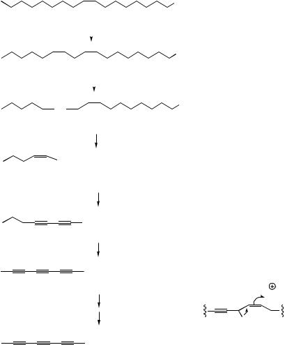
ACETYLENIC FATTY ACIDS
Many unsaturated compounds found in nature contain one or more acetylenic bonds, and these are predominantly produced by further desaturation of olefinic systems in fatty acid-derived molecules. They are surprisingly widespread in nature, and are found in many organisms, but are especially common in plants of the Compositae/Asteraceae, the Umbelliferae/Apiaceae, and fungi of the group
Basidiomycetes. These compounds tend to be highly unstable and some are even explosive if sufficient amounts are accumulated. Since only very small amounts are present in plants, this does not present any widespread hazard. Whilst fatty acids containing several double bonds usually have these in a non-conjugated array, molecules containing triple bonds tend to possess conjugated unsaturation. This gives the compounds intense and highly characteristic UV spectra which aids their detection and isolation.
The processes of desaturation are exemplified in Figure 3.9, in which oleic acid (probably as a thiol ester) features as a precursor of crepenynic acid and dehydrocrepenynic acid. The acetylenic bond is now indicated by a in the semi-systematic shorthand nomenclature. Chain shortening by β- oxidation (see page 18) is often a feature of these pathways, and formation of the C10 acetylenic acid dehydromatricaria acid proceeds through C18 intermediates, losing eight carbons, presumably via four β-oxidations. In the latter part of the pathway, the Z -double bond from oleic acid moves into conjugation with the polyacetylene chain via an allylic isomerization, giving the more favoured E -configuration. Some noteworthy acetylenic structures (though they are no longer acids and components of fats) are given in Figure 3.10. Cicutoxin from the water hemlock (Cicuta virosa; Umbelliferae/Apiaceae) and oenanthotoxin from the hemlock water dropwort (Oenanthe crocata; Umbelliferae/Apiaceae) are extremely toxic to mammals, causing persistent vomiting and convulsions, leading to respiratory paralysis. Ingestion of the roots of these plants may frequently lead to fatal poisoning. Falcarinol is a constituent of Falcaria vulgaris (Umbelliferae/Apiaceae), Oenanthe crocata, Hedera helix (Araliaceae), and several other plants, and is known to cause contact dermatitis in certain
48 |
|
|
|
|
|
|
|
|
|
|
THE ACETATE PATHWAY |
|
|
|
|
|
|
|
|
|
|
|
CO2H |
|
|
|
|
|
|
|
|
|
|
|
|
|
|
|
oleic acid |
18:1 (9c) |
|||||||
|
|
|
|
|
|
|
|
|
|
|
CO2H |
|
|
|
|
|
|
|
|
|
|
|
|
|
|
|
|
|
|
|
|
|
|
|
|
|
|
|
|
|
|
|
|
|
|
|
|
linoleic acid 18:2 (9c,12c) |
|||||||||||
|
|
|
|
|
|
|
|
|
|
|
CO2H |
|
|
|
|
|
|
|
|
|
|
|
|
|
|
|
|
|
|
|
|
|
|
|
|
|
|
|
|
|
|
|
|
|
|
|
|
|
|
|
|
|
|
|
|
|
|
|
|
|
|
|
|
|
|
||||||
crepenynic acid |
18:2 (9c,12a) |
||||||||||


 CO2H
CO2H
dehydrocrepenynic acid 18:3 (9c,12a,14c)


 CO2H 18:3 (9c,12a,14a)


 CO2H
CO2H
18:4 (9c,12a,14a,16a)
H
β-oxidations
H
allylic isomerization
 CO2H
CO2H
dehydromatricaria acid 10:4 (2t,4a,6a,8a)
Figure 3.9
individuals when the plants are handled. Falcarinol (sometimes called panaxynol) and the structurally related panaxytriol are also characteristic polyacetylene components of ginseng (Panax ginseng; Araliaceae) (see page 222). Wyerone from the broad bean (Vicia faba; Leguminosae/Fabaceae) has antifungal properties, and its structure exemplifies how the original straight chain may be crosslinked to produce a ring system. The furan ring may originate from a conjugated diyne.
The herbal preparation echinacea is derived from the roots of Echinacea purpurea (Compositae/ Asteraceae) and is used for its immunostimulant properties, particularly as a prophylactic and
treatment for the common cold. At least some of its activity arises from a series of alkylamides, amides of polyunsaturated acids with isobutylamine. These acids are predominantly C11 and C12 diene-diynes (Figure 3.11).
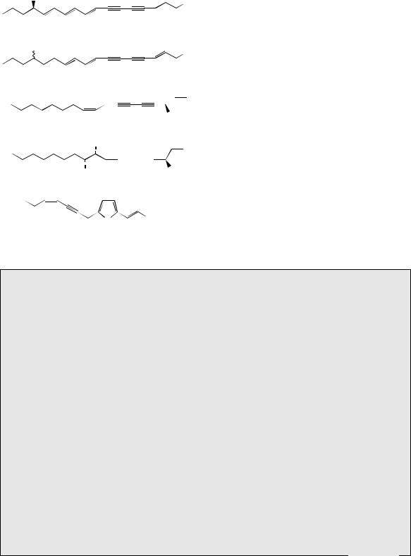
BRANCHED-CHAIN FATTY ACIDS
Whilst straight-chain fatty acids are the most common, branched-chain acids have been found to occur in mammalian systems, e.g. in wool fat and butter fat. They are also characteristic fatty acid constituents of the lipid part of cell walls in some
BRANCHED-CHAIN FATTY ACIDS |
49 |
OH
OH
cicutoxin
OH
OH
oenanthotoxin
falcarinol (panaxynol) |
|
OH |
||||
|
|
|
||||
|
OH |
|
|
|
||
|
|
|
|
|
||
|
|
|
|
|
OH |
|
|
|
|
|
|
||
|
|
|
|
|
||
OH |
|
|||||
panaxytriol |
|
|
|
|||
|
O |
CO2Me |
||||
|
||||||
|
|
|
|
|
|
|
O wyerone
Figure 3.10
pathogenic bacteria. Several mechanisms appear to operate in their formation. Thus, the structure of corynomycolic acid from Corynebacterium diphtheriae can be rationalized from a combination of two palmitic acid units (Figure 3.12). Methyl sidechains can be introduced by using methylmalonylCoA instead of malonyl-CoA as the chain
extending agent |
(Figure 3.13). |
Methylmalonyl- |
CoA arises by |
biotin-dependent carboxylation |
|
of propionyl-CoA in exactly |
the same way |
|
as malonyl-CoA was formed (see page 17).
2,4,6,8-Tetramethyldecanoic acid found in the
preen gland |
wax of the goose (Anser anser) |
is produced |
from an acetyl-CoA starter, and |
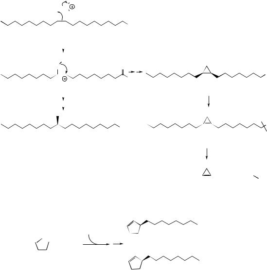
four methylmalonyl-CoA chain extender units. The incorporation of propionate as well as acetate is also a feature of many microbial antibiotic structures (see page 17). However, in other examples, methyl side-chains can be produced by a C-alkylation mechanism using S-adenosylmethionine (SAM). Tuberculostearic acid (Figure 3.14) found in Mycobacterium tuberculosis, the bacterium causing tuberculosis, is derived from oleic acid by alkylation on C-10,
Echinacea
Echinacea consists of the dried roots of Echinacea purpurea, E. angustifolia, or E. pallida
(Compositae/Asteraceae), herbaceous perennial plants indigenous to North America, and widely cultivated for their large daisy-like flowers, which are usually purple or pink. Herbal preparations containing the dried root, or extracts derived from it, are promoted as immunostimulants, particularly as prophylactics and treatments for bacterial and viral infections, e.g. the common cold. Tests have validated stimulation of the immune response, though the origins of this activity cannot be ascribed to any specific substance. Activity has variously been assigned to lipophilic alkylamides, polar caffeic acid derivatives, high molecular weight polysaccharide material, or to a combination of these. Compounds in each group have been demonstrated to possess some pertinent activity, e.g. immunostimulatory, anti-inflammatory, antibacterial or antiviral effects. The alkylamides comprise a complex mixture of unsaturated fatty acids as amides with 2-methylpropanamine (isobutylamine) or 2-methylbutanamine, amines which are probably decarboxylation products from valine and isoleucine respectively. The acid portions are predominantly C11 and C12 diene-diynes or tetraenes (Figure 3.11). These compounds are found throughout the plant though relative proportions of individual components vary considerably. The root of E. purpurea contains at least 12 alkylamides (about 0.6%), of which C12 diene-diynes predominate; levels of these compounds fall significantly during drying and storage. Caffeic acid derivatives present include caffeic acid (see page 132), chlorogenic acid (5-O-caffeoylquinic acid, see page 132), 2-O- caffeoyltartaric acid, and cichoric acid (2,3-di-O-caffeoyltartaric acid) (Figure 3.11). Cichoric acid is a major component (0.6–2.1%) in E. purpurea, but only minor in the other species.
(Continues)

50 |
THE ACETATE PATHWAY |
(Continued ) |
|
|
|
|
|
|
|
|
|
|
O |
|
|
O |
E |
|
N |
Z |
E |
|
|
|
|
N |
|
|
H |
||
R |
|
|
|
||
|
|
|
O |
||
R = H or Me |
|
H |
Z |
|
|
|
|
|
|||
|
|
|
|
||
E |
|
O |
|
|
N |
Z |
|
tetraene alkylamides |
H |
||
R |
|
N |
|
||
R = H or Me |
|
H |
|
|
OH |
|
|
|
|
||
|
|
O |
O |
CO2H |
|
|
|
|
|||
Z |
E |
HO |
O |
O |
OH |
|
|
N |
|
||
|
|
H |
|
HO2C |
O |
diene-diyne alkylamides |
HO |
|
|||
|
|
|
|||
|
|
cichoric acid |
|||
|
|
|
|
||
|
|
Figure 3.11 |
|
|
|
|
|
O |
|
|
OH |
|
|
SCoA |
|
|
|
|
|
SCoA |
|
|
CO2H |
|
|
O |
corynomycolic acid |
||
|
|
|
|
|
|
Figure 3.12
|
|
CO2, ATP |
CO2H |
||||
|
SCoA |
|
SCoA |
||||
|
biotin |
|
|||||
|
|
|
|
|
|
|
|
|
|
|
|
|
|
|
|
O |
|
|
|
O |
|||
propionyl-CoA |
|
|
methylmalonyl-CoA |
||||
|
|
|
SCoA |
|
|
CO2H |
|
|
|
|
|
|
|
|
|
|
|
|
|
|
|
|
|
|
|
O |
|
|
|
||
|
|
|
|
|
|
|
2,4,6,8-tetramethyldecanoic acid |
Figure 3.13
initiated by the double bond electrons. A postulated carbocation intermediate could then be discharged by accepting hydride from NADPH giving tuberculostearic acid. Alternatively, loss of a proton via cyclopropane ring formation could occur giving dihydrosterculic acid. This is known to be dehydrogenated to sterculic acid, an unusual fatty acid containing a highly strained cyclopropene ring. Sterculic acid is present in the seed oil from Sterculia foetida (Sterculiaceae) and with similar cyclopropene acids, e.g. malvalic acid, is present in edible cottonseed oil from Gossypium species
(Malvaceae). Malvalic acid is produced from sterculic acid by chain shortening from the carboxyl end (Figure 3.14). Sterculic acid is an inhibitor of the ∆9-desaturase which converts stearic acid into oleic acid and is potentially harmful to humans in that it can alter membrane permeability and inhibit reproduction.
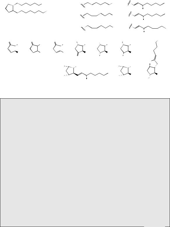
Chaulmoogric and hydnocarpic acids (Figure 3.15) are cyclopentenyl fatty acids found in chaulmoogra oil expressed from seeds of Hydnocarpus wightiana (Flacourtiaceae). These acids are known to arise by malonate chain extension of the
|
|
|
|
|
|
Ad |
|
|
H3C |
|
|
|
S |
SAM |
|
|
|
|
|||||
|
|
|
|
|
|
R |
|
|
|
|
|
|
|
||
|
oleoyl-CoA |
|
|||||
|
|
|
|
|
|||
|
|
|
|
|
|
||
|
|
|
H |
|
|||
|
H2C |
|
|||||
carbocation intermediate |
|||||||
cation discharged by |
|
|
|
|
|
NADPH |
|
|
|
|
|
|
|||
|
|
|
|
|
|||
addition of hydride |
|
|
|
|
|
||
|
|
|
|
|
|
|
|
from NADPH |
|
|
|
|
|
|
|
|
|
|
|
|
|
|
|
tuberculostearic acid
malonyl-CoA

 COSCoA
2-cyclopentenyl- carboxyl-CoA
PROSTAGLANDINS |
51 |
||
|
|
electrophilic addition: |
|
O |
SAM as alkylating agent |
|
|
|
|
||
|
|
SCoA |
|
|
|
|
|
|
|
cation discharged by |
|
|
|
formation of cyclopropane ring |
|
|
|
and loss of proton |
|
O
CO2H
SCoA
dihydrosterculic acid
desaturation
CO2H
CO2H
sterculic acid
α-oxidation


 CO2H malvalic acid
Figure 3.14

 CO2H
CO2H
hydnocarpic acid


 CO2H
CO2H
chaulmoogric acid
Figure 3.15
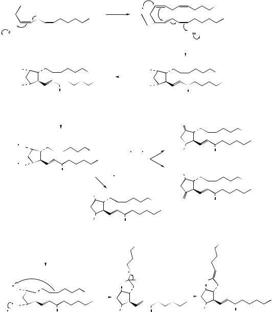
coenzyme A ester of 2-cyclopentenyl carboxylic acid as an alternative starter unit to acetate, demonstrating a further approach to unusual fatty acids. Chaulmoogra oil provided for many years the only treatment for the relief of leprosy, these two acids being strongly bactericidal towards the leprosy infective agent Mycobacterium leprae. Purified salts and esters of hydnocarpic and chaulmoogric acids were subsequently employed, until they were then themselves replaced by more effective synthetic agents.
PROSTAGLANDINS
The prostaglandins are a group of modified C20 fatty acids first isolated from human semen and initially assumed to be secreted by the prostate gland. They are now known to occur widely in animal tissues, but only in tiny amounts, and they have been found to exert a wide variety of pharmacological effects on humans and animals. They are active at very low, hormone-like concentrations and can regulate blood pressure, contractions of smooth

52 |
THE ACETATE PATHWAY |
muscle, gastric secretion, and platelet aggregation. Their potential for drug use is extremely high, but it has proved difficult to separate the various biological activities into individual agents.
The basic prostaglandin skeleton is that of a cyclized C20 fatty acid containing a cyclopentane ring, a C7 side-chain with the carboxyl function, and a C8 side-chain with the methyl terminus. Prostaglandins are biosynthesized from three essential fatty acids, ∆8,11,14- eicosatrienoic acid (dihomo-γ-linolenic acid), ∆5,8,11,14-eicosatetraenoic acid (arachidonic acid), and ∆5,8,11,14,17-eicosapentaenoic acid, which yield prostaglandins of the 1-, 2-, and 3- series, respectively (Figure 3.16) (see below for principles of nomenclature). The three precursors lead to products of similar structure, but with varying levels of unsaturation in the two side-chains. Some of the structures elaborated from arachidonic acid are shown in Figure 3.17. In the first reaction, arachidonic acid is converted into prostaglandin G2 (PGG2) by an oxygenase (cyclooxygenase; COX) enzyme, which incorporates two molecules of oxygen, liberating a compound with both cyclic and acyclic peroxide functions. In arachidonic acid the methylene group flanked by two double bonds is susceptible to oxidation, probably via a free radical process. This may lead to incorporation of oxygen giving the proposed free radical intermediate. Formation of PGG2 is then depicted as a concerted cyclization reaction, initiated by the peroxide radical, in which a second oxygen molecule is incorporated. The
acyclic peroxide group in PGG2 is then cleaved by a peroxidase to yield prostaglandin H2 (PGH2), which occupies a central role and can be modified in several different ways. These modifications can be rationally accommodated by initial cleavage of the cyclic peroxide to the diradical; alternative ionic mechanisms may also be proposed. Quenching of the free radicals by abstraction of hydrogen atoms gives rise to prostaglandin F2α (PGF2α), whilst capture and loss of hydrogen atoms would provide either prostaglandin E2 (PGE2) or prostaglandin D2 (PGD2). The bicyclic system in prostaglandin I2 (PGI2; prostacyclin) is envisaged as arising by involvement of a sidechain double bond, then loss of a hydrogen atom. Prostaglandin structures representative of the 1-series, e.g. PGE1, or of the 3-series, e.g. PGE3, can be formed in a similar way from the appropriate fatty acid precursor (Figure 3.16).
The basic skeleton of the prostaglandins is termed prostanoic acid, and derivatives of this system are collectively known as prostanoids. The term eicosanoids is also used to encompass prostaglandins, thromboxanes, and leukotrienes, which are all derived from C20 fatty acids (eicosanoic acids). Semi-systematic nomenclature of prostaglandins is based on the substitution pattern in the five-membered ring, denoted by a letter suffix (Figure 3.18), and the number of double bonds in the side-chains is given by a numerical subscript. Greek letters α and β are used to indicate the configuration at C-9, α indicating the substituent is below the plane (as
|
|
8 |
|
|
8 |
5 |
8 |
5 |
|
|
|
CO2H |
|||||||||||||
|
|
|
|
|
CO2H |
|
|
|
|
|
|
|
CO2H |
|
|
|
|
|
|
|
|
|
|
|
|
|
|
|
|
|
|
|
|
|
|
|
|
|
|
|
|
|
|
|
|
|
|
|
|||
|
|
|
|
|
|
|
|
|
|
|
|
|
|
|
|
|
|
|
|
|
|
|
|
|
|
|
|
|
|
|
|
|
|
|
|
|
|
|
|
|
|
|
|
|
|
|
|
|
|
|
|
11 |
14 |
|
11 |
|
14 |
|
|
11 |
|
14 |
|
|
17 |
|
|
||||||||||
|
|
dihomo-γ-linolenic (∆8,11,14) |
|
arachidonic (∆5,8,11,14) |
eicosapentaenoic (∆5,8,11,14,17) |
||||||||||||||||||||
O |
|
|
|
CO2H |
O |
|
|
|
|
|
O |
|
|
|
|
|
|
|
|
|
|||||
HO |
|
|
|
HO |
|
|
|
|
CO2H |
HO |
|
|
|
|
|
|
|
|
CO2H |
||||||
|
|
OH |
|
|
|
|
|
|
OH |
|
|
|
|||||||||||||
|
|
|
|
OH |
|
|
|
|
|
|
|||||||||||||||
|
|
|
|
|
|
|
|
|
|
||||||||||||||||
|
|
|
PGE1 |
|
|
|
|
PGE2 |
|
|
|
|
PGE3 |
|
|
|
|
||||||||
Figure 3.16
PROSTAGLANDINS |
53 |
methylene flanked by double bonds is susceptible to free radical oxidation; free radical reaction allows addition of O2 and formation of peroxide radical


 CO2H H
O
O |
arachidonic acid |
|
concerted formation of cyclic peroxide, cyclopentane ring, and acyclic peroxide by addition of second molecule of O2; mechanistically, this is analogous to the first step but exploits the unsaturation; the peroxide radical finally abstracts a H atom
cyclooxygenase |
CO2H |
|
(COX) |
||
O |
||
|
O |
|
|
O O |
|
|
|
|
|
|
|
|
cleavage of acyclic |
|
|
|
|
|
|
|
|||||||
|
|
|
|
|
|
|
|
|
|
|
|
|
|
|
||||||||
|
|
|
|
|
|
|
|
peroxide |
|
|
|
|
|
|
|
|
|
|
||||
O |
|
|
|
|
CO2H |
peroxidase |
O |
|
|
|
|
|
|
CO2H |
||||||||
|
|
|
|
|
|
|
|
|
|
|
|
|
|
|
|
|
|
|
|
|
||
|
|
|
|
|
|
|
|
|
|
|
|
|
|
|
|
|
|
|
|
|
|
|
O |
|
|
|
|
|
O |
|
|
|
|
|
|
|
|||||||||
|
|
|
|
OH |
|
|
|
|
|
cyclic |
|
|
O |
|
OH |
|||||||
|
|
|
|
|
|
|
|
|
peroxide |
|
|
|
||||||||||
|
|
PGH2 |
|
|
|
|
|
|
|
|
|
acyclic peroxide |
||||||||||
|
|
|
|
|
|
|
|
|
|
|
|
|
|
|||||||||
|
|
|
|
|
|
|
|
|
|
PGG2 |
|
|
|
|
||||||||
|
|
|
|
|
|
|
|
|
|
|
|
|
|
|
|
|
|
|
|
|||
radical cleavage |
|
|
|
|
|
|
|
|
|
|
|
O |
|
|
|
|
||||||
|
|
|
|
|
|
|
|
|
|
|
|
|
|
|||||||||
of cyclic peroxide |
|
|
|
|
|
|
|
|
|
|
|
|
|
|
|
|||||||
|
O |
|
|
|
|
|
|
|
|
|
|
|
|
|
|
CO2H |
||||||
|
|
|
|
|
|
|
|
|
|
|
|
|
|
|
||||||||
|
|
|
|
|
|
|
|
|
|
|
|
|
|
|
|
|||||||
|
|
|
|
|
|
|
CO2H |
|
|
|
– H , + H |
HO |
|
|
OH |
|||||||
|
|
|
|
|
|
|
|
|
||||||||||||||
|
O |
|
|
|
|
|
|
|
|
HO |
|
PGE2 |
||||||||||
|
|
|
|
|
|
|
|
|
|
|||||||||||||
|
|
|
|
|
|
|
|
|
|
|
|
|
||||||||||
|
|
|
|
OH |
|
|
|
|
|
|
|
|
|
|
|
|
||||||
|
|
|
|
|
|
|
+ 2H |
|
|
|
|
|
|
|
|
|
CO2H |
|||||
|
|
|
|
|
|
|
HO |
|
|
|
|
|
|
|
|
|
|
|
|
|
|
|
|
|
|
|
|
|
|
|
|
|
|
|
|
|
|
|
|
|
|
|
|
|
|
|
|
|
|
|
|
|
|
|
|
|
|
|
|
|
CO2H |
|
O |
|
|
OH |
||
|
|
|
|
|
|
|
|
|
|
|
|
|
|
|
|
|
||||||
|
|
|
|
|
|
|
|
|
|
|
|
|
|
|
|
|
|
|
|
PGD2 |
||
|
|
|
|
|
|
|
HO |
|
|
|
OH |
|
|
|
|
|
|
|
|
|
|
|
|
|
|
|
|
|
|
|
PGF2α |
|
|
|
|
|
|
|
|
|
|
||||
|
|
|
|
|
|
|
|
|
|
|
|
CO2H |
|
|
|
|
|
|
CO2H |
|||
|
|
|
|
|
|
|
|
|
|
|
|
|
|
|
|
|
|
|
||||
|
|
|
|
|
|
|
|
|
|
|
|
|
|
|
|
|
|
|
|
|
|
|
|
|
|
|
|
O |
H |
|
|
O |
|
|
|
|
|
|
|
|
|
|
|
|
|
O |
|
|
|
|
|
|
|||
|
|
|
CO2H |
|
|
|
|
|
|
|
|
O |
|
|
|
|
HO |
|
|||
H |
|
OH |
HO |
|
OH |
|
OH |
|||
|
|
|
|
|
|
|
|
|
|
PGI2 |
|
|
|
|
|
Figure 3.17 |
|
|
|
||
found in natural prostaglandins), and β indicating |
|
abbreviated to PG. Prostaglandins A, B, and C |
||||||||
the substituent is above the plane (as in some |
|
are inactive degradation products from the natural |
||||||||
synthetic |
analogues). ‘Prostaglandin’ is |
usually |
|
prostaglandins. |
|
|
|
|||
54 |
|
|
|
|
|
|
|
THE ACETATE PATHWAY |
|
|
|
|
|
|||||
|
|
|
|
|
|
1 |
|
|
|
|
|
|
R1 |
|
|
R2 |
||
9 |
7 |
|
5 |
3 |
|
|
|
|
|
|
|
|
|
CO2H |
|
|
|
|
6 |
|
CO |
H |
|
|
|
|
|
|
|
|
|
|
|||||
8 |
4 |
2 |
1-series |
|
|
|
|
|
|
|
|
|
||||||
|
|
|
2 |
|
|
|
|
|
|
|
|
|
|
|
||||
10 |
|
|
|
|
|
|
|
|
|
|
|
|
|
|
|
OH |
||
|
12 |
14 |
16 |
18 |
|
20 |
|
|
|
|
|
|
|
|
||||
11 |
|
|
|
|
|
|
|
|
|
|
|
|
||||||
13 |
|
15 |
17 |
|
19 |
|
2-series |
|
|
|
|
|
|
CO2H |
|
|
|
|
|
|
|
|
|
|
|
|
|
|
|
|
|||||||
|
|
|
|
|
|
|
|
|
|
|
|
|
|
|
|
|
||
|
prostanoic acid |
|
|
|
|
|
|
|
|
|
|
OH |
||||||
|
|
|
|
|
|
|
|
|
|
|
|
|
|
|
|
|||
|
|
|
|
|
|
|
|
3-series |
|
|
|
|
|
|
CO2H |
|
|
|
|
|
|
|
|
|
|
|
|
|
|
|
|
|
|
|
|
||
|
|
|
|
|
|
|
|
|
|
|
|
|
|
|
|
|
|
|
|
|
|
|
|
|
|
|
|
|
|
|
|
|
|
|
OH |
||
O |
|
|
O |
|
|
|
O |
HO |
|
|
|
|
O |
|
HO |
|
|
CO2H |
|
R1 |
|
|
|
R1 |
|
R1 |
R1 |
R1 |
|
R1 |
|||||||
|
R2 |
|
|
|
R2 |
|
R2 |
R2 |
R2 |
|
R2 |
|||||||
|
|
|
|
|
|
|||||||||||||
PGA |
PGB |
PGC |
O PGD |
|
HO PGE |
|
HO PGF |
O |
|||
|
|
|
|
|
|
|
|
|
|
|
|
|
|
O |
R1 |
|
|
|
O |
R1 |
|
||
|
|
|
|
|
|
|
|
|
|
R2 |
R2 |
|
|
O |
|
|
|
|
O |
||||
|
|
|
|
PGG |
O |
|
OH |
|
PGH |
|
HO |
|
|
|
|
|
|
|
|||||
|
|
|
|
|
|
|
|
|
PGI |
||
Figure 3.18
Prostaglandins
Prostaglandins occur in nearly all mammalian tissues, but only at very low concentrations. PGE1 and PGF1α were initially isolated from sheep seminal plasma, but these compounds and PGD2, PGE2, and PGF2α are widely distributed. Animal sources cannot supply sufficient amounts for drug usage. The soft coral Plexaura homomalla (sea whip) from the Caribbean has been identified as having very high (2–3%) levels of prostaglandin esters, predominantly the C-15 epimer of PGA2 (1–2%) with related structures. Prostaglandins of the A-, E-, and F-types are widely distributed in soft corals, especially Plexaura, but these are unlikely to provide a satisfactory and renewable natural source. Considerable effort has been exerted on the total synthesis of prostaglandins and their interconversions, and the high level of success achieved has opened up the availability of compounds for pharmacological testing and subsequent drug use. Synthetic analogues have also been developed to modify or optimize biological activity. The studies have demonstrated that biological activity is effectively confined to the natural enantiomers; the unnatural enantiomer of PGE1 had only 0.1% of the activity of the natural isomer.
The prostaglandins display a wide range of pharmacological activities, including contraction and relaxation of smooth muscle of the uterus, the cardiovascular system, the intestinal tract, and of bronchial tissue. They may also inhibit gastric acid secretion, control blood pressure and suppress blood platelet aggregation. Some of these effects are consistent with the prostaglandins acting as second messengers, modulating transmission of hormone stimulation and thus metabolic response. Some prostaglandins in the A and J series have demonstrated potent antitumour properties. Since the prostaglandins control many important physiological processes in animal tissues, their drug potential is high, but the chances of precipitating unwanted side-effects are also high, and this has so far limited their therapeutic use.
(Continues)