

De Cuyper M., Bulte J.W.M. - Physics and chemistry basis of biotechnology (Vol. 7) (2002)(en)
.pdfUrs Häfeli
6. Therapeutic uses of radioactive microspheres
Many radiolabelled particles, microspheres and liposomes are appropriate for therapy once the encapsulated diagnostic radioisotope has been exchanged for a therapeutic one from the a- or b-emitter group. Typical uses in the last 20 to 40 years include local applications for the treatment of rheumatoid arthritis, liver tumours and cystic brain tumours. However, their use remains experimental because of smaller than expected target uptake, unwanted toxicity and insufficient treatment effects that have resulted from radiochemical instability and suboptimal biodistribution of the radiopharmaceutical. In addition, there exists a general negative attitude towards the use of radioactive substances in spite of proven superior results of many radiation therapies [102-104]. What follows is a review of a few a-emitter applications as well as the more established b -emitter therapies.
6.1. THERAPY WITH ALPHA-EMITTING MICROSPHERES
Different a-emitters have been tested in ovarian cancer mouse models. Microspherebound 211At, for example, was applied in mice with ovarian cancer metastases and was found to be more effective than the b-emitting 32P- and 90Y-microspheres [63]. It was, however, also shown that the amount of radioactivity had to be tailored carefully. More than 1 MBq of 211At per animal led to shorter survival times of the treated mice. This effect is very likely due to the instability of 211At which is highly toxic to the lymphatic tissue and thyroid gland when leakage occurs. First clinical trials with the same α- emitter bound to albumin microspheres have been reported by Wunderlich et al. [105]. The authors injected the microspheres into the arteries leading to tongue and larynx tumours. After 4 hours, 80% of the radioactivity was bound to the tongue and 12% to the lungs. The rest was found in the abdomen. The tongue tumour was completely ablated, and no side effects or recurrences were observed at 2 year follow-up. Another a-emitter, 212Pb, in the form of radioactive colloids [ 106] was also investigated in an ovarian cancer mouse model. Tumour necrosis and decrease in ascites was observed in a dose-related manner, with acute gastro-intestinal toxicity developing at the highest doses. The therapeutically effective radioisotope in these experiments was 212Pb's daughter nuclide 212Bi (Table 2).
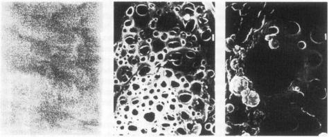
To increase the limited range of α-emitters (see general properties of α-emitters), the radiopharmaceutical should be delivered close to the tumour from where it releases the radioisotope, allowing it to diffuse into the surrounding area. Ideally, the released radioactivity binds to the tumour's cell surface and not to the surrounding normal tissue, something that could be accomplished, for example, by pre-targeting the cancer cells with an antibody metallothionein bioconjugate. This approach has been tested in vitro with the biodegradable polymer mixture of PHEA (= a,b-poly(hydroxyethyl)-D,L-
aspartamide) and Pluronic enclosing 212Bi. Within 1 hour, |
the polymer began to |
resemble Swiss cheese, with its many small holes of about 1 |
µm in diameter (Figure |
7). The size of the holes further increased, and after 2 days, |
more than 75% of the |
radioactivity had been set free [107]. The polymer tested was in the form of a paste, but microspheric radiopharmaceutical delivery forms are also possible. This approach is
234
Radioactive microspheres for medical applications
limited by the difusion distances of the radioisotope [108]. In a chopped meat model, 212Bi has been shown to diffuse a maximum distance of 10 mm. It thus may only be useful for the treatment of very small tumours, metastases or leftover tumour cells from incompletely resected tumours.
Figure 7 Scanning electron microscopy picture of the biodegradable polymer PHEAP immediately after preparation (left), after I hour (middle) and after 24 hours (right) at 37 ºC in PBSpH 7 4 The bar to the right represents 1 µm
6.2. THERAPY WITH BETA-EMITTING MICROSPHERES
One of the first applications of a-emitting microspheres was the treatment of inaccessible tumours [109]. In this approach termed radioembolization therapy, 20 to 50 µm microspheres are injected into the artery leading to the tumour. Since the microspheres are larger than the newly formed capillaries in the tumour, they are trapped and become lodged in the tumour from where they irradiate the surrounding cancerous tissue with radiation doses 20 to 30 times higher than what is achievable with external radiation therapy. This approach, pioneered with 65Znand 198Au-microspheres by Muller and Rossier in Switzerland [26], was further investigated with 198Auand 90Y-microspheres in many types of tumours by Ariel [110-112] and with 32P-resin microspheres by Caldarola and Dogliotti [113]. Turner et al. investigated 166Ho-labeled cation exchange resins [ 114] and Häfeli et al. 186Re/188Re-labeled glass microspheres for the same application [65,115]. Currently, radioembolization therapy is primarily used for the treatment of liver tumours, both hepatomas and liver metastases [116]. Since liver tumours get most of their blood supply from their hepatic artery [ 117], the radioactive microspheres injected into this artery are preferentially flushed into the tumour. Radiochemically highly stable glass 90Y microspheres sized 25 to 35 µm (Figure 4) are commercially available for the treatment of liver tumours in Canada and since June 1999 also in the United States (TheraspheresTM ; Nordion, Kanata, Ontario, Canada).
Prior to radioembolization, a diagnostic step is generally performed in order to prevent arterial shunting in the liver. Arterial shunts can divert large amounts of the
235

Urs Häfeli
highly cytotoxic microspheres to the lungs and thus lead to pneumonitis [ 118]. The diagnostic step consists of determining the "shunt index" by injecting 99mTc-labeled macro-aggregated albumin microspheres sized between 1 to 10 µm and imaging their biodistribution. If less than 5% of the radioactivity shunts to the lungs, then β-emitting microspheres such as 90Y-glass microspheres [64] or 90Y-resin microspheres [27] are injected into the hepatic artery of the patient. The first results in a disease which carries a grave prognosis with a survival rate of less than 50% after 1 year are very encouraging. After intra-hepatic injection, the microspheres are preferentially taken up by the tumour at an average ratio of about 4 to 1 (tumour to normal liver ratio) [119,120]. Very high radiation doses without side effects can thus be given. Treating 7 patients with doses of 50 to 100 Gy, Houle showed that the larger doses are necessary for successful treatment results [121], and doses between 80 and 150 Gy are now recommended. Further improvements in treatment outcome are possible by injecting the vasoconstricting agent Angiotensin II immediately after the microsphere injection. Normal hepatic vessels are able to react by constriction, but the developing tumour capillaries are not. As a result, larger amounts of the microspheres are diverted to the tumour bed [113]. The clinical results regarding radioembolization therapy have been described in detail by Harbert [ 109].
Another therapeutic application of b-emitting colloids and microspheres is the radioactive ablation of inflamed synovia in arthritic joints, which has been termed radiosynovectomy or sometimes radiosynoviorthesis. Fellinger and Schmid [122] reported in 1952 the first use of 198 Au gold colloid for the treatment of rheumatoid arthritis in knees. Their results were not very encouraging probably due to underdosing, but they did not give up and later confirmed the value of this treatment [ 123]. Therapy with 198Au has the drawback of a 41 1 keV γ -emission. To overcome this drawback other b-emitters such as 186Re [124], 90 Y [125], 165Dy [46] and 32P-colloids [126] have been investigated. Today, the choice of the radioisotope is entirely based on the size of the joint and the radioisotopes' treatment range (for example, 90Y and 188Re for knee and shoulder, and 186Re and 169Er for finger or elbow) (Table 8).
Table 8: Radioactive microspheres for therapeutic applications
Application |
Type of radioactive microspheres |
Particle size |
|
used |
|
|
|
|
Radioembolization of liver |
90Y-glass microspheres |
25-35 µm |
and spleen tumours |
(TheraspheresTM ) |
|
|
186Re/188Re-glassmicrospheres |
25-35pm |
|
188Re-Aminex A27 microspheres |
20-50 µm |
|
166Ho-Aminex A-5 microspheres |
13 µm |
236

Radioactive microspheres for medical applications
Table 8: Radioactive microspheres for therapeutic applications
Application |
Type of radioactive microspheres |
Particle size |
|
used |
|
|
|
|
Radiosynovectomy of arthritic |
35S-colloid |
0.05-0.6 pm |
joints |
90Y-resin microspheres |
20-50 pm |
|
90Y-silicate, 90Y-citrate |
0.01-1 pm |
|
I65Dy-ferric hydroxide |
2-5 pm |
|
macroaggregates |
|
|
169Er-citrate |
0.1-1 pm |
|
186Re-sulfur-colloid |
30-50 nm |
|
188Re-macro-aggregated albumin |
10-20 pm |
Local radiotherapy |
90Y-labeled poly(lactic acid) |
1-5 or 10-50 |
|
microspheres |
µm |
|
165Dy-acetylacetone poly(lactic |
1-5 or 10-50 |
|
acid) microspheres |
µm |
|
166Ho-acetylacetone poly(lactic |
1-5 or 10-50 |
|
acid) microspheres |
µm |
|
186Re/188Re-labeled poly(lactic acid) |
1-5 or 10-50 |
|
microspheres |
µm |
|
211At-microspheres |
1.8 µm, 3- |
|
212Pb-sulfur colloid |
10 µm |
|
<1 µm |
|
|
212Pb-ferrous(ferric) hydroxide |
<1 µm |
Intracavitary treatment |
chromic 32P-phosphate |
1-2 µm |
(peritoneal ovarian tumour |
90Y-silicate, 90 Y-citrate |
0.01-1µ m |
metastases, cystic brain |
198Au suspensions |
5-25 nm |
tumours) |
|
|
|
|
|
Traditionally used radioactive colloids are not ideal because their small particle size and large size distribution lead to radiation leakage from the joint [126,1271. Higher than desired leakage has also been measured in liposomes filled with 99mTc [128] and in liposomes that contain the chelating DTTA-group covalently bound to cholesterol [129]. In the second case, the chelator was incorporated into the liposomes' phospholipid-wail during preparation and was then able to bind different radioisotopes such as the β-emitter 177Lu (Table 3) and the g-emitter 67Ga (Table 4).
More radiochemically stable and better-defined microspheres of about 5 µm seem to be optimal for retention in joints. Many of the recently developed microspheres such as biodegradable glass microspheres containing 153Sm, 166Ho, 90Y, 165Dy, 186Re or 188Re
237
Urs Häfeli
[ 130], 188Re-labeled albumin microspheres [78], 166Hoor 165Dy-enclosing biodegradable poly(lactic acid) microspheres [67,131] and 90Y- or 186Re/188Re-enclosing biodegradable poly(lactic acid) microspheres [79,132] can be produced in the appropriate size, will biodegrade after complete decay and can easily be made radioactive. More information about radiosynovectomy is available in an extensive review written by Harbert [133]. It covers the medical applications and procedures in detail. The radiation dosimetry of radiosynovectomy is covered by Johnson et al. [30].
Another important area for β-emitting microspheres is their use in the local treatment of tumours. The delivery of these radioactive microspheres has been attempted in several ways. In one of them, radioactive microspheres are directly injected into the tumour. Wang et al., for example, radiolabelled ion exchange resin microspheres with 188Re and injected them directly into rat hepatomas [134]. Twelve out of 15 rats survived longer than 60 days in the treatment group, as compared to 5 out of 15 rats in the control group. In another novel method for the treatment of solid tumours, Order et al. combined embolization therapy and local radiotherapy, injecting first non-radioactive macro-aggregated albumin microspheres followed by colloidal 32P-chromic phosphate [ 135]. The blockage of the capillaries induced before the 32P- injection resulted in a 3-fold increase of colloid uptake, an effect that lasted for at least 48 hours. This technique has been tested in a first clinical phase I trial for the treatment of non-resectable pancreatic cancer [ 136]. Four patients had a complete response with a duration ranging from 2-57 weeks and 5 patients had a partial response with a duration ranging from 4-21 weeks, corresponding to an objective response of 53% (9 of 17 patients). Six of these patients were alive 33-57 weeks after treatment.
At the current time, there is only one approved application for radioactive microspheres in the United States [137]. It is the use of 32P-chromic phosphate colloid for the treatment of cystic brain tumours such as craniopharyngiomas and astrocytomas. The radiocolloid is typically instilled using stereotaxic equipment, either with or without surgical resection or drainage of the cyst. There exists persuasive evidence that this therapeutic approach is as or more efficacious than conventional methods not only for patients with recurrent malignancies, but also for patients receiving primary radiocolloid therapy [ 138]. Radioactive glassand poly(lactic acid)microspheres containing a mixture of 186Re and 188Re have recently been incorporated into a bioadhesive gel of either carboxymethylcellulose or fibrin glue and applied to the surface of growing rat 9L-glioblastomas [139]. The control group's survival was 18 days, whereas 4 out 6 of the treated animals were still alive on day 35, which represented the end of the experiment (Figure 8). The amount of radioactive 186Re and 188Re injected was less than 50 µCi combined. The surviving animals showed no signs of toxicity and had not lost any weight. Such microspheres are now planned for a clinical phase I trial of the treatment of recurrent brain tumour metastases intraoperatively after debulking. This therapeutic approach looks especially promising because the likelihood of local recurrence in these patients is very high [140], and the local radiation with g-emitters could be done in addition to chemotherapy or whole brain irradiation without risking undue toxicity.
238
Radioactive microspheres for medical applications
Figure 8. Treatment of 9L-glioblastoma brain tumours in Sprague Dawley rats The treatment and toxicity group received 50 µCi 186Re and 188Re in 0.5 mg glass microspheres contained in 30 µl of fibrin glue.
Radioactive microspheres filled with magnetite and radiolabelled with the b -emitter 90Y can also be used for targeted cancer therapy. This has been shown with 30% magnetitecontaining poly(lactic acid) microspheres sized 20 to 30 µm that were injected intraperitoneally into C57BL6/N mice and targeted to a subcutaneously growing EL-4 murine lymphoma of about 0.5 g [141]. The injection of microspheres took place inside the peritoneal cavity as far from the tumour as possible. After injection, a round, 2 mm thick rare earth magnet with a diameter of 10 mm was taped directly above the tumour. The magnetic field on top of the magnet was 0.12-0.16 Tesla. A dose dependent decrease in tumour size was observed after the 7 day treatment period (Figure 9). Close examination revealed that 3 out of 4 tumours in the 80 Gy group and 2 out of 4 tumours in the 120 Gy group were completely eradicated, but that the remaining 1 or 2 tumours, respectively, had grown. It was precisely these tumours that had initially been found to be oblong or flattened out, thus causing the magnetic microspheres to be concentrated farther than 5 mm away from the edges of the tumour. Considering that 90% of the dose of 90Y is deposited within 2.8 mm [30], it follows that the tumour cells farther away were undertreated with the applied amount of radioactivity. The tumours which were not eradicated were therefore local treatment failures.
239
Urs Häfeli
Figure 9. Treatment results of subcutaneous EL-4 lymphomas in mice after magnetic targeting of 90Y-PLA microspheres (n = 6). The numbers inside the bars represent the ratio of completely eradicated tumours to the total number of tumours.
7. Considerations for the use of radioactive microspheres
The recent surge in the evaluation and clinical testing of radiopharmaceuticals is closely related to the recent development of user-friendly kits which allow the user to prepare radioactive microspheres or other radiolabelled agents in a hospital’s radiopharmacy. These kits have served to reduce concerns about the safety, cost and handling of radioactive pharmaceuticals. Current manipulations needed in most kit preparations typically include the addition of a radioisotope, incubation for a predetermined length of time between 5 and 60 minutes, verification of the activity of the radiopharmaceutical by a simple measurement in a dose calibrator and, sometimes, a thin layer chromatogram for quality control. Ideally, the kit preparation leads to highly stable radioactive microspheres with no purification needed. Saha gives additional information in an excellent up-to-date introduction into the currently used radionuclides, radionuclide generators and radiopharmaceuticals in a nuclear pharmacy [142]. Saha covers not only all technical aspects of a nuclear pharmacy, but also the radiation regulations and radiation protection aspects.
240
Radioactive microspheres for medical applications
Increased interest in radiopharmaceuticals is also explained by easier access to generator-produced a- and b-emitting radioisotopes. The b-emitter 188Re, for example (Table 3), can now be inexpensively obtained by any hospital radiopharmacy, in the form of a 188W/188Re generator from Oak Ridge National Laboratories. This generator contains the parent nuclide 188W with a half-life of 69.4 days permanently bound to an alumina column. Because of ongoing decay into the daughter-nuclide 188Re, hundreds of mCi of a sterile 188Re-solutioncan be eluted from the column every day over the course of about 3 months [143]. Rhenium-188 is currently being tested in clinical trials for the radioactive treatment of restenosis [144], for local cancer therapy [134,145,146], for radioembolization therapy [147] and for radioimmunotherapy [148].
Two of the most important parameters for the in vivo use of radiopharmaceuticals are their stability and target specificity. The stability can be improved by using more specific chelators and by attaching the chelators to the microspheres through a linker that does not interfere with their metal-binding properties. Many of these stability issues have already been optimised for radiolabelled antibodies [149] and can thus be directly applied to radioactive microspheres and their kit preparation. The second parameter, target specificity, can be addressed by using additional surface chemistry to modify and functionalise the microspheres' surface. This allows for circulation time optimisation and foreign body response minimisation. In addition, more specific targeting of microspheres to areas other than the reticuloendothelial system (mainly liver and spleen) is possible, as well as the modification of the microspheres' adsorption behaviour to blood proteins [150].
Also important for the application of therapeutic radioactive microspheres is that the radioisotope be chosen for radiobiological and dosimetric reasons. The target size should, for example, be matched with the radiation range of the radioisotope, thus maximising the therapeutic effect and minimising the toxicity [15 11. Also, dose rates should be taken into account and radioisotopes chosen so that dose rate and total dose deposited are optimal for the target lesion [152]. These parameters are not yet well established, but are nevertheless important and should be investigated further.
Another area for the optimisation of radioactive microspheres is research into the most appropriate size of microspheres for in vivo application. It has long been known that differently sized microspheres of identical composition show different biodistribution profiles [153,154]. In addition, for both diffusion and erosion release mechanisms, the release rate of the encapsulated drugs is theoretically dependent on the available surface area. Smaller microspheres with a much larger surface area should thus release their contents at a much faster rate with all the other parameters being equal. Unfortunately, no homogenous, mono-sized microspheres made from biodegradable materials are currently available.
The use of radioactive microspheres is the basis of a large variety of wellestablished and original concepts for future biomedical, diagnostic and therapeutic applications. Optimally, radioactive microspheres should be combined with biologically active molecules such as proteins, peptides, hormones, lectins, and antibodies. This will allow for the diagnosis and treatment of many different diseases with microsurgical precision and will lead to better treatment concepts with fewer side effects.
241
Urs Häfeli
References
1.Wilder RB, DeNardo GL, and DeNardo SJ. Radioimmunotherapy: Recent results and future directions, J Clin. Oncol. 14: 1383-1400 (1996).
2.Papatheofanis FJ and Munson L. Peptide radiopharmaceutical imaging. Appl. Radiol. June: 11-17 (1994).
3. |
Cleland JL and Jones AJS. Stable formulations of recombinant human growth hormone and interferon- |
|
6 for microencapsulation in biodegradable microspheres. Pharmaceuf, Res. 13: 1464-1475 (1996). |
4.Mehta RC, Jeyanthi R, Calis S, Thanoo BC, Burton KW, and DeLuca PP. Biodegradable microspheres as depot system for parenteral delivery of peptide drugs. J. Contr. Rel. 29: 375-384 (1994).
5.Mathiowitz E, Jacob JS, Jong YS, Carino GP, Chickering DE, Chaturvedi P, Santos CA, Vijayaraghavan K, Montgomery S, Bassett M, and Morrell C. Biologically erodable microspheres as potential oral drug delivery systems. Nature 386: 410-414 (1997).
6.Langer R. Drug delivery and targeting. Nature 392: 5-10 (1998).
7.Chen H and Langer R. Oral particulate delivery: status and future trends. Adv. Drug Del. Rev. 34: 339350 (1998).
8.Muir W, Husband AJ, Gipps EM, and Bradley MP. Induction of specific IgA responses in rats after oral vaccination with biodegradable microspheres containing a recombinant protein. Immunol. Lett. 42: 203-207 (1994).
9.Hanes J, Chiba M, and Langer R. Polymer microspheres for vaccine delivery. Pharm. Biotech. 6: 389-
412 (1995).
10Smith OP, Hann IM, Cox H, and Novelli V. Visceral leishmaniasis: rapid response to AmBisome treatment. Arch. Dis. Childhood 73: 157-159 (1995).
11.Codde JP, Lumsden AJ, Napoli S, Burton MA, and Gray BN. A comparative study of the anticancer efficacy of doxorubicin carrying microspheres and liposomes using a rat liver tumour model.
Anticancer Research 13: 539-544 (1993).
12.Treleaven JG. Bone marrow purging: An appraisal of immunological and non-immunological methods. Adv. Drug Del. Rev. 2/3: 253-269 (1988).
13.Arshady R. Polymer supports, reagents and catalysts. In Arshady R (Ed.). Microspheres, microcapsules and liposomes. Citus Books, London, 1999, pp. 197-235.
14.Mikhalovsky SV. Microparticles for haemoperfusion and extracorporeal therapy. In Arshady R (Ed.). Microspheres, microcapsules and liposomes. Citus Books, London, 1999, pp. 133-169.
15.Bangs LB. Microspheres for medical diagnostics: Specific tests and assays. In Arshady R (Ed.). Microspheres, microcapsules and liposomes. Citus Books, London, 1999, pp. 7 1-96,
16.Flandroy PMJ, Grandfils C, and Jerome RJ. Clinical applications of microspheres in embolization and chemoembolisation: A comprehensive review and perspectives. In Rolland A (Ed.). Pharmaceutical particulate carriers: Therapeutic applications. Marcel Dekker Inc., New York, 1993, pp. 321-366.
17.Boschetti E and Schwarz A. Polymer microbeads: Biological applications. In Arshady R (Ed.). Microspheres, microcapsules and liposomes. Citus Books, London, 1999, pp. 191 -224.
18.Papisov MI. Modelling in vivo transfer of long-circulating polymers (two classes of long circulating polymers and factors affecting their transfer in vivo). Adv. Drug Del. Rev. 16: 127-139 (1995).
19.Torchilin VP and Trubetskoy VS. Which polymers can make nanoparticulate drug carriers longcirculating? Adv. Drug Del. Rev. 16: 141-155 (1995).
20.Ackerman NB. The blood supply of experimental liver metastases. IV. Changes in vascularity with increasing tumour growth. Surgery 75: 589-596 (1974).
21.Gupta PK. Review article: Drug targeting in cancer chemotherapy: A clinical perspective. J. Pharm. Sci. 79: 949-962 (1990).
22.Roser M, Fischer D, and Kissel T. Surface-modified biodegradable albumin nanoand microspheres. Part II: Effect of surface charges on in vitro phagocytosis and biodistribution in rats. Eur. J. Pharm. Biopharm. 46: 255-263 (1998).
23.Macklis FW, Kinsey BM, Kassis AI, Ferrara JLM, Atcher RW, Hines JJ, Coleman CN, Adelstein SJ, and Burakoff SJ. Radioimmunotherapy with alpha-particle-emitting immunoconjugates. Science 240: 1024-1026 (1988).
24.Humm JL, Macklis RM, Bump K, Cobb LM, and Chin LM. Internal dosimetry using data derived from autoradiographs. J. Nucl. Med. 34: 1811-1817 (1993).
242
Radioactive microspheres for medical applications
25.McDevitt MR, Sgouros G, Finn RD, Humm JL, Jurcic JG, Larson SM, and Scheinberg DA. Radioimmunotherapy with alpha-emitting nuclides. Eur. J. Nucl. Med. 25: 1341-1351 (1998).
26.Muller JH and Rossier PH. A new method for the treatment of cancer of the lungs by means of artificial radioactivity. Acta Radiologica 35: 449-468 (1951).
27.Rdsler H, Triller J, Baer HU, Geiger L, Beer HF, Becker C, and Blumgart LH. Superselective radioembolization of hepatocellular carcinoma: 5-year results of a prospective study. Nucl. Med. 33: 206-214 (1994).
28.Hall EJ and Brenner DJ. The dose-rate effect in interstitial brachytherapy: a controversy resolved. Brit. J, Radiology. 65: 242-247 (1992).
29.Kinsey RR National Nuclear Data Center: Nuclear Data from NuDat at Brookhaven National Laboratory [http://www.nndc.bnl.gov/nndc/nudat/]. : (1998).
30. Johnson LS, Yanch JC, Shortkroff S, Barnes CL, Spitzer AI, and Sledge CB. Beta-particle dosimetry in radiation synovectomy. Eur. J. Nucl. Med. 22: 977-988 (1995).
31.Loevinger R, Budinger TF, and Watson EE. MIRD primer for absorbed dose calculations. Society of Nuclear Medicine, New York, 1991.
32.Russell JL, Carden JL, and Herron L. Dosimetry calculations for Yttrium-90 used in the treatment of liver cancer. Endocurietherapy/Hyperthermia Oncology 4: 171-1 86 (1988).
33.Stabin MG. MIRDOSE: Personal computer software for internal dose assessment in nuclear medicine. J. Nucl. Med. 37: 538-546 (1996).
34.Harbert JC, Eckelman WC, and Neumann RD. Nuclear medicine: Diagnosis and therapy. Thieme Medical Publishers, New York, 1996.
35.Bardies M, Lame J, Myers MJ, and Simoen JP. A simplified approach to beta dosimetry for small spheres labelled on the surface. Phys. Med. Biol. 35: 1039-1050 (1990).
36. Akabani G, Poston JW, and Bolch WE. Estimates of beta absorbed fractions in small tissue volumes for selected radionuclides. J. Nucl. Med. 32: 835-839 (1991).
37.Siege1 JA and Stabin MG. Absorbed fractions for electrons and beta particles in spheres of various sizes. J. Nucl. Med. 35: 152-156 (1994).
38.Duncan R, Kopeckova-Rejmanova P, Strohalm J, Hume I, Cable HC, Pohl J, Lloyd JB, and Kopecek J. Anticancer agents coupled to N-(2-hydroxypropyl)methacrylamide copolymers. I. Evaluation of daunomycin and puromycin conjugates in vitro. Br. J. Cancer 55: 165-174 (1987).
39.Nelp WB. Evaluation of colloids for RES function studies. In Subramanian G, Rhodes B, Cooper JF, and Sodd VJ (Eds.). .Society ofNuclear Medicine, New York, NY, 1975, pp. 349-356.
40.Goodwin DA, Stern HS, and Wagner HN. Ferric hydroxide particles labelled with indium In-ll3m for
lung scanning. JAMA 206: 339-343 (1968).
41. Lin MS and Winchell HS. A "kit" method for the preparation of technetium-tin(I1) colloid and a study of its properties. J. Nucl. Med. 13: 58-65 (1972).
42.Zolle I. Method for incorporating substances into protein microspheres. US Patent No. 3937668, 1976.
43.Amersham. Guide to radioiodination techniques: Iodine-125. Amersham International, Little Chalfont, England, 1993.
44.Yang DJ, Kuang LR, Li C, Kan Z, and Wallace S. Computed tomographic liver enhancement with poly(d,l-lactide)-microencapsulated contrast media. Invest. Radiol. 29 Suppl. 2: S2674270 (1994).
45.Häfeli U, Tiefenauer LX, Schubiger PA, and Weder HG. A lipophilic complex with 186Re/188Re incorporated in liposomes suitable for radiotherapy. Nucl. Med. Biol. Int. J. Rad. Appl. Instr. Part B 18: 449-454 (1991).
46.Sledge CB, Noble J, Hnatowich DJ, Kramer RT, and Shortkroff S. Experimental radiation synovectomy by 165Dy ferric hydroxide macroaggregate. Arthritis Rheum. 20: 1334-1342 (1977).
47.Howson MP, Shepard NL, and Mitchell NS. Colloidal chromic phosphate P-32 synovectomy in antigen-induced arthritis in the rabbit. Clin. Orthopaed. Rel. Res. 229: 283-293 (1988).
48.Gürkan H, Yalabik-Kas HS, Hincal AA, and Ercan MT. Streptomycin sulphate microspheres. Formulation and in vivo distribution. J. Microencapsulation 3: 101-108 (1986).
49.Reza MS and Whateley TL. Iodo-2'-deoxyuridine (IUdR) and I-125-IUdR loaded biodegradable microspheres for controlled delivery to the brain, J. Microencapsulation 15: 789-801 (1998).
50.Senyei AE and Widder KJ. Drug Targeting: Magnetically responsive albumin microspheres - a review ofthe system to date. Gynecol. Oncol. 12: 1-13 (1981).
243