

De Cuyper M., Bulte J.W.M. - Physics and chemistry basis of biotechnology (Vol. 7) (2002)(en)
.pdfUrs Häfeli
A relatively recent development in the preparation of radioactive particles is the in situ production of 99mTc-particles in a Technegas generator [52]. The 99mTc-pertechnetate is pyrolised together with carbon at 2500 °C and forms not only Buckminster fullerenes (C60 to C80) each enclosing a technetium atom, but also agglomerates of graphite and technetium.
4.2 RADIOLABELING AFTER THE MICROSPHERE PREPARATION
Compared to radiolabelling during microsphere preparation, methods of radiolabelling already prepared microspheres are conceptually more straightforward. Spherical anion or cation exchange resins of different sizes are examples of microspheres which can be radiolabelled with ionic radionuclides (Table 6). The resins can be loaded with labelling efficiencies generally exceeding 95% by simple incubation in saline or aqueous buffer solutions containing the radioisotope. Their stability, however, has to be evaluated carefully, since not all resins have the capacity or the binding affinity necessary to bind radioisotopes such as 90 Y3+. Yttrium is a radioisotope that will, in its ionic form, be taken up easily by the bone marrow where it will remain until complete decay, leading to severe toxicity (myelosuppression). It is thus of utmost importance that bound 90Y not be released in vivo. It has been found that of the cation-exchange resins Bio-Rex 70, Sephadex SP, Chelex 100, AG 50W-X8 or Cellex-P, only Bio-Rex 70 was able to provide the stability needed for 90Y-radioembolization in vivo [53]. Other ion-exchange resins have been used for the adsorption of negatively charged radioisotopes. Pertechnetate, 99mTcO4-, for example, has been adsorbed to 300 µm -large Dowex 1-X4 beads [54]. Even larger 1 mm Amberlite 410 resin pellets were labelled with pertechnetate in the same way [55] and have been used for GI transit studies. Chromate, 51CrO43-, has been adsorbed to Dowex 1-X8 sized 10 to 50 µm and used for the measurement of mucociliary functions [56].
Many different functional groups such as -OH, -NH2, -SH and -COOH are used to bind specific drugs, radiolabelled chemicals, and chelators to microspheres, and to introduce other functional groups for further derivatisation. These chemical modifications are possible before microsphere preparation, but are more commonly performed afterwards. For example, the chelator DTPA (Figure 3) has been bound via an amide bond to albumin microspheres using one of the DTPA's carboxyl groups [58]. Such microspheres are quite versatile, since DTPA is able to chelate not only "'In,but also 90 Y, 99mTc, 166Ho and many other lanthanides. Currently, the two most stable and most often used chelators able to bind diagnostic and therapeutic radioisotopes are DOTA and MAG3 (see Figure 3). DOTA (= 1,4,7,10-tetra- azacyclododecane N,N',N",N"'-tetraacetic acid) is able to complex 212Bi [72] and has also been shown to chelate 90Y and 111In with better than 99% stability over 2 weeks [73]. MAG3 (= mercaptoacetylglycylglycylglycine) is able to complex the radioisotopes from group VIIB, 186Re, 188Re and 99mTc [74] at almost 100% stability in serum over 24 hours [75].
224

Radioactive microspheres for medical applications
Table 6. Methods of preparing radioactive microspheres from preformed, nonradioactive microspheres
Method of Labelling |
Examples |
Ref. |
|
|
|
Radiolabelling by ion exchange |
Anionand cation-exchange resins: |
|
|
BioRex 70 loaded with 90Y |
[57] |
|
Dowex 1-X4 loaded with 99mTcO4- |
[54,55] |
|
Dowex I-X8 loadedwith 56CrO43- |
[56] |
Chelation (complex formation) |
111In-DTPA-albumin microspheres |
[58] |
of the radioisotope |
68Ga-DTPA-albumin microspheres |
[59] |
|
99mTc-polystyrene latex microspheres |
[60] |
|
186Re-polycysteine/polylysine |
[61] |
|
microspheres |
|
Isotope exchange with 131I, 125I |
131I-Mitomycin C gelatine |
[62] |
and 211At |
microspheres |
|
|
131I-albumin microspheres |
[58] |
|
211 At-methacrylate microspheres |
[63] |
Neutron activation (typically |
90Y-glass and 32P-glass microspheres |
[64] |
n,g-reaction) |
186Re/188Re-glass microspheres |
[65] |
|
166Ho-glass microspheres |
[66] |
|
166Ho-PLA microspheres |
[67,68] |
|
186Re/188Re-PLA microspheres |
[69] |
Reduction to insoluble, |
99mTc-Sn PLA microspheres |
[70] |
colloidal compounds |
|
|
Affinity to microsphere material |
186Re-HEDP bound to hydroxyapatite |
[7 1] |
|
microspheres |
|
153Sm-citrate bound to hydroxyapatite
[71]
microspheres
Abbreviations: DTPA = Diethylenetriamine pentaacetic acid
225
Urs
COOH
HOOC
OH
DTPA |
DOTA |
Figure 3. Typical chelators used to complex diagnostic and therapeutic radioisotopes
111In, 90Y: 212Bi, 186Re, 188Re , and 99mTc among many others.
The radiolabelling of microspheres with chelator-groups on the surface typically involves an incubation with the radioisotope of between 5 and 60 minutes, at temperatures of 20 to 100 °C. The labelling of DOTA or DTPA with 90Y,111In or many other 3+-charged ions occurs directly at the optimal pH. The complexation of the +VII pertechnetate or perrhenate, however, additionally involves the reduction of technetium and rhenium to the +V or +IV state. Many different reducing agents such as NaBH4, Na2S2O4, H3PO2, hydrazine, ascorbic acid or electric reduction have been used, but the most common method is the use of SnCI2. The reduction and complexation of technetium, together with ways of developing it into kit form, has been well reviewed by Eckelman et al. and can be directly applied to many chelator-containing microspheres [76].
Microspheres made from appropriate materials can also be labelled using functional groups such as reduced sulfhydryl-groups, alone or in combination with nearby carboxyland amine-groups. This method has been termed the ”direct method” by chemists using it for the radioactive labelling of antibodies [77] and works especially well for microspheres made from proteins, such as the human serum albumin microspheres labelled with 188Re after reduction using Sn(II) [78]. Other microspheres that bind radioactivity with sufficient stability for therapy are 90Y+3-labeled magnetic PLA microspheres with native carboxylic groups [79] and 99mTc-labeled polystyrene microspheres derivatised with poly(acryclic acid) in order to introduce carboxylic groups [60].
4.3. RADIOLABELING BY NEUTRON ACTIVATION OF PRE-MADE MICROSPHERES
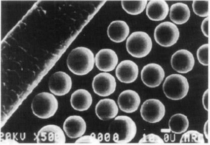
A very effective way of preventing leakage of the radioactive isotope(s) from the microsphere is to seal the radioisotope into the microsphere matrix. The pre-made microspheres enclose the non-radioactive precursor of the radioisotope and are activated in a nuclear reactor by bombardment with thermal neutrons shortly before use (Table 6). The most stable matrix for this kind of microsphere activation is glass. Day and Ehrhardt pioneered such therapeutic radioactive microspheres (Figure 4) from aluminosilicate glass containing 17 mol% Y2O3 [80]. The glass mixture was melted in a platinum crucible at 1600 °C the annealed glass crushed and the splinters spheroidised
226
Radioactive microspheres for medical applications
by sprinkling them from above through an oxygen flame. During neutron-activation in the reactor, the non-radioactive 89 Y captured a neutron and became the radioactive β- emitter 90Y. The leakage rate of the 90Y enclosed in the glass matrix was extremely low. Not more than 92 Bq were released from 50 mg of microspheres when activated to therapeutic activities of 11.1 GBq. Very similar glass microspheres have also been prepared enclosing rhenium, resulting in 186Re/188Re microspheres after neutron activation [65]. Advantages of glass microspheres are their excellent stability, radiation resistance, insolubility and non-toxicity. Disadvantages include their high density (3.3 g/ml) which makes the complete injection through syringes and intravenous lines difficult, and their non-biodegradability which can lead to immunologic reactions. Research is ongoing, however, in the preparation of glass microspheres from biodegradable glass material such as lithium boride [81].
Figure 4 Yttrium glass microspheres for neutron activation in a nuclear reactor in comparison with the size ofa hair
The disadvantages of glass were overcome by the preparation of PLA-microspheres containing either an acetylacetone-complex of 161Ho [67,68] or small particles of metallic rhenium in its native form, 185Re and 187Re [69] (Table 6). The stability of the activated 166Ho and 186Rel188Re-microspheres was sufficient for therapy (less than 1% of activity released within a week). The activation time of these poly(lactic acid) microspheres, however, is limited due to the radiolytic breakdown of ester bonds and must be characterised for each polymer-microsphere composition. Specifically, it has been found that activation of rhenium microspheres made from PLA with a molecular weight of 2000 for 1 hour at a neutron-flux of 5-1012 n/cm2/sec produced 450 MBq 188Re and 78 MBq of 186Re. Longer activation times led to melting and polymer
227
Urs Häfeli
breakdown [69]. The therapy of liver tumours which requires high specific activities is thus not possible with these 188Re/186Re-PLA-microspheres, but they could be used in local treatment of brain metastases or applied to incompletely resected tumour tissue after surgery. Recently, 166Ho-acetylacetonate-microspheres made from PLA with a molecular weight of 20,000 have been described [68]. The authors reported that up to 1 hour of neutron-activation at a flux of 5 1013 n/cm2/sec was possible, yielding an activity of 20 GBq in 400 mg of microspheres. This activity would be sufficient to allow for their transport to the hospital and use in liver tumour patients on the following day.
4.4. IN SITU NEUTRON CAPTURE THERAPY USING NON-RADIOACTIVE MICROSPHERES
Neutron capture therapy is an exciting bimodal tumour treatment concept originally proposed in 1936 by Locher [82]. The first component of this therapy is the delivery to tumour cells of non-radioactive atoms or molecules either alone or packed into carriers such as liposomes or microspheres. The target nuclei have large thermal and/or epithermal neutron capture cross-sections and a resulting reaction having a large positive Q value. The aim is to attain a higher concentration of these nuclei in the tumour than in the surrounding normal tissue cells. The second component is the exposure of a selected patient volume to a neutron beam. During neutron capture in situ, excessive energy between the initial and final state of the reactive nuclei (the positive Q-value) is released either as the recoil energy of heavy particles (6Li, 10B) and a-particles, or as g-rays (155Gd, 157Gd) (see Table 7). In the case of boron or lithium neutron capture, most of this energy is deposited in the tumour cell, since the range of the produced particles is less than 10 µm. In the case of gadolinium neutron capture, energy is spread out more because of photonic interactions. Although the first clinical trials with neutron capture therapy were completed in the 1950's, it took developments of the next 40 years to make this therapeutic approach very promising for cancer therapy [83]. Developments included the synthesis of superior targeting compounds such as the sulphur containing boron compounds mercaptoundecahydrododecaborate (= BSH) or boronated porphyrins (= BOPP).
In neutron capture therapy, boron and gadolinium are generally delivered intravenously, although their delivery is also possible via microparticles. Tokumitsu et al developed Gd-DTPA loaded chitosan microspheres of 4.1 1.3 µm for intratumoural injection [84,85]. Boron can be delivered in a similar way by packaging BSH into liposomes with anti-carcinoembryonic antigen antibodies on their surface [86], Different boron compounds have been encapsulated not only in the aqueous compartment of liposomes, but also in their bilayer and attached to their surface and work in this area is ongoing [87].
228

Radioactive microspheres for medical applications
Table 7, Isotopes useful for neutron capture therapy. Their half-life is zero.
Isoto |
Reaction |
Q |
Cross |
Modeof |
Range |
Delivery |
Pe |
|
[MeV] |
section |
energy |
in |
vehicles |
|
|
|
Sth |
deposition |
tissue |
|
|
|
|
[barn] |
|
in µm |
|
|
|
|
|
|
|
|
6Li |
6Li(n,a)3H |
4.784 |
940 |
α - 2.105 |
< 10 |
|
|
|
|
|
MeV |
|
|
|
|
|
|
3H- 2.734 |
|
|
|
|
|
|
MeV |
|
|
10B |
10B(n,a)7Li |
2.790 |
3837 |
α 0- 1.775 |
7.2, |
Liposomes |
|
|
|
|
MeV |
8.9 |
containing |
|
|
|
|
α 1 - 1 . 4 7 |
|
B S H [ 8 6 , 8 8 ] |
|
|
|
|
MeV |
|
|
|
|
|
|
7Li0 - 1.015 |
|
|
|
|
|
|
MeV |
|
|
|
|
|
|
7Li1 - 0.84 |
|
|
|
|
|
|
MeV |
|
|
155Gd |
155Gd(n,y)15 |
11.452 |
61000 |
|
|
|
|
6Gd |
|
|
|
|
|
157Gd |
157Gd(n,g)15 |
7.937 |
254000 |
|
|
Chitosan |
|
8Gd |
|
|
|
|
microspheres; |
|
|
|
|
|
|
microcapsules |
containing Gd-DTPA [84,85]
5. Diagnostic uses of radioactive microspheres
Diagnostic studies with radiopharmaceuticals include dynamic and static imaging and in vivo function tests. Dynamic imaging provides information about the biodistribution and pharmacokinetics of drugs in organs. Performed with a g-camera, dynamic studies are generally carried out over a pre-set length of time and provide clues to the functioning of the organ being examined. Static imaging, on the other hand, provides morphological information about an organ such as its shape, location and size. Furthermore it allows the exact location of tumours to be determined. Static imaging, unlike dynamic imaging, is normally done at a single point in time, with the imaging time being dependent on the organ activity. In contrast to dynamic and static imaging, in vivo function tests do not require imaging. Instead they are evaluated by comparing an injected or swallowed amount of radioactivity to the measured radioactivity in urine or blood.
229
Urs Häfeli
All three types of diagnostic studies can be performed with radioactive microspheres which contain one or several -emitters that can be detected by a g-camera. The first such "microspheres" in clinical use were red and white blood cells, which were taken from a patient, labelled with 111In or 51Cr, and then re-injected. Red blood cells labelled with 51Cr are commonly used for the measurement of red blood cell mass and for imaging of the spleen. For the latter purpose, the red blood cells are denatured by heating, which renders them spheroidal and nondeformable, and makes them easy to take up by the spleen. Another common application of radiolabelled red blood cells is the accurate determination of total systemic arterial blood flow or venous return, as well as, for blood flow determination within specific organs [89]. These blood flow parameters are important when drugs for the treatment of cardiovascular diseases are evaluated. White blood cells labelled with 111In-oxine are used for the detection of inflammatory diseases, abscesses or other infections. A less expensive method has been developed in which the neutral and lipophilic 99m Tc-HMPA0 complex is prepared from a kit and then incubated with the leukocytes [90]. Platelets labelled with "'In are also used to detect actively forming deep vein thrombi, to measure blood flow, and to detect regions of infection [91]. Radiolabelled blood cells are still used today, although premade radioactive microspheres containing several different γ -emitters (see Table 7) are easier to use and do not require time-consuming labelling procedures [92]. Unfortunately, the radioactive microspheres of homogenous size are made from polystyrene and thus are not biodegradable, making them inappropriate for clinical use.
Figure 5. Diagnostic lung imaging obtained after the injection of 99mTc-labeled macroaggregated albumin in different projections. The top row shows a normal lung and the bottom row the lung of a patient with multiple pulmonary emboli in both lobes of the lungs.
For the diagnosis of pulmonary embolism, both the inhalation of small, radioactive 99mTc-carbon particles (Technegas) and the perfusion of the lung with 99mTc-labeled
230

Radioactive microspheres for medical applications
albumin particles are used. In the first case, Technegas behaves, due to the small particle size of less than 100 nm, much more like a gas than a radioaerosol and diffuses into the entire accessible lung volume. In the second case, macroaggregated albumin is mainly used for the quantification of shunts associated with intrapulmonary arteriovenous malformations and the diagnosis of pulmonary diseases such as cancer and hypertension (Figure 5). The diagnostic determination of shunts within an organ is generally done prior to using radioactive microspheres in radioembolization therapy (see below) [93] in order to prevent radiotoxicity to the lungs [94]. The biological halflife of the albumin particles is only 1 to 3 hours, so any therapeutic interventions can easily be performed afterwards.
Table 7: Radioactive microspheres for diagnostic applications
Application |
Type of radioactive microspheres used |
Particle size |
|
|
|
Gated blood pool study |
111Inor 51Cr-labeled red blood cells |
6-8 µm |
Thrombus imaging in |
111In-labeled platelets |
0.5-1 µm |
deep vein thrombosis |
99mTc-macro-aggregated human serum |
10-90 µm |
|
albumin (MAA) |
|
|
99mTc-sulfur colloid |
0.05-0.6µm |
Blood flow |
Polystyrene-microspheres labelled with |
10, 15 µm |
measurements |
thegg-emitters 141Ce, 57Co, 114mIn, 85Sr, |
(other sizes) |
|
51Cr, and others (animal experiments) |
|
Investigation of biodistribution and fate of (drug-loaded) microspheres
Lung scintigraphy
3H, 14 C-labeled microspheres (animal |
all sizes |
experiments) |
|
141Ce-polystyrene microspheres |
11.4 µm |
99mTc-impregnated carbon particles (= |
50 nm |
Technegas) |
|
99m Tc-macro-aggregated human serum |
10-90 µm |
albumin (MAA)
Diagnostic |
99mTc-macro-aggregated human serum |
10-90 µm |
radioembolization |
albumin (MAA) |
|
Liver and spleen |
99mTc-macro-aggregated human serum |
10-90 µm |
imaging |
albumin (MAA) |
|
|
99m Tc-sulfur colloid |
0.05-0.6µm |
|
99mTc-tin colloid |
0.05-0.6µm |
Bone marrow imaging |
99mTc-sulfur colloid |
0.05-0.6µm |
|
99mTc-antimony sulphide colloid |
0.05-0.6µm |
231

Urs Häfeli
Table 7: Radioactive microspheresfor diagnostic applications
Application |
Type of radioactive microspheres used |
Particle size |
|
|
|
Infection localisation |
111In-labeled leukocytes |
12-20µm |
|
111In-labeled liposomes |
20 nm-1µm |
|
99mTc-labeled liposomes |
20 nm-1µm |
|
99mTc-albumin nanocolloid |
<80 nm |
Tumour imaging |
99mTc-labeled liposomes |
20 nm-1 µm |
|
67Ga-NTA- or 111In-NTA-labeled |
65 nm |
|
liposomes |
|
Gastrointestinal transit |
99m Tc-sulfur colloid |
0.05-0.6 µm |
studies |
111In-labeled ion exchange resins |
|
Local restenosis |
141Ce microspheres (preliminary imaging |
11.4 µm |
prevention in coronary |
tests) |
|
arteries |
|
|
|
|
|
For liver, spleen, bone marrow and lymphatic system imaging, colloidal microparticles, such as 99mTc-sulfur colloids, are most useful (Table 7). To illustrate, Figure 6 shows the changes in a cirrhotic patient made visible by 99mTc-sulfur colloid. The lymphatic system can also be imaged or targeted with drugs through the use of the poly(lysine) nanospheres [95]. The ideal nanospheres for this purpose are 10-30 nm, contain carbohydrate groups on the surface, and are able to bind the g-emitter 111In via the covalently bound chelator DTPA.
Figure 6. Liver scintigraphy performed with 99mTc-sulfur colloid in different projections. The top row shows a normal liver, the bottom row the corresponding views of a liver from a patient with cirrhosis.
232
Radioactive microspheres for medical applications
The radiopharmaceutical 99mTc-sulfur colloid is also used for gastrointestinal blood loss studies, for the preparation of a 99mTc-labeled egg sandwich for gastric emptying studies [39], and for the determination of oesophageal transit and gastro-oesophageal reflux. For colonic transit studies, radioisotopes with a half-life longer than 99mTc are more appropriate, and 111 In-labeled ion exchange resins, but also 131l-cellulose are utilised [96]. Latex-particles of 2.5 µm size and labelled with 99mTc have also been shown to give excellent abdominal images [97]. In all these gastrointestinal transit time studies, the size of the radiolabelled microspheres does not influence the measured times.
Radiolabelled liposomes, another diagnostic class of radioactive particles, has been used for tumour imaging since 1977. In order to prolong the blood residence time and maximise tumour uptake, neutral, positively and negatively charged small unilamellar vesicles (= SUV's) of 65 nm encapsulating 111In were made and their biodistribution measured in mice [98]. The highest uptake of 18.5% of injected dose per gram of tumour was measured with the neutral liposomes. A further attempt to minimise the high blood-background radioactivity levels was to inject 67Gaor 111In-labeled liposomes containing biotin groups on their surface and then chasing the non-tumour bound liposomes 2 hours later with avidin [99]. This chase removed the unbound liposomes effectively from the circulation and the blood concentration of the radioactive liposomes dropped to a tumour-to-blood ratio of about 15 to 1 shortly after the avidin chase. The avidin-biotin-liposome conglomerates accumulated in the liver and increased the liver activity about 2.5 fold.
Radiolabelled microspheres can also be used to image cancer lesions. An interesting application is the use of PLA-microspheres labelled with 131T-iopanoic acid derivatives for the imaging of liver tumours [44]. The normal liver parenchyma lined with Kupffer cells takes up the microspheres, but the cancer lesions do not possess fixed macrophages and therefore exclude the radioactive microspheres, showing the focal lesions as defects. The recently introduced 99mTc-PLA microspheres which were radiolabelled in a SnCl2-containing kit could be used for the same application [100], although the stability described as "more than 80% bound after 6 hours" is not optimal yet.
The in vivo faith of microspheres after intra-arterial catheter-mediated delivery through a porous balloon to a rabbit's femoral artery has been investigated with radioactive 141Ce-microspheres [ 101]. Although only 0.14% to 0.16% of the microspheres were delivered to the vessel wall, an average of 92% of these microspheres was still present 7 days later. In addition, much higher amounts of the microspheres were found in the periadventitia (the vessel's "outside") and the overlying musculature and are believed to be caused by the increase of vaso vasora present in atherosclerotic patients. Although the targeted amounts of microspheres are small, they can lead to drug concentrations a few hundred times higher than the serum concentrations, allowing for effective restenosis therapy with microspheres containing cytostatic or antiproliferative agents, especially from the periadventitia side.
233