

SURGICAL ANATOMY by Joseph Maclise
.pdfCOMMENTARY ON PLATES 61 & 62. |
135 |
Fig. 9, Plate 61.--The lateral lobes, b b, of the prostate are enlarged. The third lobe, a, projects at the neck of the bladder, distorting the vesical outlet. A small calculus occupies the prostatic urethra, and being closely impacted in this part of the canal, would arrest the progress of a catheter, and probably lead to the supposition that the instrument grated against a stone in the interior of the bladder, in which case it would be inferred that since the urine did not flow through the catheter no retention existed.
Plate 61.--Figure 9. |
Plate 61.--Figure 10. |
Fig. 10, Plate 61.--Both lateral lobes, b c, of the prostate appear much increased in size. A large irregular shaped mass, a, grows from the base of the right lobe, and distorts the prostatic canal and vesical orifice. When the lobes of the prostate increase in size in this direction, the prostatic canal becomes much more elongated than natural, and hence the instrument which is to be passed for relieving the existing retention of urine should have a wide and long curve to correspond with the form of this part of the urethra. [Footnote]
[Footnote: Both lobes of the prostate are equally liable to chronic enlargement. Home believed the left lobe to be oftener increased in size than the right. Wilson (on the Male Urinary and Genital Organs) mentions several instances of the enlargement of the right lobe. No reason can be assigned why one lobe should be more prone to hypertrophy than the other, even supposing it to be matter of fact, which it is not. But the observations made by Cruveilhier (Anat. Pathol.), that the lobulated projections of the prostate always take place internally at its vesical aspect, is as true as the manner in which he accounts for the fact is plausible. The dense fibrous envelope of the prostate is sufficient to repress its irregular growth externally.]

Fig. 11, Plate 61.--Both lobes of the prostate are enlarged, and from the base of each a mass projects prominently around the vesical orifice, a b. The prostatic urethra has been moulded to the shape of the instrument, which was retained in it for a considerable time.
Plate 61.--Figure 11.
Fig. 12, Plate 61.--The prostate, c b, is enlarged and dilated, like a sac. Across the neck, a, of the bladder the prostate projects in an arched form, and is transfixed by the instrument, d. The prostate may assume this appearance, as well from instruments having been forced against it, as from an abscess cavity formed in its substance having received, from time to time, a certain amount of the urine, and retained this fluid under the pressure of strong efforts, made to void the bladder while the vesical orifice was closed above.
Plate 61.--Figure 12.

Fig. 13, Plate 61.--The lateral lobes, d e, of the prostate are enlarged; and, occupying the position of the third lobe, appear as three masses, a b c, plicated upon each other, and directed towards the vesical orifice, which they close like valves. The prostatic urethra branches upwards into three canals, formed by the relative position of the parts, e, c, b, a, d, at the neck of the bladder. The ureters are dilated, in consequence of the regurgitation of the contents of the bladder during the retention which existed ..
Plate 61.--Figure 13.
Fig. 1, Plate 62, exhibits the lobes of the prostate greatly increased in size. The part, a b, girds irregularly, and obstructs the vesical outlet, while the lateral lobes, c d, encroach upon the space of the prostatic canal. The walls of the bladder are much thickened.
Plate 62.--Figure 1.

136 |
COMMENTARY ON PLATES 61 & 62. |
Fig. 2, Plate 62.--The three lobes, a, d, c, of the prostate are enlarged and of equal size, moulded against each other in such a way that the prostatic canal and vesical orifice appear as mere clefts between them. The three lobes are encrusted on their vesical surfaces with a thick calcareous deposit. The surface of the third lobe, a, which has been half denuded of the calcareous crust, b, in order to show its real character, appeared at first to be a stone impacted in the neck of the bladder, and of such a nature it certainly would seem to the touch, on striking it with the point of a sound or other instrument.
Plate 62.--Figure 2. |
Plate 62.--Figure 3. |
Fig. 3, Plate 62, represents the prostate with its three lobes enlarged, and the prostatic canal and vesical orifice narrowed. The walls of the bladder are thickened, fasciculated, and sacculated; the two former appearances being caused by a hypertrophy of the vesical fibres, while the latter is in general owing to a protrusion of the mucous membrane between the fasciculi.

Fig. 4, Plate 62.--The prostate presents four lobes, a, b, c, d, each being of large size, and projecting far into the interior of the bladder, from around the vesical orifice which they obstruct. The bladder is thickened, and the prostatic canal is elongated. The urethra and the lobes of the prostate have been perforated by instruments, passed for the retention of urine which existed. A stricturing band, e, is seen to cross the membranous part of the canal.
Plate 62.--Figure 4. |
Plate 62.--Figure 5. |
Fig. 5, Plate 62.--The prostate, a a, is greatly enlarged, and projects high in the bladder, the walls of the latter, b b, being very much thickened. The ureters, c, are dilated, and perforations made by instruments are seen in the prostate. The prostatic canal being directed almost vertically, and the neck of the bladder being raised nearly as high as the upper border of the pubic symphysis, it must appear that if a stone rest in the bas fond of the bladder, a sound or staff cannot reach the stone, unless by perforating the prostate; and if, while the staff occupies this position, lithotomy be performed, the incisions will not be required to be made of a greater depth than if the prostate were of its ordinary proportions. On the contrary, if the staff happen to have surmounted the prostate, the incision, in order to divide the whole vertical thickness of this body, will require to be made very deeply from the perinaeal surface, and this circumstance occasions what is termed a "deep perinaeum."

Fig. 6, Plate 62.--The lower half, c, b, f, of the prostate, having become the seat of abscess, appears hollowed out in the form of a sac. This sac is separated from the bladder by a horizontal septum, e e, the proper base of the bladder, g g. The prostatic urethra, between a e, has become vertical in respect to the membranous part of the canal, in consequence of the upward pressure of the abscess. The sac opens into the urethra, near the apex of the prostate, at the point c; and a catheter passed along the urethra has entered the orifice of the sac, the interior of which the instrument traverses, and the posterior wall of which it perforates. The bladder contains a large calculus, i. The bladder and sac do not communicate, but the urethra is a canal common to both. In a case of this sort it becomes evident that, although symptoms may strongly indicate either a retention of urine, or the presence of a stone in the bladder, any instrument taking the position and direction of d d, cannot relieve the one or detect the other; and such is the direction in which the instrument must of necessity pass, while the sac presents its orifice more in a line with the membranous part of the urethra than the neck of the bladder is. The sac will intervene between the rectum and the bladder; and on examination of the parts through the bowel, an instrument in the sac will readily be mistaken for being in the bladder, while neither a calculus in the bladder, nor this organ in a state of even extreme distention, can be detected by the touch any more than by the sound or catheter. If, while performing lithotomy in such a state of the parts, the staff occupy the situation of d d d, then the knife, following the staff, will open, not the bladder which contains the stone, but the sac, which, moreover, if it happen to be filled with urine regurgigated from the urethra, will render the deception more complete.
Plate 62.--Figure 6.

Fig. 7, Plate 62.--The walls, a a, of the bladder, appear greatly thickened, and the ureters, b, dilated. The sides, c c c, of the prostate are thinned; and in the prostatic canal are two calculi, d d, closely impacted. In such a state of the parts it would be impossible to pass a catheter into the bladder for the relief of a retention of urine, or to introduce a staff as a guide to the knife in lithotomy. If, however, the staff can be passed as far as the situation of the stone, the parts may be held with a sufficient degree of steadiness to enable the operator to incise the prostate upon the stone.
Plate 62.--Figure 7.
COMMENTARY ON PLATES 63 & 64.
DEFORMITIES OF THE URINARY BLADDER.--THE OPERATIONS OF SOUNDING FOR STONE, OF CATHETERISM AND OF PUNCTURING THE BLADDER ABOVE THE PUBES.
The urinary bladder presents two kinds of deformity--viz., congenital and pathological. As examples of the former may be mentioned that in which the organ is deficient in front, and has become everted and protruded like a fungous mass through an opening at the median line of the hypogastrium; that in which the rectum terminates in the bladder posteriorly; and that in which the foetal urachus remains pervious as a uniform canal, or assumes a sacculated shape between the summit of the bladder and the umbilicus. The pathological deformities are, those in which vesical fistulae, opening either above the pubes, at the perinaeum, or into the rectum, have followed abscesses or the operation of puncturing the bladder in these situations, and those in which the walls of the organ appear thickened and contracted, or thinned and expanded, or sacculated externally, or ridged internally, in consequence of its having been subjected to abdominal pressure while overdistended with its contents, and while incapable of voiding these from some permanent obstruction in the urethral canal.[Footnote] The bladder is liable to become sacculated from two causes--from a hernial protrusion of its mucous membrane through the separated fasciculi of its fibrous coat, or from the cyst of an abscess which has formed a communication with the bladder, and received the contents of this organ. Sacs, when produced in the former way, may be of any number, or size, or in any situation; when caused by an abscess, the sac is single, is generally formed in the prostate, or corresponds to the base of the bladder, and may attain to a size equalling, or even exceeding, that of the bladder itself. The sac, however formed, will be found lined by mucous membrane. The cyst of an abscess, when become a recipient for the urine, assumes after a time a lining membrane similar to that of the bladder. If the sac be situated at the summit or back of the bladder, it will be found invested by peritonaeum; but, whatever be its size, structure, or position, it may be always distinguished from the bladder by being devoid of the fibrous tunic, and by having but an indirect relation to the vesical orifice.
[Footnote: On considering these cases of physical impediments to the passage of urine from the vesical reservoir through the urethral conduit, it seems to me as if these were sufficient to account for the formation of stone in the bladder, or any other part of the urinary apparatus, without the necessity of ascribing it to a constitutional disease, such as that named the lithic diathesis by the humoral pathologists.
The urinary apparatus (consisting of the kidneys, ureters, bladder, and urethra) is known to be the principal emunctory for eliminating and voiding the detritus formed by the continual decay of the parts comprising the animal economy. The urine is this detritus in a state of solution. The components of urine are chemically similar to those of calculi, and as the components of the one vary according to the disintegration occurring at the time in the vital alembic, so do those of the other. While, therefore, a calculus is only as urine precipitated and solidified, and this fluid only as calculous matter suspended in a menstruum, it must appear that the lithic diathesis is as natural and universal as structural disintegration is constant and general in operation. As every individual, therefore, may be said to void day by day a dissolved calculus, it must follow that its form of precipitation within some part of the urinary apparatus alone constitutes the disease, since in this form it cannot be passed. On viewing the subject in this light, the question that springs directly is, (while the lithic diathesis is common to individuals of all ages and both sexes,) why the lithic sediment should present in the form of concrement in some and not in others? The principal, if not the sole, cause of this seems to me to be obstruction to the free egress of the urine along the natural passage. Aged individuals of the male sex, in whom the prostate is prone to enlargement, and the urethra to organic stricture, are hence more subject to the formation of stone in the bladder, than youths, in whom these causes of obstruction are less frequent, or than females of any age, in whom the prostate is absent, and the urethra simple, short, readily dilatable, and seldom or never strictured. When an obstruction exists, lithic concretions take place in the urinary apparatus in the same manner as sedimentary particles cohere or crystallize elsewhere. The urine becoming pent up and stagnant while charged with saline matter, either deposits this around a nucleus introduced into it, or as a surplus when the menstruum is insufficient to suspend it. The most depending part of the bladder is that where lithic concretions take place; and if a sacculus exist here, this, becoming a recipient for the matter, will favour the formation of stone.]
(Page 137)
138 |
COMMENTARY ON PLATES 63 & 64. |
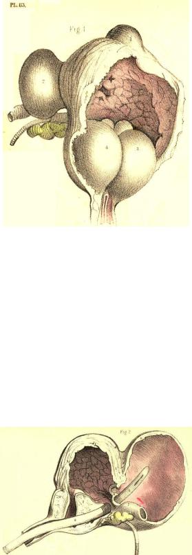
FIG. 1, Plate 63.--The lateral lobes of the prostate, 3, 4, are enlarged, and contract the prostatic canal. Behind them the third lobe of smaller size occupies the vesical orifice, and completes the obstruction. The walls of the bladder have hence become fasciculated and sacculated. One sac, 1, projects from the summit of the bladder; another, 2, containing a stone, projects laterally. When a stone occupies a sac, it does not give rise to the usual symptoms as indicating its presence, nor can it be always detected by the sound.
Plate 63,--Figure 1.
FIG. 2, Plate 63.--The prostate, 2, 3, is enlarged, and the middle lobe, 2, appears bending the prostatic canal to an almost vertical position, and obstructing the vesical orifice. The bladder, 1, 1, 1, is thickened; the ureters, 7, are dilated; and a large sac, 6, 6, projects from the base of the bladder backwards, and occupies the recto-vesical fossa. The sac, equal in size to the bladder, communicates with this organ by a small circular opening, 8, situated between the orifices of the ureters. The peritonaeum is reflected from the summit of the bladder to that of the sac. A catheter, 4, appears perforating the third lobe of the prostate, 2, and entering the sac, 5, through the base of the bladder, below the opening, 8. In a case of this kind, a catheter occupying the position 4, 5, would, while voiding the bladder through the sac, make it seem as if it really traversed the vesical orifice. If a stone occupied the bladder, the point of the instrument in the sac could not detect it, whereas, if a stone lay within the sac, the instrument, on striking it here, would give the impression as if it lay within the bladder.
Plate 63,--Figure 2.