

SURGICAL ANATOMY by Joseph Maclise
.pdfCOMMENTARY ON PLATES 48 & 49. |
111 |
The pelvic fascia, in being reflected to the bladder from the front and sides of the pelvis, at a lower level than that of the peritonaeum, forms the "true ligaments." In addition to these ligaments, which serve to keep the base and front of the bladder fixed in the pelvis, other structures, such as the ureters, K, the vasa deferentia, I, the hypogastric cords, the urachus, and the bloodvessels, embrace the organ in various directions, and act as bridles, to limit its expansion more or less in all directions, but least so towards its summit, which is always comparatively free.
The neck and outlet of the bladder, V, are situated at the anterior part of its base, and point towards the subpubic space. The prostate gland, V, surrounds its neck, and occupies a position behind and below the pubic arch, D, and in front of the rectum, W. The gland, V, being of a rounded form and dense structure, can be felt in this situation by the finger, passed upwards through the bowel. The prostate is suspended from the back of the pubic arch by the anterior true ligament of the bladder, and at its forepart, where the membranous portion of the urethra commences, this passes through the deep perinaeal fascia, X. The anterior fibres of the levator ani muscle embrace the prostate on both its sides. Behind the base of the prostate, the ureter, K, is seen to enter the coats of the bladder obliquely, whilst the vas deferens, I, joined by the vesicula seminalis, L, penetrates the substance of the prostate, V, at its lower and back part, which lies in apposition with the rectum.
The rectum, W C, at its middle and upper parts, occupies the hollow of the sacrum, A Q, and is behind the bladder. The lower third of the rectum, W, not being covered by the peritonaeum, is that part on which the various surgical operations are performed. At its upper three-fifths, the rectum describes a curve corresponding to that of the sacrum; and if the bladder be full, its convex back part presses the bowel against the bone, causing its curve to be greater than if the bladder were empty and collapsed. This fact requires to be borne in mind, for, in order to introduce a bougie, or to allow a large injection to pass with freedom into the bowel, the bladder should be first evacuated. The coccygeal bones, Q, continuing in the curve of the sacrum, bear the rectum, W, forwards against the base of the bladder, and give to this part a degree of obliquity upwards and backwards, in respect to the perinaeum and anus. From the point where the prostate, V, lies in contact with the rectum, W, this latter curves downwards, and slightly backwards, to the anus, P. The prostate is situated at a distance of about an inch and a half or two inches from the anus--the distance varying according to whether the bladder and bowel be distended or not. [Footnote]
[Footnote: The distance between any two given parts is found to vary in different cases. "In subjects of an advanced age," Mr. Stanley remarks, "a deep perinaeum, as it is termed, is frequently met with. This may be occasioned either by an unusual quantity of fat in the perinaeum, or by an enlarged prostate, or by the dilatation of that part of the rectum which is contiguous to the prostate and bladder. Under either of these circumstances, the prostate and bladder become situated higher in the pelvis than naturally, and consequently at a greater distance from the perinaeum."--On the Lateral Operation of Lithotomy.]
The arteries of the bladder are derived from the branches of the internal iliac, S. The rectum receives its arteries from the inferior mesenteric and pudic. The veins which course upwards from the rectum are large and numerous, and devoid of valves. When these veins become varicose, owing to a stagnation of their circulation, produced from whatever cause, the bowel is liable to be affected with haemorrhoids or to assume a haemorrhagic tendency.
The pudic artery, S s, is a branch of the internal iliac. It passes from the pelvis by the great sciatic foramen, below the pyriformis muscle, and in company with the sciatic artery. The pudic artery and vein wind around the spine, E, of the ischium, where they are joined by the pudic nerve, derived from, T, the sacral plexus.
112 |
COMMENTARY ON PLATES 49 & 49. |
The artery, in company with the nerve and vein, re-enters the pelvis by the small sciatic foramen, and gets under cover of a dense fibrous membrane (obturator fascia), between which and the obturator muscle, it courses obliquely downwards and forwards to the forepart of the perinaeum. At the place where the vessel re-enters the pelvis, it lies removed at an interval of an inch and a half from the perinaeum, but becomes more superficial as it approaches the subpubic space, N. The levator ani muscle separates the pudic vessels and nerves from the sides of the rectum and bladder. The principal branches given off from the pudic artery of either side, are (1st), the inferior hemorrhoidal, to supply the lower end of the rectum; (2nd), the transverse and superficial perinaeal; (3rd), the artery of the bulb; (4th), that which enters the corpus cavernosum of the penis, N; and (5th), the dorsal artery of the penis. [Footnote] The branches given off from the pudic nerve correspond in number and place to those of the artery. Having now considered the relations of the pelvic organs in a lateral view, we are better prepared to understand these relations when seen at their perinaeal aspect.
[Footnote: The pudic artery, or some one of its branches, occasionally undergoes marked deviations from the ordinary course. In Mr. Quain's work, ("Anatomy of the Arteries,") a case is represented in which the artery of the bulb arose from the pudic as far back as the tuber ischii, and crossed the line of incision made in the lateral operation of lithotomy. In another figure is seen a vessel ("accessory pudic"), which, passing between the base of the bladder and the levator ani muscle, crosses in contact with the left lobe of the prostate.]
DESCRIPTION OF PLATES 48 & 49.
PLATE 48.
A.The anterior superior iliac spine.
B.The anterior inferior iliac spine.
C.The acetabulum; c, the ligamentum teres.
D.The tuber ischii.
E.The spine of the ischium.
F.The pubic horizontal ramus.
G.The summit of the bladder covered by the peritonaeum.
H.The femoral artery.
I.The femoral vein.
K.The anterior crural nerve.
L.The thyroid ligament.
M.The spermatic cord.
N.The corpus cavernosum penis; n, its artery.
O.The urethra; o, the bulbus urethrae.
P.The sphincter ani muscle.
Q.The coccyx.
R.The sacro-sciatic ligament.
S.The pudic artery and nerve.
T.The sacral nerves.
U.The pyriformis muscle, cut.
V.The gluteal artery.
W.The small gluteus muscle.
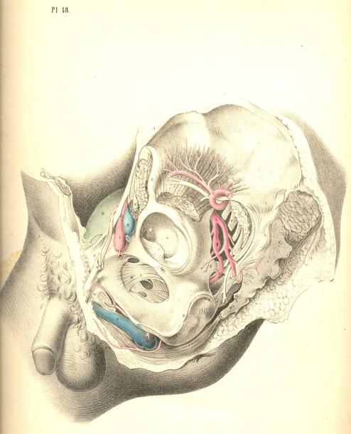
Plate 48

PLATE 49.
A.The part of the sacrum which joins the ilium.
B.The external iliac artery, cut across.
C.The upper part of the rectum.
D.The ascending pubic ramus.
E.The spine of the ischium, cut.
F.The horizontal pubic ramus, cut.
G.The summit of the bladder covered by the peritonaeum; G *, its side, not covered by the membrane.
H H. The recto-vesical peritonaeal pouch,
I. The vas deferens.
K.The ureter.
L.The vesicula seminalis.
M, N, O, P, Q, R, S, T, U, refer to the same parts as in Plate 48.
V.The prostate.
W.The lower part of the rectum.
X.The deep perinaeal fascia.
Plate 49
COMMENTARY ON PLATES 50 & 51.
THE SURGICAL DISSECTION OF THE SUPERFICIAL STRUCTURES OF THE
MALE PERINAEUM.
The median line of the body is marked as the situation where the opposite halves unite and constitute a perfect symmetrical figure. Every structure--superficial as well as deep--which occupies the median line is either single, by the union of halves, or dual, by the cleavage and partition of halves. The two sides of the body being absolutely similar, the median line at which they unite is therefore common to both. Union along the median line is an occlusion taking place by the junction of sides; and every hiatus or opening, whether normal or abnormal, which happens at this line, signifies an omission in the process of central union. The sexual peculiarities are the results of the operation of this law, and all forms which are anomalous to either sex, may be interpreted as gradations in the same process of development; a few of these latter occasionally come under the notice of the surgeon.
The region which extends from the umbilicus to the point of the coccyx is marked upon the cutaneous surface by a central raphe dividing the hypogastrium, the penis, the scrotum, and the perinaeum respectively into equal and similar sides. The umbilicus is a cicatrix formed after the metamorphosis of a median foetal structure--the placental cord, &c. In the normal form, the meatus urinarius and the anus coincide with the line of the median raphe, and signify omissions at stated intervals along the line of central union. When between these intervals the process of union happens likewise to be arrested, malformations are the result; and of these the following are examples:--Extrusion of the bladder at the hypogastrium is caused by a congenital hiatus at the lower part of the linea alba, which is in the median line; Epispadias, which is an urethral opening on the dorsum of the penis; and Hypospadias, which is a similar opening on its under surface, are of the same nature--namely, omissions in median union. Hermaphrodism may be interpreted simply as a structural defect, compared to the normal form of the male, and as a structural excess compared to that of the female. Spina bifida is a congenital malformation or hiatus in union along the median line of the sacrum or loins. As the process of union along the median line may err by a defect or omission, so may it, on the other hand, err by an excess of fulfilment, as, for example, when the urethra, the vagina, or the anus are found to be imperforate. As the median line of union thus seems to influence the form of the hypogastrium, the genitals, and the perinaeum, the dissection of these parts has been conducted accordingly.
By removing the skin and subjacent adipose membrane from the hypogastrium, we expose the superficial fascia. This membrane, E E E*, Fig. 1, Plate 50, is, in the middle line, adherent to B, the linea alba, and thereby contributes to form the central depression which extends from the navel to the pubes. The adipose tissue, which in some subjects accumulates on either side of the linea alba, renders this depression more marked in them. At the folds of the groin the fascia is found adherent to Poupart's ligament, and this also accounts for the depressions in both these localities. From the central linea alba to which the fascia adheres, outwards on either side to the folds of both groins, the membrane forms two distinct sacs, which droop down in front, so as to invest the testicles, E**, and penis in a manner similar to that of the skin covering these parts. As the two sacs of the superficial fascia join each other at the line B, coinciding with the linea alba, they form by that union the suspensory ligament of the penis, which is a structure precisely median.
(Page 113)
114 |
COMMENTARY ON PLATES 50 & 51. |
The superficial fascia having invested the testicles each in a distinct sac, the adjacent sides of both these sacs, by joining together, form the median septum scroti, E, Fig. 2, Plate 50. In the perinaeum, Fig. 1, Plate 51, the fascia, A, may be traced from the back of the scrotum to the anus. In this region the membrane is found to adhere laterally to the rami of the ischium and pubes; whilst along the median perinaeal line the two sacs of which the membrane is composed unite, as in the scrotum, and form an imperfect septum. In front of the anus, beneath the sphincter ani, the fascia degenerates into cellular membrane, one layer of which is spread over the adipose tissue in the ischiorectal space, whilst its deeper and stronger layer unites with the deep perinaeal fascia, and by this connexion separates the urethral from the anal spaces. The superficial fascia of the hypogastrium, the scrotum, and the perinaeum forming a continuous membrane, and being adherent to the several parts above noticed, may be regarded as a general double sac, which isolates the inguino-perinaeal region from the femoral and anal regions, and hence it happens that when the urethra becomes ruptured, the urine which is extravasated in the perinaeum, is allowed to pass over the scrotum and the abdomen, involving these parts in consequent inflammation, whilst the thighs and anal space are exempt. The tunicae vaginales, which form the immediate coverings of the testicles, cannot be entered by the urine, as they are distinct sacs originally protruded from the abdomen. It is in consequence of the imperfect state of the inguino-perinaeal septum of the fascia, that urine effused into one of the sacs is allowed to enter the other.
Like all the other structures which join on either side of the median line, the penis appears as a symmetrical organ, D D, Fig. 2, Plate 50. While viewed in section, its two corpora cavernosa are seen to unite anteriorly, and by this union to form a septum "pectiniforme;" posteriorly they remain distinct and lateral, F F, Fig. 2, Plate 51, being attached to the ischio-pubic rami as the crura penis. The urethra, B, Fig. 2, Plate 50, is also composed of two sides, united along the median line, but forming between them a canal by the cleavage and partition of the urethral septum. All the other structures of the perinaeum will be seen to be either double and lateral, or single and median, according as they stand apart from, or approach, or occupy the central line.
The perinaeum, Figs. 1, 2, Plate 51, is that space which is bounded above by the arch of the pubes, behind by C, the os coccygis, and the lower borders of, I I, the glutaei muscles and sacro-sciatic ligaments, and laterally by D D, the ischiatic tuberosities. The osseous boundaries can be felt through the integuments. Between the back of the scrotum and the anus the perinaeum swells on both sides of the raphe, A B, Fig. 3, Plate 50, and assumes a form corresponding with the bag of the superficial fascia which encloses the structures connected with the urethra. The anus is centrally situated in the depression formed between D D, the ischiatic tuberosities, and the double folds of the nates.
The perinaeum, Fig. 3, Plate 50, is, for surgical purposes, described as divisible into two spaces (anterior and posterior) by a transverse line drawn from one tuber ischii, D, to the other, D, and crossing in front of the anus. The anterior space, A D D, contains the urethra; the posterior space, D D C, contains the rectum. The central raphe, A B C, traverses both these spaces. The anterior or urethral space is (while viewed in reference to its osseous boundaries) triangular in shape, the apex being formed by the pubic symphysis beneath A, whilst two lines drawn from A to D D, would coincide with the ischio-pubic rami which form its sides. The raphe in the anterior space indicates the central position of the urethra, as may be ascertained by passing a sound into the bladder, when the shaft of the instrument will be felt prominently between the points A B. Behind the point B, the sound or staff sinks deeper in the perinaeum as it follows the curve of the urethra towards the bladder, and becomes overlaid by the bulb, &c.
The ischiatic tuberosities, D D, Fig. 3, Plate 50, are, in all subjects, sufficiently prominent to be felt through the integuments, &c.; and the line which, when drawn from one to the other, serves to divide the two perinaeal spaces, forms the base of the anterior one.
COMMENTARY ON PLATES 50 & 51. |
115 |
In well-formed subjects, the anterior space is equiangular, the base being equal to each side; but according as the tuberosities approach the median line, the base becomes narrowed, and the triangle is thereby rendered acute. These circumstances influence the direction in which the first incision in the lateral operation of lithotomy should be made. When the tuberosity of the left ischium stands well apart from the perinaeal centre, the line of incision, B E, Fig. 3, Plate 50, is carried obliquely from above downwards and outwards; but in cases where the tuberosity approaches the centre, the incision must necessarily be made more vertical. The posterior perinaeal space may be described on the surface by two lines drawn from D D, the ischiatic tuberosities, to C, the point of the coccyx, whilst the transverse line between D and D bounds it above.
By removing the integument and superficial fascia, we expose the superficial vessels and nerves, together with the muscles in the neighbourhood of the urethra and the anus. The accelerator urinae, E, Fig. 2, Plate 51, which embraces the urethra, and the sphincter ani, B C, which surrounds the anus, H, occupy the median line, and are divided each into halves by a central tendon, E B C, which traverses the perinaeum from before backwards, to the point of the coccyx. On either side of the anus, in the ischio-rectal space, D D, Fig. 1, Plate 51, is found a considerable quantity of granular adipose tissue, traversed by the inferior haemorrhoidal arteries and nerves-branches of the pudic artery and nerve.
In front of the anus are seen two small muscles (transversae perinaei), G G, Fig. 2, Plate 51, each arising from the tuber ischii of its own side, and the two becoming inserted into, B, the central tendon. These transverse muscles serve to mark the boundary between the anterior and posterior perinaeal spaces. Behind each muscle is found a small artery, crossing to the median line. The left transverse muscle and artery are always divided in the lateral operation of lithotomy. On the outer sides of the anterior perinaeal space are seen the erectores penis muscles, F F, overlaying the crura penis. Between each muscle and the accelerator urinae, the superficialis perinaei artery and nerve course forwards to the scrotum, &c.
The perinaeal muscles having been brought fully into view, Plate 52, Fig. 1, their symmetrical arrangement on both sides of the median line at once strikes the attention. On either side of the anterior space appears a small angular interval, L, formed between B, the accelerator urinae, D, the erector penis, and E, the transverse muscle. Along the surface of this interval, the superficial perinaeal artery and nerve are seen to pass forwards; and deep in it, beneath these, may also be observed, L, the artery of the bulb, arising from the pudic, and crossing inwards, under cover of the anterior layer of the membrane named the deep perinaeal fascia. The first incision in the lateral operation of lithotomy is commenced over the inferior inner angle of this interval.
The muscles occupying the anterior perinaeal space require to be removed, Fig. 1, Plate 53, in order to expose the urethra, B M, the crus penis, D, and the deep perinaeal fascia. The fascia will be now seen stretched across the subpubic triangular space, reaching from one ischio-pubic ramus to the other, whilst by its lower border, corresponding with the line of the transversae perinaei muscles, it becomes continuous with the superficial fascia, in the manner before described. The deep perinaeal fascia (triangular ligament) encloses between its two layers, C E, on either side of the urethra, the pudic artery, the artery of the bulb, Cowper's glands, and some muscular fibres occasionally to be met with, to which the name "Compressor urethrae" has been assigned. At this stage of the dissection, as the principal vessels and parts composed of erectile tissue are now in view, their relative situations should be well noticed, so as to avoid wounding them in the several cutting operations required to be performed in their vicinity.
Along the median line (marked by the raphe) from the scrotum to the coccyx, and close to this line on either side, the vessels are unimportant as to size. The urethra lies along the middle line in the anterior perinaeal space; the rectum occupies the middle in the posterior space. When either of these parts specially requires to be incised--the urethra for impassable stricture, &c., and the lower part of the rectum for fistula in ano-- the operation may be performed without fear of inducing dangerous arterial haemorrhage.
116 |
COMMENTARY ON PLATES 50 & 51. |
With the object of preserving from injury these important parts, deep incisions at, or approaching to, the middle line must be avoided. The outer (ischio-pubic) boundary of the perinaeum is the line along which the pudic artery passes. The anterior half of this boundary supports also the crus penis; hence, therefore, in order to avoid these, all deep incisions should be made parallel to, but removed to a proper distance from this situation. The structures placed at the middle line, B M F, Fig. 2, Plate 52, and those in connexion with the left perinaeal boundary, D G L, require (in order to insure the safety of these parts) that the line of incision necessary to gain access to the neck of the bladder in lithotomy should be made through the left side of the perinaeum from a point midway between M, the bulb, and D, crus penis above, to a point, K, midway between the anus, F, and tuber ischii, G, below. As the upper end of this incision is commenced over the situation of the superficial perinaeal artery and the artery of the bulb, the knife at this place should only divide the skin and superficial fascia. The lower end, K, just clears the outer side of the dilated lower part of the rectum. The middle of the incision is over the left lobe of the prostate gland and neck of the bladder, which parts, together with the membranous portion of the urethra, are still concealed by the deep perinaeal fascia, the structures between its layers, and the anterior fibres of K, the levator ani muscle. The incision, if made in due reference to the relative situation of the parts above noticed, will leave them untouched; but when the pudic artery, or some one of its branches, deviates from its ordinary course and crosses the line of incision, a serious haemorrhage will ensue, despite the anatomical knowledge of the most experienced operator. When it is requisite to divide the superficial and deep sphincter ani as in the operation for complete fistula in ano, if the incision be made transversely in the ischio-rectal fossa, the haemorrhoidal arteries and nerves converging towards the anus will be the more likely to escape being wounded.
DESCRIPTION OF THE FIGURES OF PLATES 50 & 51.
PLATE 50.
FIGURE 1.
A.The umbilicus.
B.The linea alba.
C.The suspensory ligament of the penis. D D. The two corpora cavernosa penis.
E E**. The hypogastric and scrotal superficial fascia. F F. The spermatic cords.
FIGURE 2.
A.The umbilicus.
B.The urethra.
C*. The tunica vaginalis; c, the testicle invested by the tunic.
D D. The corpora cavernosa seen in section.
E. The scrotal raphe and septum scroti.
FIGURE 3.
A B. The perinaeal raphe.
C. The place of the coccyx.
D D. The projections of the ischiatic tuberosities.
BE. The line of section in lithotomy.

Plate 50
Figure 2 |
Figure 3 |
Figure 1 |
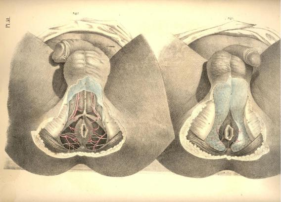
PLATE 51.
FIGURE 1.
A.The superficial fascia covering the urethral space.
B.The sphincter ani.
C.The coccyx.
D D. The right and left ischiatic tuberosities.
H. The anus.
I I. The glutei muscles.
FIGURE 2.
A, B, C, D, H, I. The same parts as in Fig. 1.
E. The accelerator urinae muscle.
F F. Right and left erector penis muscle.
G G. Right and left transverse muscle.
Plate 51
Figure 2 |
Figure 1 |