

SURGICAL ANATOMY by Joseph Maclise
.pdf68 |
COMMENTARY ON PLATE 26. |
An abscess or other tumour of the liver may, by pressing on the vena portae, cause serous effusion into the peritonaeal sac; or by pressure on the inferior vena cava, which is connected with the posterior thick border of the liver, may cause anasarca of the lower limbs. Matter accumulating habitually in the sigmoid flexure of the colon may cause a hydrocele, or a varicocele, by pressing on the spermatic veins of the left side. It is quite true that these two last-named affections appear more frequently on the left side than on the right; and it seems to me much more rational to attribute them to the abovementioned circumstance than to the fact that the left spermatic veins open, at a disadvantageous right angle, into the left renal vein.
DESCRIPTION OF PLATE 26.
A.The systemic aorta. Owing to the body being inclined forwards, the root of the aorta appears to approach too near the lower boundary (N) of the thorax.
B.The left brachio-cephalic vein.
C.Left subclavian vein.
D.Right brachia-cephalic vein.
E.Left common carotid artery.
F.Brachio-cephalic artery.
G G*. The first pair of ribs.
H.Superior vena cava.
I.Left bronchus.
K K*. Fourth pair of ribs.
L.Descending thoracic aorta.
M.Oesophagus.
N.Epigastrium.
O.Left kidney.
P.Umbilicus.
Q.Abdominal aorta, at its bifurcation. R R*. Right and left iliac fossae.
S.Left common iliac vein.
T.Inferior vena cava.
U.Psoas muscle, supporting the right spermatic vessels.
V.Left external iliac artery crossed by the left ureter.
W.Right external iliac artery crossed by the right ureter.
X.The rectum.
Y.The urinary bladder, which being fully distended, and viewed from above, gives it the appearance of being higher than usual above the pubic symphysis.
Z.Pubic symphysis.
2.The left internal abdominal ring complicated with the epigastric vessels, the vas deferens, and the spermatic vessels.
3.The right internal abdominal ring in connection with the like vessels and duct as that of left side.
4.Superior mesenteric artery.
5, 6. Right and left external iliac veins.
7, 8. Situations of the anterior superior iliac spinous processes. 9, 10. Situations of the coracoid processes.
11, 12. Right and left hypochondriac regions.

Plate 26
COMMENTARY ON PLATE 27.
THE SURGICAL DISSECTION OF THE SUPERFICIAL BLOODVESSELS ETC. OF
THE INGUINO-FEMORAL REGION.
Hernial protrusions are very liable to occur at the inguino-femoral region; and this fact has led the surgeon to study the anatomical relations of this part with more than ordinary care and patience. So minutely has he dissected every structure proper to this locality, and so closely has he investigated every possible condition of it as being the seat of hernial, that the only novelty which now remains to be sought for is that of a simplification of the facts, already known to be far too much obscured by an unwieldy nomenclature, and a useless detail of trifling evidence. And it would seem that nothing can more directly tend to this simplification, than that of viewing the inguinal and femoral regions, not separately, but as a relationary whole. For as both regions are blended together by structures which are common to both, so do the herniae which are described as being proper to either region, occur in such close connexion as at times to render it very difficult to distinguish between them.
The human species is, of all others, most subject to hernial in the groin. The erect attitude of the human form, and the fact that many of its more powerful muscular efforts are performed in this posture, cause its more frequent liability to the accidents called abdominal herniae or ruptures.
The viscera of the abdomen occupy this cavity completely, and indeed they naturally, at all times, subject the abdominal parietes to a state of constant pressure, as may be proved by their escape from the abdomen in cases of large wounds of this region. In the erect posture of the body this pressure is increased, for the viscera now gravitate and force downwards and forwards against the abdominal parietes. In addition to this gravitating force, another power impels the viscera from above downwards--namely, that of the muscles of the trunk, and the principal agent amongst these is the diaphragm. The lungs, again, expanding above the diaphragm, add also to the gravitation of the abdominal contents, and these, under the pressure thus accumulated, occasionally make an exit for themselves at the groins, which are the weakest and most depending parts of the abdomen.
Herniae are variously named in accordance with the following circumstances--viz., the precise locality at which they occur--the size and form of the tumour--the time of life at which they happen. Sexual peculiarities do not serve to distinguish herniae, though it is true that the inguinal form, at the part D F, occurs more commonly in the male, whilst the crural form, at the opening E, happens more frequently in the female.
The most common forms of herniae happen at those localities where the abdominal walls are traversed by the bloodvessels on their way to the outstanding organs, and where, in consequence, the walls of the abdomen have become weakened. It also happens, that at these very situations the visceral pressure is greatest whilst the body stands erect. These localities are, A, the umbilicus, a point characterized as having given passage (in the foetal state) to the umbilical vessels; D, the place where the spermatic vessels and duct pass from the abdomen to the testicle; and immediately beneath this, the crural arch, which gives exit to the crural vessels.
(Page 69)
70 |
COMMENTARY ON PLATE 27. |
Herniae may happen at other localities, such as at the thyroid aperture, which transmits the thyroid vessels; and at the greater sacrosciatic notch, through which the gluteal vessels pass; and all regions of the abdominal walls may give exit to intestinal protrusion in consequence of malformations, disease, or injury. But as the more frequent varieties of herniae are those which traverse the localities, A, D, E, and as these, fortunately, are the most manageable under the care of the surgical anatomist, we proceed to examine the structures concerned in their occurrence.
A direct opening from within outwards does not exist in the walls of the abdomen; and anatomy demonstrates to us the fact, that where the spermatic cord, D F, and the femoral vessels, pass from the abdomen to the external parts, they carry with them a covering of the several layers of structures, both muscular and membranous, which they encounter in their passage. The inguinal and crural forms of herniae which follow the passages made by the spermatic cord, and the crural vessels, must necessarily carry with them the like investments, and these are what constitute the coverings of the herniae themselves.
The groin in its undissected state is marked by certain elevations and depressions which indicate the general relations of the subcutaneous parts. The abdomen is separated from the thigh by an undulating grooved line, extending from C*, the point of the iliac bone, to B, the symphysis pubis This line or fold of the groin coincides exactly with the situation of that fibrous band of the external oblique muscle named Poupart's ligament. From below the middle of this abdomino-femoral groove, C B, another curved line, D, b, springs, and courses obliquely, inwards and downwards, between the upper part of the thigh and the pubis, to terminate in the scrotum. The external border of this line indicates the course of the spermatic cord, D F, which can be readily felt beneath the skin. In all subjects, however gross or emaciated they may happen to be, these two lines are readily distinguishable, and as they bear relations to the several kinds of rupture taking place in these parts, the surgeon should consider them with keen regard. A comparison of the two sides of the figure, PLATE 27, will show that the spermatic cord, D F, and Poupart's ligament, C B, determine the shape of the inguino-femoral region. When the integument with the subcutaneous adipose tissue is removed from the inguino-femoral region, we expose that common investing membrane called the superficial fascia. This fascia, a a a, stretches over the lower part of the abdomen and the upper part of the thigh. It becomes intimately attached to Poupart's ligament along the ilio-pubic line, C B; it invests the spermatic cord, as shown at b, and descends into the scrotum, so as to encase this part. Where this superficial fascia overlies the saphenous opening, E, of the fascia lata, it assumes a "cribriform" character, owing to its being pierced by numerous lymphatic vessels and some veins. As this superficial fascia invests all parts of the inguino-femoral region, as it forms an envelope for the spermatic cord, D F, and sheathes over the saphenous opening, E, it must follow of course that wherever the hernial protrusion takes place in this region, whether at D, or F, or E, or adjacent parts, this membrane forms the external subcutaneous covering of the bowel.
There is another circumstance respecting the form and attachments of the superficial fascia, which, in a pathological point of view, is worthy of notice--viz., that owing to the fact of its enveloping the scrotum, penis, spermatic cord, and abdominal parietes, whilst it becomes firmly attached to Poupart's ligament along the abdomino-femoral fold, B C, it isolates these parts, in some degree, from the thigh; and when urine happens to be from any cause extravasated through this abdominal-scrotal bag of the superficial fascia, the thighs do not in general participate in the inflammation superinduced upon such accident.
The spermatic cord, D, emerges from the abdomen and becomes definable through the fibres of the sheathing tendon of the external oblique muscle, H, at a point midway between the extremities of the ilio-pubic line or fold. In some cases, this place, whereat the cord first manifests itself in the groin, lies nearer the pubic symphysis; but however much it may vary in this particular, we may safely regard the femoro-pubic fold, D, b, as containing the cord, and also that the place where this fold meets the iliopubic line, C B, at the point D, marks the exit of the cord from the abdomen.
COMMENTARY ON PLATE 27 |
71 |
The spermatic cord does not actually pierce the sheathing tendon of the external oblique muscle at the point D, and there does not, in fact, exist naturally such an opening as the "external abdominal ring," for the cord carries with it a production of the tendon of the external oblique muscle, and this has been named by surgical anatomists the "intercolumnar fascia,"[Footnote] the "spermatic fascia." The fibres of this spermatic fascia are seen at D F, crossing the cord obliquely, and encasing it. This covering of the cord lies beneath the spermatic envelope formed by, a b, the superficial fascia; and when a hernial protrusion descends through the cord, both these investing membranes form the two outermost envelopes for the intestine in its new and abnormal situation.
[Footnote: On referring to the works of Sir Astley Cooper, Hesselbach, Scarpa, and, others, I find attempts made to establish a distinction between what is called the "intercolumnar fascia" and the "spermatic fascia," and just as if these were structures separable from each other or from the aponeurotic sheath of the external oblique muscle. I find, in like manner, in these and other works, a tediously-laboured account of the superficial fascia, as being divisible into two layers of membrane, and that this has given rise to considerable difference of opinion as to whether or not we should regard the deeper layer as being a production of the fascia lata, ascending from the thigh to the abdomen, or rather of the membrane of the abdomen descending to the thigh, &c. These and such like considerations I omit to discuss here; for, with all proper deference to the high authority of the authors cited, I dare to maintain, that, in a practical point of view, they arc absolutely of no moment, and in a purely scientific view, they are, so far as regards the substance of the truth which they would reveal, wholly beneath the notice of the rational mind. The practitioner who would arm his judgment with the knowledge of a broad fact or principle, should not allow his serious attention to be diverted by a pursuit after any such useless and trifling details, for not only are they unallied to the stern requirements of surgical skill, but they serve to degrade it from the rank and roll of the sciences. Whilst operating for the reduction of inguinal hernia by the "taxis" or the bistoury, who is there that feels anxiety concerning the origin or the distinctiveness of the "spermatic fascia?" Or, knowing it to be present, who concerns himself about the better propriety of naming it "tunica vaginalis communis," "tunique fibreuse du cordon spermatique," "fascia cremasterica," or "tunica aponeurotica?"]
The close relations which the cord, D F, bears to the saphenous opening, E, of the fascia lata, should be closely considered, forasmuch as when an oblique inguinal hernia descends from D to F, it approaches the situation of the saphenous opening, E, which is the seat of the femoral or crural hernia, and both varieties of hernia may hence be confounded. But with a moderate degree of judgment, based upon the habit of referring the anatomy to the surface, such error may always be avoided. This important subject shall be more fully treated of further on.
The superficial bloodvessels of the inguino-femoral region are, e e, the saphenous vein, which, ascending from the inner side of the leg and thigh, pierces the saphenous opening, E, to unite with the femoral vein. The saphenous vein, previously to entering the saphenous opening, receives the epigastric vein, i, the external circumflex ilii vein, h, and another venous branch, d, coming from the fore part of the thigh. In the living body the course of the distended saphenous vein may be traced beneath the skin, and easily avoided in surgical operations upon the parts contained in this region. Small branches of the femoral artery pierce the fascia lata, and accompany these superficial veins. Both these orders of vessels are generally divided in the operation required for the reduction of either the inguinal or the femoral strangulated hernia; but they are, for the most part, unimportant in size. Some branches of nerves, such as, k, the external cutaneous, which is given off from the lumbar nerves, and, f, the middle cutaneous, which is derived from the crural nerve, pierce the fascia lata, and appear upon the external side and middle of the thigh.
72 |
COMMENTARY ON PLATE 27. |
Numerous lymphatic glands occupy the inguino-femoral region; these can be felt, lying subcutaneous, even in the undissected state of the parts. These glands form two principal groups, one of which, c, lies along the middle of the inguinal fold, C B; the other, G g, lies scattered in the neighbourhood of the saphenous opening. The former group receive the lymphatic vessels of the generative organs; and the glands of which it is composed are those which suppurate in, syphilitic or other affections of these parts.
The general relations which the larger vessels of the inguino-femoral region bear to each other and to the superficies, may be referred to in PLATE 27, with practical advantage. The umbilicus, A, indicates pretty generally the level at which the aorta bifurcates on the forepart of the lumbar vertebrae. In the erect, and even in the recumbent posture, the aorta may (especially in emaciated subjects) be felt pulsating under the pressure of the hand; for the vertebrae bear forward the vessel to a level nearly equal with, C C, the anterior superior spinous processes of the iliac bones. If a gunshot were to pass through the abdomen, transversely, from these points, and through B, it would penetrate the aorta at its bifurcation. The line A B coincides with the linea alba. The oblique lines, A D, A D,* indicate the course of the iliac vessels. The point D marks the situation where the spermatic vessels enter the abdomen; and also where the epigastric artery is given off from the external iliac. The most convenient line of incision that can be made for reaching the situation of either of the iliac arteries, is that which ranges from C, the iliac spine, to D, the point where the spermatic cord enters the abdomen. The direct line drawn between D and G marks the course of the femoral artery, and this ranges along the outer border, E, of the saphenous opening.
DESCRIPTION OF PLATE 27.
A.The umbilicus.
B.The upper margin of the pubic symphysis.
C.The anterior superior spine of the left iliac bone. C*, the situation of the corresponding part on the right side.
D.The point where, in this subject, the cord manifested itself beneath the fibres of the external oblique muscle. D*, a corresponding part on the opposite side.
E.The saphenous opening in the fascia lata, receiving e, the saphenous vein.
F.The lax and pendulous cord, which in this case, overlies the upper part of the saphenous opening.
G.Lymphatic glands lying on the fascia lata in the neighbourhood of the saphenous opening.
H.The fleshy part of the external oblique muscle.
a a a. The superficial fascia of the abdomen.
b.The same fascia forming an envelope for the spermatic cord and scrotum.
c.Inguinal glands lying near Poupart's ligament.
d.A common venous trunk, formed by branches from the thigh and abdomen, and joining--
e e. The saphenous vein.
f.The middle cutaneous nerve, derived from the anterior crural nerve.
g.Femoral lymphatic glands.
h. Superficial external iliac vein.
i. Superficial epigastric vein.
k. External cutaneous branches of nerves from the lumbar plexus.
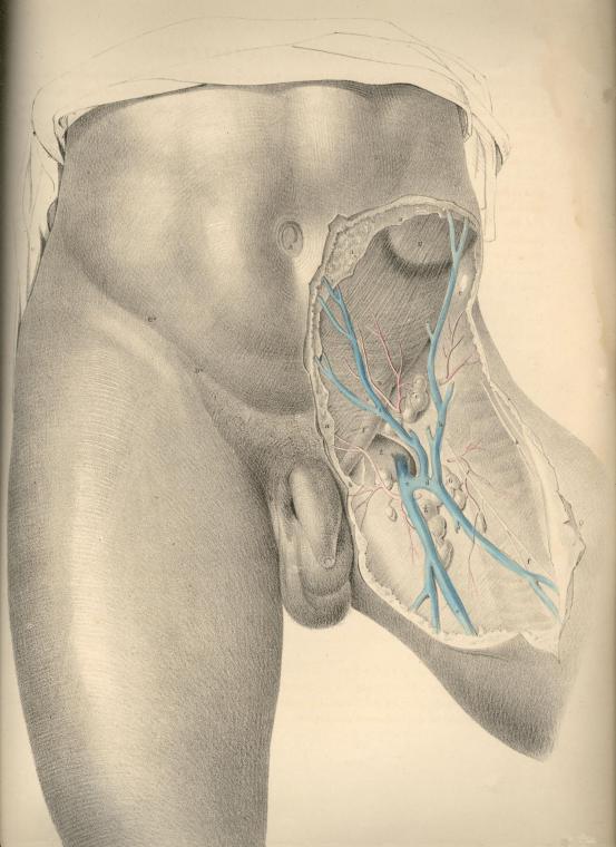
PLATE 27
COMMENTARY ON PLATES 28 & 29.
THE SURGICAL DISSECTION OF THE FIRST, SECOND, THIRD, AND FOURTH LAYERS OF THE INGUINAL REGION IN CONNEXION WITH THOSE OF THE THIGH.
The common integument or first layer of the inguino-femoral region being removed, we expose the superficial fascia constituting the second layer. The connexion of this fascia with Poupart's ligament along the line C D, together with the facts, that corresponding with this line the fascia is devoid of adipous substance, and the integument thin and delicate, whilst above over the abdomen, and below over the upper part of the thigh, the meshes of the fascia are generally loaded with a considerable quantity of adipous tissue, will account for the permanency and distinctness of the fold of the groin. As this fold corresponds with Poupart's ligament, it is taken as a guide to distinguish between the inguinal and femoral forms of herniae.
The general relations of the superficial fascia are well described by Camper in the following sentence: "Musculus obliquus igitur externus abdominis, qua parte carneus est, membrana quadam propria, quali omnes musculi, tegitur, quae sensim in aponeurosin mutata, ac cum tendineis hujus musculi partibus unita, externe ac anteriore parte abdomen tegit; finem vero nullibi habere perspicuum est, ad pubem enim miscet cellulosa membrana, cum ligamento penis in viris ac clitoridis in feminis, involucrum dat musculo cremasteri, ac aponeuroseos speciem musculis anterioribus femoris, qua glandulae inguinales, ac cruris vasa majora obteguntur." (Icones Herniarum.)
Owing to the varied thickness of the adipous tissue contained in the superficial fascia at several regions of the same body, and at some corresponding regions of different individuals, it will be evident that the depth of the incision required to divide it, so as to expose subjacent structures, must vary accordingly. Where the superficial fascia, after encasing the cord, descends into the scrotum, it is also devoid of the fatty tissu.
By the removal of the superficial fascia and glands we expose the aponeurosis of the external oblique muscle, A a, Pl. 28, (constituting the third layer of the groin,) and also the fascia of the thigh, H L. These strong fibrous structures will be observed to hold still in situ the other parts, and to be the chief agents in determining the normal form of this region.
The inguino-femoral region, as being the seat of hernial protrusions, may in this stage of the dissection be conveniently described as a space formed of two triangles--the one inguinal, the other femoral, placed base to base. The inguinal triangle may be drawn between the points, B C D, Pl. 28, while the femoral triangle may be marked by the points, C D N. The conjoined bases of these triangles correspond to Poupart's ligament along the line, C D. The inguinal varieties of herniae occur immediately above the line, C D, while the femoral varieties of herniae take place below this line. The herniae of the inguinal triangle are, therefore, distinguishable from those of the femoral triangle by a reference to the line, C D, or Poupart's ligament.
The aponeurosis of the external oblique muscle occupies the whole of that space which I have marked as the inguinal triangle, B C D, Pl. 28. The fleshy fibres of the muscle, A, after forming the lateral wall of the abdomen, descend to the level of C, the iliac spinous process, and here give off the inguinal part of their broad tendon, a. The fibres of this part of the tendon descend obliquely downwards and forwards to become inserted at the median line of the abdomen into the linea alba, B D, as also into the symphysis and crista of the os pubis. The lower band of the fibres of this tendinous sheath--viz., that which is stretched between C, the iliac spine, and D, the crista pubis, is named Poupart's ligament; and this is strongly connected with H, the iliac portion of the fascia lata of the thigh.
(Page 73)
74 |
COMMENTARY ON PLATES 28 & 29. |
Poupart's ligament is not stretched tensely in a right line, like the string of a bow, between the points, C and D. With regard to these points it is lax, and curves down towards the thigh like the arc of a circle. The degree of tension which it manifests when the thigh is in the extended position is chiefly owing to its connexion with the fascia lata. If in this position of the limb we sever the connexion between the ligament and fascia, the former becomes relaxed in the same degree as it does when we flex the thigh upon the abdomen. The utmost degree of relaxation which can be given to Poupart's ligament is effected by flexing the thigh towards the abdomen, at the same time that we support the body forwards. This fact has its practical application in connexion with the reduction of herniae.
Immediately above the middle of Poupart's ligament, at the point E, Pl. 28, we observe the commencement of a separation taking place among the fibres of the aponeurosis. These divide into two bands, which, gradually widening from each other as they proceed inwards, become inserted, the upper one into the symphysis pubis, the lower into the spine and pectineal ridge of this bone. The lower band identifies itself with Poupart's ligament. The interval which is thus formed by the separation of these fibres assumes the appearance of an acute triangle, the apex of which is at E, and the base at D. But the outer end of this interval is rounded off by certain fibres which cross those of the bands at varying angles. At this place, the aponeurosis, thus constituted of fibres disposed crossways, is elongated into a canal, forming an envelope for the cord, K. This elongation is named the "external spermatic fascia," and is continued over the cord as far as the testicle. In the female, a similar canal encloses the round ligament of the uterus. From the above-mentioned facts, it will appear that the so-called "external abdominal ring" does not exist as an aperture with defined margins formed in the tendon of the external oblique muscle. It is only when we divide the spermatic fascia upon the cord at K, that we form the external ring, and then it must be regarded as an artificial opening, as at D, Pl. 29.
The part of the groin where the spermatic fascia is first derived from the aponeurosis, so as to envelope the cord, varies in several individuals; and thereupon depends, in great measure, the strength or weakness of the groin. In some cases, the cord becomes pendulous as far outwards as the point E, Pl. 28, which corresponds to the internal ring, thereby offering a direct passage for the hernial protrusion. In other instances, the two bands of the aponeurosis, known as the "pillars of the ring," together with the transverse fibres, or "intercolumnar fascia," firmly embrace and support the cord as far inwards as the point K, and by the oblique direction thus given to the cord in traversing the inguinal parietes, these parts are fortified against the occurrence of hernia. In Pl. 28, the cord, K, will be observed to drop over the lower band of fibres, ("external pillar of the ring,") and to have D, the crista pubis, on its inner side. In Pl. 29, the upper band of fibres ("internal pillar of the ring") may be seen proceeding to its insertion into the symphysis pubis. When a hernial tumour protrudes at the situation K, it is invested, in the same manner as the cord, by the spermatic fascia, and holds in respect to the fibrous bands or pillars the same relations also as this part.
After removing the tendon of the external oblique muscle, A a, Pl. 28, together with its spermatic elongation, E, we expose the internal oblique, F E, Pl. 29, and the cremaster, constituting the fourth inguinal layer. The fleshy part of this muscle, F E, occupies a much greater extent of the inguinal region than does that of the external oblique. Whilst the fleshy fibres of the latter terminate on a level with C, the iliac spine, those of the internal oblique are continued down as far as the external abdominal ring, E D h, and even protrude through this place in the form of a cremaster. The muscular fibres of the internal oblique terminate internally at the linea semilunaris, g; while Poupart's ligament, the spinous process and crest of the ilium, give origin to them externally.
COMMENTARY ON PLATES 28 & 29. |
75 |
At the linea semilunaris, the tendon of the internal oblique is described as dividing into two layers, which passing, one before and the other behind the rectus abdominis, thus enclose this muscle in a sheath, after which they are inserted into the linea alba, G. The direction of the fibres of the inguinal portion of the muscle, F E, is obliquely downwards and forwards, and here they are firmly overlaid by the aponeurosis of the external oblique.
The cremaster muscle manifests itself as being a part of the internal oblique, viewing this in its totality. Cloquet (Recherches anatomiques sur les Hernies de l'Abdomen) first demonstrated the correctness of this idea.
The oblique and serial arrangement of the muscular fibres of the internal oblique, F, Pl. 29, is seen to be continued upon the spermatic cord by the fibres of the cremaster, E e. These fibres, like those of the lower border of the internal oblique, arise from the middle of Poupart's ligament, and after descending over the cord as far as the testicle in the form of a series of inverted loops, e, again ascend to join the tendon of the internal oblique, by which they become inserted into the crest and pectineal ridge of the os pubis. The peculiar looping arrangement exhibited by the cremasteric fibres indicates the fact that the testicle, during its descent from the loins to the scrotum, carried with it a muscular covering, at the expense of the internal oblique muscle. The cremaster, therefore, is to be interpreted as a production of the internal oblique, just as the spermatic fascia is an elongation of the external oblique. The hernia, which follows the course of the spermatic vessels, must therefore necessarily become invested by cremasteric fibres.
The fascia lata, H, Pl. 28, being strongly connected and continuous with Poupart's ligament along its inferior border, the boundary line, which Poupart's ligament is described as drawing between the abdomen and thigh, must be considered as merely an artificial one.
In the upper region of the thigh the fascia lata is divided into two parts--viz., H, the iliac part, and L, the pubic. The iliac part, H, which is external, and occupying a higher plane than the pubic part, is attached to Poupart's ligament along its whole extent, from C to D, Pl. 28; that is, from the anterior iliac spinous process to the crista pubis. From this latter point over the upper and inner part of the thigh, the iliac division of the fascia appears to terminate in an edge of crescentic shape, h; but this appearance is only given to it by our separating the superficial fascia with which it is, in the natural state of the parts, blended. The pubic part of the fascia, L, Pl. 28, which is much thinner than the iliac part, covers the pectineus muscle, and is attached to the crest and pectineal ridge of the os pubis, occupying a plane, therefore, below the iliac part, and in this way passes outwards beneath the sheath of the femoral vessels, K I, Pl. 29. These two divisions of the fascia lata, although separated above, are united and continuous on the same plane below. An interval is thus formed between them for the space of about two inches below the inner third of Poupart's ligament; and this interval is known as the "saphenous opening," L h, Pl. 28. Through this opening, the saphena vein, O, Pl. 29, enters the femoral vein, I.
From the foregoing remarks it will appear that no such aperture as that which is named "saphenous," and described as being shaped in the manner of L h, Pl. 28, with its "upper and lower cornua," and its "falciform process," or edge, h, exists naturally. Nor need we be surprised, therefore, that so accurate an observer as Soemmering (de Corporis Humani Fabrica) appears to have taken no notice of it.
Whilst the pubic part of the fascia lata passes beneath the sheath of the femoral vessels, K I, Pl. 29, the iliac part, H h, blends by its falciform margin with the superficial fascia, and also with N n, the sheath of the femoral vessels. The so-called saphenous opening, therefore, is naturally masked by the superficial fascia; and this membrane being here perforated for the passage of the saphena vein, and its tributary branches, as also the efferent vessels of the lymphatic glands, is termed "cribriform."