

Color Atlas of Neurology (Thieme 2004)
.pdfPeripheral Paralysis
|
Myopathy |
|
|
|
|
|
ism), and chronic toxic myopathies (alcohol, |
|||||||
|
Problems such as muscle weakness, fatigue, |
corticosteroids, chloroquine). |
|
|||||||||||
|
|
|
|
|
|
|||||||||
|
stiffness, cramps, tension, atrophy, pain, and in- |
! Disorders of Muscle Function |
|
|||||||||||
|
voluntary movement do not necessarily signify |
In these disorders, weakness is due to impaired |
||||||||||||
|
disease of the muscle itself. Myopathy must be |
|||||||||||||
|
function of the muscle fibers. Persistent weak- |
|||||||||||||
|
distinguished |
from |
neurogenic weakness of |
|||||||||||
|
ness can lead to muscle atrophy. The episodic |
|||||||||||||
|
UMN or LMN type. Weakness may accompany |
|||||||||||||
|
occurrence or worsening of muscle weakness is |
|||||||||||||
|
systemic disease because of a generalized cata- |
|||||||||||||
|
typical. |
|
|
|
||||||||||
|
bolic state or through a specific disease-related |
|
|
|
||||||||||
|
Primary myopathies. Hypokalemiaand hyper- |
|||||||||||||
|
impairment of muscle function. Myopathy may |
|||||||||||||
|
kalemia-related forms of paralysis belong to this |
|||||||||||||
|
be either primary or secondary, i.e., the product |
|||||||||||||
|
group. |
|
|
|
||||||||||
|
of another underlying disease. Different types of |
|
|
|
||||||||||
|
Myasthenic syndromes. Myasthenia gravis and |
|||||||||||||
|
myopathy affect different muscle groups: some |
|||||||||||||
|
Lambert–Eaton syndrome are characterized by |
|||||||||||||
|
are |
generalized (congenital |
myopathy), |
while |
||||||||||
|
abnormal fatigability of the muscles. |
|
||||||||||||
|
others are mainly either proximal (Duchenne |
|
||||||||||||
Function |
Postviral fatigue syndrome. Mildly |
increased |
||||||||||||
type muscular dystrophy, polymyositis) or distal |
||||||||||||||
! Muscle Pain and Stiffness |
|
|||||||||||||
chondrial myopathy). Myasthenia gravis, strictly |
|
|||||||||||||
|
(myotonic dystrophy, inclusion body myositis), |
fatigability of the muscles may persist for weeks |
||||||||||||
|
or mainly affect the head and face (mito- |
after recovery from a viral illness. |
|
|||||||||||
|
|
|
|
|
|
|||||||||
Motor |
speaking a disorder of neuromuscular transmis- |
Muscle pain and stiffness restrict movement, |
||||||||||||
sion |
rather than a |
form of |
myopathy, |
most |
||||||||||
causing weakness as a secondary consequence. |
||||||||||||||
prominently |
affects the |
orbicularis |
oculi |
|||||||||||
Muscle pain. Muscle pain (myalgia, p. 346) at |
||||||||||||||
|
muscle; weakness |
increases with exercise. |
||||||||||||
|
rest |
and |
on exertion |
accompanies muscle |
||||||||||
|
Muscle power is commonly graded according to |
|||||||||||||
|
trauma (muscle rupture, strain, soreness, com- |
|||||||||||||
|
the scale proposed by the British Medical Re- |
|||||||||||||
|
partment syndromes), viral myositis (influenza, |
|||||||||||||
|
search Council (MRC) (1976): |
|
|
|
||||||||||
|
|
|
|
Coxsackie virus, herpes simplex virus), fibromy- |
||||||||||
|
|
|
|
|
|
|
|
|
||||||
|
|
|
|
|
|
|
|
|
algia, polymyalgia rheumatica, and muscle |
|||||
|
|
0 |
No muscle contraction |
|
|
|
cramps and spasms of various causes (malig- |
|||||||
|
|
1 |
Visible or palpable contraction, but no move- |
|
nant |
hyperthermia, |
carnitine |
palmitoyl- |
||||||
|
|
|
ment |
|
|
|
|
|
transferase |
deficiency, |
phosphorylase defi- |
|||
|
|
2 |
Movement occurs, but not against gravity |
|
||||||||||
|
|
|
ciency/glycogen storage disease type V). |
|||||||||||
|
|
3 |
Movement against gravity |
|
|
|
||||||||
|
|
|
|
|
Muscle stiffness. Stiffness is prominent in con- |
|||||||||
|
|
4 |
Movement against gravity and additional re- |
|
||||||||||
|
|
|
sistance |
|
|
|
|
|
genital myotonia, neuromyotonia, and cold- |
|||||
|
|
5 |
Normal muscle power |
|
|
|
induced paramyotonia. |
|
|
|||||
! Muscle Atrophy
Myopathy produces atrophy through the impaired development, the destruction, and the impaired regeneration of muscle fibers.
Primary (genetic) myopathies include the progressive muscular dystrophies, myotonic muscular dystrophies, congenital myopathies (e. g., central core disease, nemaline myopathy), and metabolic myopathies (Pompe disease/glycogen storage disease type II, Kearns–Sayre syndrome, carnitine deficiency).
52Secondary myopathies include myositis, myopathy due to endocrine disorders (hyperthy-
roidism and hypothyroidism, hyperparathyroid-
Rohkamm, Color Atlas of Neurology © 2004 Thieme
All rights reserved. Usage subject to terms and conditions of license.

Peripheral Paralysis
Artery
Striated muscle fiber
Mitochondrion 
Three primary
bundles of muscle fibers
Muscle fascia with epimysium
Motor end plate region
Progressive Duchenne muscular dystrophy |
Artery |
(proximal leg weakness, patients use arms to raise themselves to standing position = Gowers’s sign, calf hypertrophy, lumbar hyperlordosis)
Structure of skeletal muscle
Myotonic response |
Myasthenic response |
|
(exercise-induced muscle |
||
(delayed fist opening) |
||
weakness; here in eyes) |
||
|
Muscle pain and stiffness
(exercise-induced; here due to ischemia)
External ophthalmoplegia
(here mitochondrial myopathy)
Motor Function
53
Rohkamm, Color Atlas of Neurology © 2004 Thieme
All rights reserved. Usage subject to terms and conditions of license.

Cerebellum
The functions of the cerebellum include the control of balance, posture, gait, and goal-directed movement, and the regulation of muscle tone.
Neural Pathways
|
Afferent connections. The three large white- |
||
|
matter tracts (peduncles) of the cerebellum con- |
||
|
vey afferent input to the cerebellar cortex from |
||
|
the cerebral cortex (especially visual areas), pon- |
||
|
tine nuclei, the brain stem nuclei of the trigemi- |
||
|
nal, vestibular, and cochlear nerves, and the spi- |
||
|
nal cord. The superior cerebellar peduncle con- |
||
|
veys ipsilateral proprioceptive input (p. 104) |
||
|
from the anterior spinocerebellar tract of the |
||
Function |
spinal cord. The middle cerebellar peduncle car- |
||
ries fibers of pontine origin (p. 45). The inferior |
|||
|
|||
|
cerebellar peduncle carries fibers from the vesti- |
||
|
bular nerve and nucleus to the flocculonodular |
||
Motor |
lobe and fastigial nucleus, and from the con- |
||
tralateral inferior olive to the cerebellar hemi- |
|||
|
|||
|
spheres (olivocerebellar tract), as well as propri- |
||
|
oceptive input from the posterior spinocerebel- |
||
|
lar tract (derived from muscle spindles and |
||
|
destined for the anterior and posterior portions |
||
|
of the paramedian cerebellar cortex) and fibers |
||
|
from the brain stem reticular formation. |
||
|
Efferent connections. The cerebellar nuclei |
||
|
(fastigial, |
globose, emboliform, and dentate; |
|
|
p. 43) project via the (contralateral) superior |
||
|
cerebellar peduncle to the red nucleus, thalamus, |
||
|
and reticular formation. The thalamus projects |
||
|
in turn to the premotor and primary motor cor- |
||
|
tex, whose output travels down to the pons, |
||
|
which projects back to the cerebellum, forming |
||
|
a neuroanatomical circuit. Cerebellar output in- |
||
|
fluences (ipsilateral) spinal motor neurons by |
||
|
way of the red nucleus and rubrospinal tract. |
||
|
The inferior cerebellar peduncle projects to the |
||
|
vestibular nuclei and brain stem reticular for- |
||
|
mation |
(completing the vestibulocerebellar |
|
feedback loop) and influences spinal motor neurons by way of the vestibulospinal and reticulospinal tracts.
Functional Systems
The cerebellum can be thought of as containing
54three separate functional components.
Vestibulocerebellum (archeocerebellum). Struc-
tures: Flocculonodular lobe and lingula. Afferent
connections: From the semicircular canals and maculae (p. 56), vestibular nucleus, visual system (lateral geniculate body), superior colliculus, and striate area to the vermis. Efferent connections: From the fastigial nucleus to the vestibular nucleus and reticular formation. Functions: Control of balance, axial and proximal muscle groups, respiratory movements, and head and eye movements (stabilization of gaze). Effects of lesions: Loss of balance (truncal ataxia, postural ataxia ! gait ataxia), nystagmus on lateral gaze, and absence of visual fixation suppression (p. 26) resulting in oscillopsia (stationary objects seem to move).
Spinocerebellum (paleocerebellum). Structures:
Parts of the superior vermis (culmen, central lobule) and inferior vermis (uvula, pyramis), parts of the cerebellar hemispheres (wing of central lobule, quadrangular lobule, paraflocculus). Afferent connections: The pars intermedia receives the spinocerebellar tracts, projections from the primary motor and somatosensory cortex, and projections conveying auditory, visual, and vestibular information. Efferent connections: From the nucleus interpositus to the reticular formation, red nucleus, and ventrolateral nucleus of the thalamus, which projects in turn to area 4 of the cortex. Functions: Coordination of distal muscles, muscle tone (postural control), balance, and velocity and amplitude of saccades. Effects of lesions: Gait ataxia ! postural ataxia, muscular hypotonia, dysmetria.
Pontocerebellum (neocerebellum). Structures:
Most of the cerebellar hemispheres, including the declive, folium, and tuber of the vermis. Afferent connections: From sensory and motor cortical areas, premotor cortex, and parietal lobes via pontine nuclei and the inferior olive. Efferent connections: From the dentate nucleus to the red nucleus and the ventrolateral nucleus of the thalamus, and from these structures onward to motor and premotor cortex. Functions: Coordination, speed, and precision of body movement and speech. Effects of lesions: Delayed initiation and termination of movement, mistiming of agonist and antagonist contraction in movement sequences, intention tremor, limb ataxia.
Rohkamm, Color Atlas of Neurology © 2004 Thieme
All rights reserved. Usage subject to terms and conditions of license.

|
|
|
Cerebellum |
|
|
|
|
Fastigial nucleus |
|
|
|
|
Reticular formation |
|
|
|
|
Reticulospinal |
|
|
|
|
tract |
|
|
|
|
Vestibular |
|
|
|
|
nucleus |
|
|
|
|
Vestibular n. |
|
|
|
|
Vestibulospinal |
|
|
|
|
tract |
|
Uvula |
|
|
|
|
Nodulus |
Vestibulocerebellum |
|
|
|
|
|
Postural and gait ataxia |
Function |
|
|
|
|
||
Pyramis |
|
Culmen |
Thalamocortical tract |
|
|
|
|
||
|
|
|
Central lobule |
|
|
|
|
|
|
|
|
|
Thalamus |
Motor |
|
|
|
(ventral lateral nucleus) |
|
|
|
|
|
|
|
|
|
Emboliform and globose |
|
|
|
|
nuclei |
|
|
|
|
Red nucleus |
|
|
|
|
Reticular formation |
|
Spinocerebellum |
|
Rubrospinal tract |
|
|
|
|
|
||
Areas 5 and 7 |
Area 4 |
|
Reticulospinal tract |
|
|
|
|
||
|
|
|
Spinocerebellar tract |
|
|
|
Area 6 |
|
|
|
|
|
Thalamus |
|
|
|
|
Spinocerebellum |
|
|
|
|
Red |
|
|
|
|
nucleus |
|
|
|
|
Pontocere- |
|
|
|
|
bellum |
|
|
|
|
Pontine |
|
|
|
|
nuclei |
|
|
Dentate |
|
Vestibulocerebellum |
|
|
nucleus |
|
|
|
Hemisphere |
|
|
|
|
Olive |
|
Spinocerebellum |
|
|
|
|
|
||
|
Rubrospinal tract |
|
|
|
|
Pontocerebellum |
|
Structure of cerebellum |
55 |
|
|
|
(overview; median section of vermis right) |
|
Rohkamm, Color Atlas of Neurology © 2004 Thieme
All rights reserved. Usage subject to terms and conditions of license.

Vestibular System
Labyrinth
|
The vestibular apparatus (labyrinth) consists of |
|
|
the saccule, the utricle, and three semicircular |
|
|
canals, each in a plane approximately at right |
|
|
angles to the others. The labyrinth is filled with |
|
|
fluid (endolymph) and has five receptor organs: |
|
|
the ampullary crests, which lie in a dilatation |
|
|
(ampulla) in front of the utricle at the end of |
|
|
each semicircular canal; the saccular macula |
|
|
(macula sacculi), a vertically oriented sensory |
|
|
field on the medial wall of the saccule; and the |
|
|
utricular macula (macula utriculi), a horizon- |
|
|
tally oriented sensory field on the floor of the |
|
|
utricle. |
|
Function |
Semicircular canals. Angular acceleration is |
|
sensed by the hair cells of the ampullary crests |
||
|
||
|
and the gelatinous bodies (cupulae) suspended |
|
|
in the endolymph above them. Rotation about |
|
Motor |
the axis of one of the semicircular canals causes |
|
its cupula to deflect in the opposite direction, |
||
|
||
|
because it is held back by the more slowly |
|
|
moving endolymph. With persistent rotation at |
|
|
a constant angular velocity (i.e., zero angular ac- |
|
|
celeration), the cupula returns to its neutral |
|
|
position; but if the rotation should suddenly |
|
|
stop, the cupula is deflected once again, this |
|
|
time in the direction of the original rotation, be- |
|
|
cause it is carried along by the still moving en- |
|
|
dolymph. The subject feels as if he were rotating |
|
|
counter to the original direction of rotation and |
|
|
also tends to fall in the original direction of rota- |
|
|
tion. |
|
|
Maculae. The otolithic membrane of the saccu- |
|
|
lar and utricular maculae is denser than the sur- |
|
|
rounding endolymph because of the calcite |
|
|
crystals (otoliths) embedded in it. Linear accel- |
|
|
eration of the head thus causes relative motion |
|
|
of the otolithic membrane and endolymph, re- |
|
|
sulting in activation of the macular receptor |
|
|
cells (hair cells). The resultant forces lead to ac- |
|
|
tivation of the sensory receptors of the maculae. |
|
|
Neural Pathways |
|
|
Afferent connections. The semicircular organs |
|
|
project mainly to the superior and medial vesti- |
|
|
bular nuclei, the macular organs to the inferior |
56vestibular nuclei. The vestibulocerebellum maintains both afferent and efferent connections with the vestibular nuclei; in particular,
the lateral vestibular nucleus receives its major
input from the paramedian region of the cerebellar cortex. Fibers reach the vestibular nucleus from the spinal cord ipsilaterally, and also bilaterally by way of the fastigial nucleus. The oculomotor nuclei project to the ipsilateral vestibular nuclei through the medial longitudinal fasciculus. The vestibular nuclei are interconnected by internuclear and commissural fibers. Efferent connections. The vestibulocerebellum projects to the ipsilateral nodulus, uvula, and anterior lobe of the vermis, and to the flocculi bilaterally. The lateral vestibulospinal tract projects ipsilaterally to the motor neurons of the spinal cord and also gives off fibers to cranial nerves X and XI. Fibers to the motor neurons of the contralateral cervical spinal cord decussate in the medial vestibulospinal tract. The medial longitudinal fasciculus (p. 86) gives off caudal fibers to the motor neurons of the cervical cord, and rostral fibers bilaterally to the nuclei subserving eye movement. Other fibers cross the midline to the contralateral thalamus, which projects in turn to cortical areas 2 and 3 (primary somatosensory area).
Functional Systems
The vestibular system provides vestibulocochlear input to the cerebellum, spinal cord, and oculomotor apparatus to enable the coordination of head, body, and eye movements. It influences extensor muscle tone and reflexes via the lateral vestibulospinal tract (postural motor system). The medial longitudinal fasciculus permits simultaneous, integrated control of neck muscle tone and eye movements. The oculomotor system (p. 86) communicates with the vestibular nuclei, the cerebellum, and the spinal cord via the medial longitudinal fasciculus and pontine projection fibers; thus the control of eye movements is coordinated with that of body movements. Proprioceptive input concerning joint position and muscle tone reaches the vestibular system from the cerebellum (p. 54). Thalamocortical connections permit spatial orientation. Phenomena such as nausea, vomiting, and sweating arise through interaction with the hypothalamus, the medullary “vomiting center,” and the vagus nerve, while the emotional component of vestibular sensation (pleasure and discomfort) arises through interaction with the limbic system.
Rohkamm, Color Atlas of Neurology © 2004 Thieme
All rights reserved. Usage subject to terms and conditions of license.

Vestibular System
Cupula |
|
|
Otoliths |
|
|
|
|
|
|
|
Otolithic membrane |
|
|
|
|
Hair cells with villi |
|
|
|
Ampullary crest |
Thalamocortical tracts |
|
Utricular macule |
|
Ampulla |
(to areas 2, 3) |
|
|
|
|
|
|
|
|
|
Thalamus |
|
|
|
Visual |
|
|
|
|
information |
|
|
|
|
(area 8) |
|
|
|
|
Visual |
|
|
Visual information |
|
|
|
(areas 17, 18, 19) |
Function |
|
information |
|
|
||
|
|
|
||
(CN II) |
|
|
Vestibular ganglion |
|
|
|
|
||
|
|
|
Spinocerebellar tract |
|
|
|
|
Cerebellum |
Motor |
|
|
Vestibular portion of CN VIII |
||
|
|
|
||
|
|
|
Vestibular apparatus |
|
Medial |
Vestibulocerebellar |
|
|
|
longitudinal |
tracts |
|
|
|
fasciculus |
|
|
|
|
|
Joint afferent fibers |
Endolymph |
|
|
Cuneate nucleus |
|
|
|
|
|
|
|
|
|
Nucleus of vagus n. |
Neck muscle |
|
|
|
|
|
|
|
|
|
Effect of |
|
|
|
|
endolymph |
|
|
|
|
pressure on cupula |
|
Cupula |
|
Posterior |
Anatomic |
|
|
|
Horizontal semicircular |
|
|||
spinocerebellar tract |
|
|||
pathways and |
|
|||
|
|
canal |
|
|
Motor neuron |
functional |
|
|
|
|
|
|
||
systems |
|
|
|
|
|
|
|
|
|
|
Otolithic membrane |
|
|
|
|
Hair cells with villi |
Left |
Right |
|
|
|
|
||
|
|
|
Head rotation |
|
Linear acceleration |
Deflection of hair cells |
|
(above, rotation to right; |
57 |
to side |
(effect of gravity) |
|
below, sudden stop) |
|
Rohkamm, Color Atlas of Neurology © 2004 Thieme
All rights reserved. Usage subject to terms and conditions of license.

Vertigo
|
Patients often use the word “dizziness” non- |
|
|
specifically to mean lightheadedness, unsteadi- |
|
|
ness, reeling, staggering, or a feeling of rotation. |
|
|
Dizziness in this broad sense has many possible |
|
|
causes. Vertigo, or dizziness in the narrow sense, |
|
|
is the unpleasant illusion that one is moving or |
|
|
that the external world is moving (so-called |
|
|
subjective and objective vertigo, respectively). |
|
|
Pathogenesis. Vertigo arises from a mismatch |
|
|
between expected and received sensory input |
|
|
(vestibular, visual, and somatosensory) regard- |
|
|
ing spatial orientation and movement. |
|
|
Cause. Vertigo occurs as a normal response to |
|
|
certain stimuli (physiological vertigo) or as the |
|
|
result of diseases (pathological vertigo) affect- |
|
Function |
ing the labyrinth (peripheral vestibular vertigo), |
|
central vestibular system (central vestibular |
||
|
||
|
vertigo), or other functional systems (nonvesti- |
|
|
bular vertigo). |
|
Motor |
Symptoms and signs. The manifestations of ver- |
|
tigo are the same regardless of etiology. They fall |
||
|
||
|
into the following categories: autonomic (drow- |
|
|
siness, yawning, pallor, sialorrhea, increased |
|
|
sensitivity to smell, nausea, vomiting), mental |
|
|
(decreased drive, lack of concentration, apathy, |
|
|
sense of impending doom), visual (oscillopsia = |
|
|
illusory movement of stationary objects), and |
|
|
motor (tendency to fall, staggering and swaying |
|
|
gait). |
|
|
Physiological Vertigo |
|
|
Healthy persons may experience vertigo when |
|
|
traveling by car, boat, or spaceship (kinetosis = |
|
|
motion sickness) or on looking down from a |
|
|
mountain or tall building (height vertigo). |
|
|
Peripheral Vestibular (Labyrinthine) |
|
|
Vertigo (p. 88) |
There is usually an acute, severe rotatory vertigo directed away from side of the labyrinthine lesion, with a tendency to fall toward the side of the lesion, horizontal nystagmus away from the side of lesion, nausea, and vomiting. Peripheral vestibular vertigo may depend on position, being triggered, for example, when the patient turns over in bed or stands up (positional ver-
58tigo), or it may be independent of position (persistent vertigo). It may also occur in attacks as
episodic vertigo.
Positional vertigo. Benign, paroxysmal positional vertigo (BPPV) of peripheral origin is usually due to detached otoliths of the utricular macula floating in the posterior semicircular canal (canalolithiasis). With every bodily movement, the freely floating otoliths move within the canal, under the effect of gravity. An abnormal cupular deflection results, starting 1–5 seconds after movement and lasting up to 30 seconds. The Dix– Hallpike maneuver is a provocative test for BPPV: the patient is rapidly taken from a sitting to a supine position while the head is kept turned 45° to one side. If nystagmus and vertigo ensue, they are due to canalolithiasis on the side of the ear nearer the ground. The canalith repositioning procedure (CRP), by which particles can be removed from the semicircular canal, involves repeatedly turning the patient’s head to the opposite side, then back upright.
Episodic and persistent vertigo may be due to viral infection of the vestibular apparatus (vestibular neuritis, labyrinthitis) or to Ménière disease, which is characterized by attacks of rotatory vertigo, tinnitus, hearing loss, and ear pressure. Other causes include labyrinthine fistula and vestibular paroxysm.
Central Vestibular Vertigo
(pp. 70 ff and 88)
This type of vertigo is caused by a lesion of the vestibular nuclei, vestibulocerebellum, thalamus, or vestibular cortex, or their interconnecting fibers. Depending on the etiology (e. g., hemorrhage, ischemia, tumor, malformation, infection, multiple sclerosis, “vestibular” epilepsy, basilar migraine), vertigo may be transient or persistent, acute, episodic, or slowly progressive. It may be associated with other neurological deficits depending on the location and extent of the responsible lesion.
Nonvestibular Vertigo
Episodic or persistent nonvestibular vertigo often manifests itself as staggering, unsteady gait, and loss of balance. The possible causes include disturbances of the oculomotor apparatus, cerebellum, or spinal cord; peripheral neuropathy; intoxication; anxiety (phobic attacks of vertigo); hyperventilation; metabolic disorders; and cardiovascular disease.
Rohkamm, Color Atlas of Neurology © 2004 Thieme
All rights reserved. Usage subject to terms and conditions of license.

Vertigo
Motor Function
Rotatory vertigo |
|
|
Nonvestibular vertigo |
(positional, chronic) |
|
|
|
|
|
(unsteady posture/gait; nondirectional |
|
|
|
|
|
|
|
|
vertigo) |
|
|
|
Utricle |
|
|
|
|
|
|
|
Cupula |
|
|
|
|
|
|
|
Otolith in posterior |
|
|
|
semicircular canal |
Semicircular canal after repositioning
Benign peripheral paroxysmal positional vertigo
59
Rohkamm, Color Atlas of Neurology © 2004 Thieme
All rights reserved. Usage subject to terms and conditions of license.

Motor Function
Gait Disturbances
Normal Gait
Posture. The assumption of an upright posture and the maintenance of balance (postural reflexes) are essential for walking upright. Locomotion requires the unimpaired function of the motor, visual, vestibular, and somatosensory systems. The elderly cannot stand up as quickly and tend to walk somewhat unsteadily, with stooped posture and broader steps, leading to an elevated risk of falling.
Locomotion. Normally, walking can be initiated without hesitation. The gait cycle (time between two successive contacts of the heel of one foot with the ground = 2 steps) is characterized by the gait rhythm (number of steps per unit time), the step length (actually the length of an entire cycle, i.e. 2 steps), and the step width (distance
between the lines of movement of the two heels, roughly 5–10 cm). Touchdown is with the heel of the foot. Each leg alternately functions as the supporting leg (stance phase, roughly 65% of the gait cycle), and as the advancing leg (swing phase, roughly 35% of the gait cycle). During the shifting phase, both feet are briefly in contact with the ground (double-stance phase, roughly 25% of the stance phase). Because the body’s center of gravity shifts slightly to the side with each step, the upper body makes small compensatory movements to maintain balance. The arms swing alternately and opposite to the direction of leg movement. Normally, the speed of gait can be changed instantaneously. In old age, the gait sequence is less energetic and more hesitant, and turns tend to be carried out en bloc.
Gait Disturbances
Description |
Related Terms |
Site of Lesion |
Possible Cause |
|
|
|
|
Antalgic gait |
Limping gait, leg differ- |
Foot, leg, pelvis, spinal |
Lumbar root lesion, bone dis- |
|
ence, limp |
column |
ease, peripheral nerve com- |
|
|
|
pression |
Steppage gait |
Foot-drop gait |
Sciatic or peroneal nerve, |
Polyneuropathy, peroneal |
|
|
spinal root L4/5, motor |
paresis; lesions of motor neu- |
|
|
neuron |
ron, sciatic nerve, or L4/5 root |
Waddling gait |
Duchenne gait, Trendelen- |
Paresis of pelvic girdle |
Myopathy, osteomalacia; le- |
|
burg gait, gluteal gait |
muscles (Duchenne) or of |
sions of the hip joint or super- |
|
|
gluteal abductors (Tren- |
ior gluteal nerve; L5 lesion |
|
|
delenburg) |
|
Toe-walking |
|
Talipes equinus, spasticity |
Foot deformity, cerebral palsy, |
|
|
|
Duchenne muscular dystrophy, |
|
|
|
habit |
Spastic gait |
Paraspastic gait, leg cir- |
Pyramidal tract, extrapy- |
Unilateral or bilateral central |
|
cumduction, spastic-ataxic |
ramidal motor system (su- |
paralysis with spasticity, stiff- |
|
gait, Wernicke–Mann gait |
pratentorial, infratentorial, |
man syndrome |
|
|
spinal) |
|
Ataxia of gait |
Gait ataxia, staggering |
|
gait, unsteady gait, ta- |
|
betic gait, reeling gait |
Peripheral nerves, posterior |
Polyneuropathy, disease af- |
column of spinal cord, |
fecting posterior columns, |
spinocerebellar tracts, cere- |
tabes dorsalis, cerebellar le- |
bellum, thalamus, postcen- |
sion, intoxication, progressive |
tral cortex |
supranuclear palsy |
|
Dystonic gait |
Choreiform gait |
Basal ganglia |
Torsion dystonia, dopa-respon- |
|
|
|
|
sive dystonia, kinesiogenic |
|
|
|
|
paroxysmal dystonia, Hunting- |
|
|
|
|
ton disease |
|
Start delay |
Hypokinetic rigid gait, gait |
Frontal lobe, basal ganglia, |
Parkinson disease, frontal lobe |
|
|
apraxia, festinating gait |
extensive white matter le- |
lesion, normal-pressure hydro- |
|
|
|
sions |
cephalus, Binswanger disease |
60 |
Psychogenic |
Functional gait distur- |
|
Mental illness, malingering |
|
gait distur- |
bance |
|
|
bance
Rohkamm, Color Atlas of Neurology © 2004 Thieme
All rights reserved. Usage subject to terms and conditions of license.
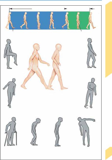
Gait Disturbances
Stance phase |
|
|
|
Swing phase |
|
||||
|
|
|||
|
|
|
|
|
Right leg supports |
Right leg |
|
advances |
|
Gait cycle |
Steppage gait |
Knee instability |
|
(quadriceps paresis, leg dorsally |
||
|
||
|
angulated) |
Posture and gait in youth
(left) and old age (right)
Ataxic gait |
Spastic gait |
|
(right hemiparesis) |
Spastic gait |
Hypokinetic-rigid gait |
Psychogenic gait disturbances |
(spastic paraparesis) |
(left, Parkinson disease; right, start |
(histrionic movements) |
|
delay/gait apraxia) |
|
Motor Function
61
Rohkamm, Color Atlas of Neurology © 2004 Thieme
All rights reserved. Usage subject to terms and conditions of license.