

Color Atlas of Neurology (Thieme 2004)
.pdfCerebral Circulation
12
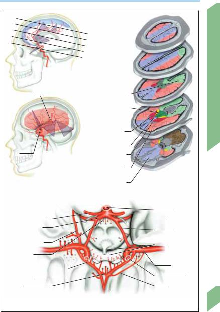
Anterior Circulation of the Brain
The anterior and middle cerebral arteries are the terminal branches of the internal carotid artery. They originate at the ICA bifurcation, located in the circle of Willis at the level of the anterior clinoid process, between the optic chiasm and the temporal pole.
Anterior Cerebral Artery (ACA)
The ACA is the more medial of the two arteries arising from the ICA bifurcation. It ascends lateral to the anterior clinoid process and past the the optic nerve and optic chiasm, giving off a small branch, the anterior communicating artery (ACommA), which crosses the midline to join the contralateral ACA. The segment of ACA proximal to the origin of the ACommA is its precommunicating segment (segment A1). The A1 segments on either side and the ACommA together form the anterior half of the circle of Willis. Segment A1 gives off an average of eight basal perforating arteries that enter the brain through the anterior perforated substance. The recurrent artery of Heubner arises from the ACA near the origin of the ACommA, either from the distal part of A1 or from the proximal part of A2.
The postcommunicating segment of the ACA (segments A2 to A5) ascends between the frontal lobes and runs toward the occiput in the interhemispheric fissure, along the corpus callosum and below the free border of the falx cerebri, as thepericallosal artery.SegmentA2,whichusually gives off the frontopolar artery, ends where the artery turns forward to become apposed to the genu of the corpus callosum; segment A3 is the frontally convex arch of the vessel along the genu. TheA4andA5segmentsrunroughlyhorizontally overthecallosalsurfaceandgiveoffsupracallosal branches that run in a posterior direction.
Distribution. The basal perforating arteries arisingfromA1supplytheventralhypothalamusand a portion of the pituitary stalk. Heubner’s artery supplies the head of the caudate nucleus, the rostral four-fifths of the putamen, the globus pallidus, and the internal capsule. The blood supply of the inferior portion of the genu of the corpus callosum, and of the olfactory bulb, tract, and trigone, is variable.
The ACommA gives off a few small branches (anteromedial central branches) to the hypothalamus.
Branches from the postcommunicating segment of the ACA supply the inferior surface of the frontal lobe (frontobasilar artery), the medial and parasagittal surfaces of the frontal lobe (callosomarginal artery), the paracentral lobule (paracentral artery), the medial and parasagittal surfaces of the parietal lobe (precuneal artery), and the cortex in the region of the parieto-occipital sulcus (parieto-occipital artery).
Middle Cerebral Artery (MCA)
The MCA is the more lateral of the two arteries arising from the ICA bifurcation. Its first segment (M1, sphenoidal segment) follows the anterior clinoid process for a distance of 1 to 2 cm. The MCA then turns laterally to enter the depths of the Sylvian fissure (i.e., the Sylvian cistern), where it lies on the surface of the insula and gives off branches to it (M2, insular segment). It bends back sharply to travel along the surface of the operculum (M3, opercular segment) and then finally emerges through the Sylvian fissure onto the lateral convexity of the brain (M4 and M5, terminal segments).
Distribution. Small branches of M1 (the thalamostriate and lenticulostriate arteries) supply the basal ganglia, the claustrum, and the internal, external, and extreme capsules. M2 and M3 branches supply the insula (insular arteries), lateral portions of the orbital and inferior frontal gyri (frontobasal artery), and the temporal operculum, including the transverse gyrus of Heschl (temporal arteries). M4 and M5 branches supply most of the cortex of the lateral cerebral convexity, including portions of the frontal lobe (arteries of the precentral and triangular sulci), the parietal lobe (anterior and posterior parietal arteries), and the temporal lobe (arteries of central and postcentral sulci). In particular, important cortical areas supplied by M4 and M5 branches include the primary motor and sensory areas (precentral and postcentral gyri) and the language areas of Broca and Wernicke.
Rohkamm, Color Atlas of Neurology © 2004 Thieme
All rights reserved. Usage subject to terms and conditions of license.
Anterior Circulation of the Brain
|
A 4 |
A 5 |
|
|
A |
|
|
|
|
|
|
|
|
|
|
|
|
A |
|
|
|
|
A 3 |
|
B |
B |
|
|
|
|
C |
|
||
|
|
|
|
|
|
|
|
|
|
|
D |
|
|
A 2 |
|
|
|
E |
|
|
|
Anterior cerebral artery |
C |
|
|||
|
|
|
||||
|
(blue: ACA distribution, sections A-E) |
|
|
|||
A. of central |
|
|
|
Posterior cerebral a. |
D |
Circulation |
sulcus (rolandic a.) |
|
|
(peripheral branches) |
|
||
|
|
|
|
|||
M2 and M3 |
|
|
|
Posterior cerebral a. |
|
|
|
|
|
|
(central branches) + |
|
|
|
|
|
|
posterior |
|
|
|
|
|
|
communicating a. |
E |
Cerebral |
|
|
|
|
Middle cerebral a. |
||
|
|
|
|
|
||
|
|
M 4 |
M 5 |
(central branches) |
|
|
|
|
Anterior choroidal a. |
|
|||
|
|
|
|
|
|
|
Insular |
Internal carotid a. |
Anterior cerebral a. |
|
|
||
arteries |
(central branches) |
|
|
|||
|
|
|
|
|
||
|
|
|
|
Middle cerebral a. |
|
|
|
|
|
|
(peripheral branches) |
|
|
Middle cerebral artery |
|
|
Anterior cerebral a. |
Horizontal sections A-E |
|
|
|
|
|
|
|||
(red: MCA distribution) |
|
|
(peripheral branches) |
|
|
|
|
|
|
|
|
Basilar a. |
|
Superior cerebellar a. |
|
|
|
Oculomotor a. |
|
|
Posterior cerebral a. |
|
|
|
Posterior |
|
|
(precommunicating segment) |
|
|
|
|||
|
|
communicating a. |
|
|||
|
|
|
|
|
|
|
Anterior choroidal a. |
|
|
|
Optic chiasm, |
|
|
|
|
|
|
|
|
|
Posteromedial |
|
|
|
|
pituitary stalk |
|
|
|
|
|
|
|
|
central arteries |
|
|
|
|
|
|
A1 (precommunicating segment) |
|
M2 and M3 |
|
|||
|
|
|
||||
Olfactory tract |
|
|
|
|
M1 |
|
|
|
|
|
|
|
|
Anterior |
|
|
|
A2 |
recurrent a. of |
|
communicating a. |
|
|
Heubner |
|
||
|
|
|
|
|||
|
|
|
|
Circle of Willis |
|
13 |
|
|
|
|
|
|
|
Rohkamm, Color Atlas of Neurology © 2004 Thieme
All rights reserved. Usage subject to terms and conditions of license.

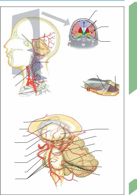
Vertebral and Basilar Arteries
Extracranial Portion
|
The vertebral artery arises from the arch of the |
|
|
subclavian artery at a point designated V0. The |
|
|
prevertebral or V1 segment extends from V0 to |
|
|
the foramen transversarium of the transverse |
|
|
process of C6. The transversarial or V2 segment |
|
|
passes vertically through the foramina transver- |
|
|
saria of C6 through C2, accompanied by venous |
|
|
plexuses and sympathetic nerves derived from |
|
|
the cervical ganglia. It gives off branches to the |
|
|
cervical nerves, vertebrae and intervertebral |
|
|
joints, neck muscles, and cervical spinal cord. |
|
Circulation |
Often, a prominent branch at the C5 level anas- |
|
tomoses with the anterior spinal artery. The V3 |
||
|
||
|
segment, also called the atlas (C1) loop, runs |
|
|
laterally and then vertically to the foramen |
|
|
transversarium of C1, which it passes through, |
|
Cerebral |
winds medially along the lateral mass of C1, |
|
pierces the posterior atlanto-occipital mem- |
||
|
||
|
brane behind the atlanto-occipital joint, and |
|
|
then enters the dura mater and arachnoid mem- |
|
|
brane at the level of the foramen magnum. The |
|
|
two vertebral arteries are unequal in size in |
|
|
about 75% of persons, and one of them is ex- |
|
|
tremely narrow (hypoplastic) in about 10%, usu- |
|
|
ally on the right side. |
|
|
Intracranial Portion |
|
|
The V4 segment of the vertebral artery lies en- |
|
|
tirely within the subarachnoid space. It termi- |
|
|
nates at the junction of the two vertebral arter- |
|
|
ies to form the basilar artery, at the level of the |
|
|
lower border of the pons. Proximal to the junc- |
|
|
tion, each vertebral artery gives off a mediobasal |
|
|
branch; these two branches run for ca. 2 cm and |
|
|
then unite in the midline to form a single ante- |
|
|
rior spinal artery, which descends along the |
|
|
anterior surface of the medulla and spinal cord |
|
|
(see p. 23). The posterior inferior cerebellar artery |
|
|
(PICA), which originates from the V4 segment at |
|
|
a highly variable level, curves around the infe- |
|
|
rior olive and extends dorsally through the root |
|
|
filaments of the accessory nerve. It then ascends |
|
|
behind the fibers of the hypoglossus and vagus |
|
|
nerves, forms a loop on the posterior wall of the |
|
|
fourth ventricle, and gives off terminal branches |
14to the inferior surface of the cerebellar hemisphere, the tonsils, and the vermis. It provides
most of the blood supply to the dorsolateral
medulla and the posteroinferior surface of the cerebellum. The posterior spinal artery (there is one on each side) arises from either the vertebral artery or the PICA.
The basilar artery runs in the prepontine cistern along the entire length of the pons and then bifurcates to form the posterior cerebral arteries. Its inferior portion is closely related to the abducens nerves, its superior portion to the oculomotor nerves. Its paramedian, short circumferential, and long circumferential branches supply the pons and the superior and middle cerebellar peduncles.
The anterior inferior cerebellar artery (AICA) arises from the lower third of the basilar artery. It runs laterally and caudally toward the cerebellopontine angle, passes near the internal acoustic meatus, and reaches the flocculus, where it gives off terminal branches that supply the anteroinferior portion of the cerebellar cortex and part of the cerebellar nuclei. The AICA lies basal to the abducens nerve and ventromedial to the facial and auditory nerves in the cerebellopontine cistern. It often gives rise to a labyrinthine branch that enters the internal acoustic meatus.
The superior cerebellar arteries (SCA) of both sides originate from the basilar trunk just below its bifurcation. Each SCA travels through the perimesencephalic cistern dorsal to the oculomotor nerve, curves around the cerebral peduncle caudal and medial to the trochlear nerve, and then enters the ambient cistern, where it gives off its terminal branches. The SCA supplies the upper pons, part of the mid brain, the upper surface of the cerebellar hemispheres, the upper portion of the vermis, and the cerebellar nuclei.
Rohkamm, Color Atlas of Neurology © 2004 Thieme
All rights reserved. Usage subject to terms and conditions of license.
Vertebral and Basilar Arteries
|
|
|
|
Anterior cerebral a. |
|
|
|
|
|
Middle cerebral a. |
|
|
|
|
|
(peripheral + central |
|
|
|
|
|
branches) |
|
|
Posterior |
|
|
Posterior |
|
|
|
|
cerebral a. |
|
|
|
cerebral a. |
|
|
|
|
Basilar a. |
|
|
(peripheral + |
|
|
|
|
|
|
||
|
|
|
|
central branches) |
|
|
|
|
|
Anterior |
|
|
|
|
|
choroidal a. |
|
V4 |
|
|
|
|
|
|
V3 |
|
Coronal section |
|
|
|
|
|
|
Circulation |
|
|
Occipital a. |
|
|
||
|
V2 |
|
|
|
|
|
|
|
Mediolateral branches |
Lateral branches |
|
External |
|
|
Cerebral |
||
V1 |
|
|
|
||
carotid a. |
|
Medial branches |
|
||
Common |
V0 |
|
Basilar a. |
|
|
carotid a. |
|
|
|
|
|
|
|
|
|
|
|
Subclavian a. |
|
|
|
|
|
Vertebrobasilar system |
|
|
Brainstem vessels, |
|
|
(extracranial; plane of coronal section) |
|
|
territories |
|
|
|
|
|
|
(pons) |
|
Caudate nucleus |
|
|
|
|
|
Pericallosal a. |
|
|
|
Thalamus |
|
Internal capsule |
|
|
|
Posterior cerebral a. |
|
Putamen |
|
|
Superior cerebellar a. |
|
|
Anterior cerebral a. |
III |
|
|
Labyrinthine a. |
|
|
IV |
|
|
||
Middle cerebral a. |
|
|
|
|
|
|
|
|
|
|
|
Posterior |
V |
|
|
|
|
|
|
|
|
|
|
communicating a. |
VI |
|
|
VIII |
|
|
IX |
|
|
||
|
|
|
|
||
Internal carotid a. |
|
|
|
|
|
AICA |
X |
|
VII |
|
|
|
|
|
|
||
|
|
|
|
|
|
Basilar a., |
|
|
PICA |
|
|
pontine branches |
|
XI |
|
|
|
|
|
|
|
|
|
|
Vertebrobasilar system (intracranial) |
|
15 |
||
|
|
|
|||
Rohkamm, Color Atlas of Neurology © 2004 Thieme
All rights reserved. Usage subject to terms and conditions of license.

Cerebral Circulation
16
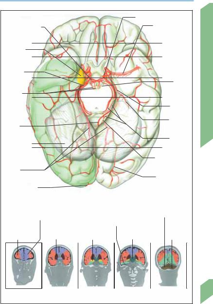
Posterior Circulation of the Brain
Posterior Cerebral Artery (PCA)
The precommunicating segment of the PCA (P1) extends from the basilar bifurcation to the origin of the posterior communicating artery
(PCommA). Its course lies within the interpeduncular cistern, which is demarcated by the clivus and the two cerebral peduncles. The oculomotor nerve, after its emergence from the brain stem, runs between the PCA and the superior cerebellar artery. The postcommunicating segment (P2) curves laterally and backward around the crus cerebri and reaches the posterior surface of the midbrain at an intercollicular level.
The precommunicating and postcommunicating segments are together referred to as the pars circularis of the PCA. (Alternatively, the pars circularis may be divided into three segments— interpeduncular, ambient, and quadrigeminal— named after the cisterns they traverse.)
Distal to the pars circularis of the PCA is the pars terminalis, which divides above the tentorium and caudal to the lateral geniculate body to form its terminal branches, the medial and lateral occipital arteries.
Pars circularis. The precommunicating segment gives off fine branches (posteromedial central arteries) that pierce the interpeduncular perforated substance to supply the anterior thalamus, the wall of the third ventricle, and the globus pallidus. The postcommunicating segment gives off fine branches (posterolateral central arteries) to the cerebral peduncles, the posterior portion of the thalamus, the colliculi of the mid brain, the medial geniculate body, and the pineal body. Further branches supply the posterior portion of the thalamus (thalamic branches), the cerebral peduncle (peduncular branches), and the lateral geniculate body and choroid plexus of the third and lateral ventricles (posterior choroidal branches).
Pars terminalis. Of the two terminal branches of this terminal portion of the PCA, the lateral occipital artery (together with its temporal branches) supplies the uncus, the hippocampal gyrus, and the undersurface of the occipital lobe. The medial occipital artery passes under the splenium of the corpus callosum, giving off branches that supply it (dorsal branch to the corpus callosum) as well as the cuneus and pre-
cuneus (parieto-occipital branch), the striate cortex (calcarine branch), and the medial surfaces of the occipital and temporal lobes (occipitotemporal and temporal banches), including the parasagittal portion of the occipital lobe.
Rohkamm, Color Atlas of Neurology © 2004 Thieme
All rights reserved. Usage subject to terms and conditions of license.
Posterior Circulation of the Brain
|
|
|
Posterior communicating a. |
|
||
Precommunicating |
|
|
Middle cerebral a. |
|
||
|
|
|
|
|
|
|
segment (P1) |
|
|
|
|
|
|
|
|
|
|
A |
|
|
Basal area |
|
|
|
|
|
|
of anterior |
|
|
|
B |
|
|
choroidal a. |
|
|
|
|
|
|
|
|
|
|
|
|
|
Postcom- |
|
|
|
|
|
|
municating |
|
|
|
C |
Oculo- |
|
segment (P2) |
|
|
|
|
|
|
|
|
|
|
|
motor n. |
Circulation |
Postero- |
|
|
|
D |
|
|
medial |
|
|
|
|
||
central |
|
|
|
|
Anterior |
|
arteries |
|
|
|
|
||
|
|
|
|
choroidal a. |
||
|
|
|
|
|
||
|
|
|
|
|
|
|
|
|
|
|
|
Posterior |
Cerebral |
Medial |
|
|
|
|
choroidal |
|
|
|
|
|
branch |
||
occipital a. |
|
|
|
|
||
|
|
|
|
|
||
|
|
|
|
|
Thalamic |
|
Undersurface |
|
|
|
|
|
|
|
|
|
E |
branch |
|
|
of cerebellum |
|
|
|
|
|
|
(showing arteries) |
|
|
Branch to corpus |
|
||
|
|
|
|
|||
|
|
|
|
|
callosum |
|
Lateral |
|
|
|
|
|
|
occipital a. |
|
|
Temporal branch |
|
||
|
|
|
|
|||
Calcarine branch |
|
|
|
|
|
|
|
Posterior cerebral artery |
|
|
|
|
|
|
(green = peripheral branches) |
|
|
|
|
|
Anterior cerebral a. |
|
|
Superior cerebellar a. |
|
||
|
Posterior cerebral a. |
|
|
|
||
|
|
|
|
|
||
Middle cerebral a. |
|
(peripheral branches) |
|
Posterior |
|
|
Middle cerebral a. |
Anterior |
Posterior cerebral a. |
|
|
||
(peripheral |
|
inferior |
|
|||
branches) |
(central branches) |
choroidal a. |
(central branches) |
|
cerebellar a. |
|
A |
B |
C |
D |
|
E |
|
Regional arterial blood flow (frontal and coronal planes A-E) |
|
|
17 |
|||
|
|
|
||||
Rohkamm, Color Atlas of Neurology © 2004 Thieme
All rights reserved. Usage subject to terms and conditions of license.

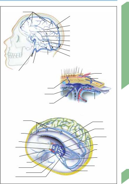
Intracranial Veins
Cerebral Veins
|
The superficial cerebral veins (cortical veins) |
|
|
carry blood from the outer 1–2 cm of the brain |
|
|
surface to large drainage channels such as the |
|
|
superior and inferior sagittal sinuses, the great |
|
|
cerebral vein of Galen, the straight sinus, and |
|
|
the tentorial veins. Thus, the cerebellar veins |
|
|
drain blood from the cerebellar surface into the |
|
|
superior vermian vein and thence into the great |
|
|
cerebral vein, straight sinus, and transverse |
|
|
sinuses. The deep cerebral veins (central veins) |
|
|
drain blood from the inner regions of the brain |
|
Circulation |
(hemispheric white matter, basal ganglia, cor- |
|
pus callosum, choroid plexus) and from a few |
||
|
||
|
cortical areas as well. |
|
|
Superficial cerebral veins (cortical veins). The |
|
|
superficial cerebral veins are classified by their |
|
Cerebral |
location as prefrontal, frontal, parietal, and |
|
occipital. Except for the occipital veins, which |
||
|
||
|
empty into the transverse sinus, these veins all |
|
|
travel over the cerebral convexity to join the su- |
|
|
perior sagittal sinus. They are termed bridging |
|
|
veins at their distal end, where they pierce the |
|
|
arachnoid membrane and bridge the sub- |
|
|
arachnoid space to join the sinus. The superficial |
|
|
middle cerebral vein (not shown) usually follows |
|
|
the posterior ramus of the Sylvian fissure and |
|
|
the fissure itself to the cavernous sinus. The infe- |
|
|
rior cerebral veins drain into the cavernous sinus, |
|
|
superior petrosal sinus, and transverse sinus. |
|
|
The superior cerebral veins drain into the super- |
|
|
ior sagittal sinus. |
|
|
Deep cerebral veins (central veins). The internal |
|
|
cerebral vein arises bilaterally at the level of the |
|
|
interventricular foramen (of Monro). It traverses |
|
|
the transverse cerebral fissure to a point just in- |
|
|
ferior to the splenium of the corpus callosum. |
|
|
The venous angle at its junction with the super- |
|
|
ior thalamostriate vein can be seen in a laterally |
|
|
projected angiogram. The two internal cerebral |
|
|
veins join under the splenium to form the great |
|
|
cerebral vein (of Galen), which receives the basal |
|
|
vein (of Rosenthal) and then empties into the |
|
|
straight sinus at the anterior tentorial edge at |
|
|
the level of the quadrigeminal plate. The basal |
|
|
vein of Rosenthal is formed by the union of the |
|
|
anterior cerebral vein, the deep middle cerebral |
18vein, and the striate veins. It passes posteromedial to the optic tract, curves around the cerebral
peduncle, and empties into the internal vein or
the great cerebral vein posterior to the brain stem.
Posterior fossa. The anterior, middle, and posterior veins of the posterior fossa drain into the great cerebral vein, the petrosal vein, and the tentorial and straight sinuses, respectively.
Extracerebral Veins
The extracerebral veins—most prominently, the dural venous sinuses—drain venous blood from the brain into the sigmoid sinuses and jugular veins.
The diploic veins drain into the extracranial veins of the scalp and the intracranial veins (dural venous sinuses).
The emissary veins connect the sinuses, diploic veins, and superficial veins of the skull. Infections sometimes travel along the emissary veins from the extracranial to the intracranial compartment.
The veins of the brain empty into the superior and inferior groups of dural venous sinuses. The sinuses of the superior group (the superior and inferior sagittal, straight, and occipital sinuses) join at the confluence of the sinuses (torcular Herophili), which drains into both transverse sinuses and thence into the sigmoid sinuses and internal jugular veins. The sinuses of the inferior group (superior and inferior petrosal sinuses) join at the cavernous sinus, which drains into the sigmoid sinus and internal jugular vein via the inferior petrosal sinus, or into the internal vertebral plexus via the basilar plexus.
Rohkamm, Color Atlas of Neurology © 2004 Thieme
All rights reserved. Usage subject to terms and conditions of license.
Intracranial Veins
Superior cerebral veins, bridging veins
Cavernous sinus 
Inferior petrosal 
sinus
Transverse sinus
Sigmoid sinus
Superior sagittal sinus
Inferior sagittal sinus Venous angle Internal cerebral v.
Great cerebral v. (Galen) Basal v. (Rosenthal) Straight sinus Confluence of sinuses
Internal jugular v.
Cerebral veins
Emissary v.
Superior sagittal sinus
Cerebral vein
Superior cerebral veins, bridging veins
Inferior sagittal sinus
Venous angle
Cavernous sinus
Ophthalmic v. 
Sphenoparietal sinus
Basilar plexus
Middle meningeal v.
Petrosal v.
Scalp vein
Diploic veins
Superior cerebral v., bridging vein
Extracerebral veins
Superior sagittal sinus
Basal v. (Rosenthal)
Great cerebral v.
Straight sinus
Confluence of sinuses
Sigmoid sinus
Transverse sinus
Superior petrosal sinus
Cerebral veins and sinuses
Cerebral Circulation
19
Rohkamm, Color Atlas of Neurology © 2004 Thieme
All rights reserved. Usage subject to terms and conditions of license.

Cerebral Circulation
Extracranial Veins
Craniocervical Veins
Anastomotic channels connect the cutaneous veins of the two sides of the head. Venous blood from the facial, temporal, and frontal regions drains into the facial and retromandibular veins and thence into the internal jugular vein. Some blood from the forehead drains via the nasofrontal, angular, and superior ophthalmic veins into the cavernous sinus. The occipital vein carries blood from the posterior portion of the scalp into the deep cervical vein and thence into the external jugular vein. Blood from the jugular veins continues to the brachiocephalic vein, superior vena cava, and right atrium. The venous channels in the spinal canal and the transcranial emissary veins play no more than a minor role in venous drainage. The pterygoid plexus links the cavernous sinus, the facial vein, and the internal jugular vein.
The numerous anastomoses between the extracranial and intracranial venous systems provide a pathway for the spread of infection from the scalp or face to the intracranial compartment. For example, periorbital infection may extend inward and produce septic thrombosis of the cavernous sinus.
which anastomoses with the occipital venous plexus and finally drains into the external jugular vein.
The pterygoid plexus lies between the temporalis, medial pterygoid, and lateral pterygoid muscles and receives blood from deep portions of the face, the external ear, the parotid gland, and the cavernous sinus, which it carries by way of the maxillary and retromandibular veins to the internal jugular vein.
Cervical Veins
The deep cervical vein originates from the occipital vein and suboccipital plexus. It follows the course of the deep cervical artery and vertebral artery to arrive at the brachiocephalic vein, which it joins.
The vertebral vein, which also originates from the occipital vein and suboccipital plexus, envelops the vertebral artery like a net and accompanies it through the foramina transversaria of the cervical vertebrae, collecting blood along the way from the cervical spinal cord, meninges, and deep neck muscles through the vertebral venous plexus, and finally joining the brachiocephalic vein.
Cranial Veins
The facial vein drains the venous blood from the face and anterior portion of the scalp. It begins at the inner canthus as the angular vein and communicates with the cavernous sinus via the superior ophthalmic vein. Below the angle of the mandible, it merges with the retromandibular vein and branches of the superior thyroid and superior laryngeal veins. It then drains into the internal jugular vein in the carotid triangle. The veins of the temporal region, external ear, temporomandibular joint, and lateral aspect of the face join in front of the ear to form the retromandibular vein, which either joins the facial vein or drains directly into the internal jugular vein. Its upper portion gives off a prominent dorsocaudal branch that joins the posterior auricular vein over the sternocleidomastoid muscle to communicate with the external jugu-
20lar vein. Venous blood from the posterior portion of the scalp and the mastoid and occipital emissary veins drains into the occipital vein,
Rohkamm, Color Atlas of Neurology © 2004 Thieme
All rights reserved. Usage subject to terms and conditions of license.

Extracranial Veins
Superficial temporal veins
Supratrochlear v.
Nasofrontal v.
Angular v.
Infraorbital v.
Facial v.
Submental v.
Anterior jugular v.
Left brachiocephalic v.
Lymph vessels joining to form thoracic duct
Extracranial veins
 Occipital v.
Suboccipital venous plexus
Pterygoid plexus
Retromandibular v.
Deep cervical v.
External jugular v.
Internal jugular v.
Transverse
cervical v.
Suprascapular v.
Subclavian v.
Cerebral Circulation
21
Rohkamm, Color Atlas of Neurology © 2004 Thieme
All rights reserved. Usage subject to terms and conditions of license.