

Fundamentals of Biomedical Engineering
.pdf112 |
|
|
|
|
|
FUNDAMENTALS OF BIOMEDICAL ENGINEERING |
||||||||
|
|
|
|
|
|
|
|
|
|
|
|
|
|
|
|
|
|
|
|
|
|
|
|
|
|
|
|
|
|
|
|
|
|
|
|
|
|
|
|
|
|
|
|
|
|
|
|
Nasal cavity |
|
|
|
|
|
|
|
|
|
|
|
|
|
|
|
|
|
|
|
|
|
|
|
|
|
|
|
|
|
|
|
|
|
|
|
|
Soft plate |
||||||||||||||||||||||||||||||||||||||||||||||||||
|
|
|
|
|
|
|
|
|
|
|
|
|
|
|
|
|
|
|
|
|
|
|
|
|
|
|
|
|
|
|
|
|
|
|
||||||||||||||||||||||||||||||||||||||||||||||||||||||||
Hard plate |
|
|
|
|
|
|
|
|
|
|
|
|
|
|
|
|
|
|
|
|
|
|
|
|
|
|
|
|
|
|
|
|
|
|
|
|
|
|
|
|
|
|
|
|
|
|
|
|
|
|
|
|
|
|
|
|
|
|
|
|
|
|
|
|
|
Epiglottis valve |
||||||||||||||||||||||||
|
|
|
|
|
|
|
|
|
|
|
|
|
|
|
|
|
|
|
|
|
|
|
|
|
|
|
|
|
|
|
|
|
|
|
|
|
|
|
|
|
|
|
|
|
|
|
|
|
|
|
||||||||||||||||||||||||||||||||||||||||
|
|
|
|
|
|
|
|
|
|
|
|
|
|
|
|
|
|
|
|
|
|
|
|
|
|
|
|
|
|
|
|
|
|
|
|
|
|
|
|
|
|
|
|
|
|
|
|
|
|
|
|
|
|
|
|
|
|
|
|
|
|
|
|
|
|
|
|
|
|
|
|
|
|
|||||||||||||||||
Oral cavity |
|
|
|
|
|
|
|
|
|
|
|
|
|
|
|
|
|
|
|
|
|
|
|
|
|
|
|
|
|
|
|
|
|
|
|
|
|
|
|
|
|
|
|
|
|
|
|
|
|
|
|
|
|
|
|
|
|
|
|
|
|
|
|
|
|
|
|
|||||||||||||||||||||||
|
|
|
|
|
|
|
|
|
|
|
|
|
|
|
|
|
|
|
|
|
|
|
|
|
|
|
|
|
|
|
|
|
|
|
|
|
|
|
|
|
|
|
|
|
|
|
|
|
|
|
|
|
|
|
|
|
|
|
|
|
|
|
|
|
Larynx (vocal cord) |
|||||||||||||||||||||||||
|
|
|
|
|
|
|
|
|
|
|
|
|
|
|
|
|
|
|
|
|
|
|
|
|
|
|
|
|
|
|
|
|
|
|
|
|
|
|
|
|
|
|
|
|
|
|
|
|
|
|
|
|
|
|
|
|
|
|
|
|
|
|
|
|
||||||||||||||||||||||||||
|
|
|
|
|
|
|
|
|
|
|
|
|
|
|
|
|
|
|
|
|
|
|
|
|
|
|
|
|
|
|
|
|
|
|
|
|
|
|
|
|
|
|
|
|
|
|
|
|
|
|
|
|
|
|
|
|
|
|
|
|
|
|
|
|
||||||||||||||||||||||||||
|
|
|
Pharynx |
|
|
|
|
|
|
|
|
|
|
|
|
|
|
|
|
|
|
|
|
|
|
|
|
|
|
|
|
|
|
|
|
|
|
|
|
|
|
|
|
|
|
|
|
|
|
|
|
|
|
|
|
|
|
Trachea (wind pipe) |
||||||||||||||||||||||||||||||||
|
|
|
(throat) |
|
|
|
|
|
|
|
|
|
|
|
|
|
|
|
|
|
|
|
|
|
|
|
|
|
|
|
|
|
|
|
|
|
|
|
|
|
|
|
|
|
|
|
|
|
|
|
||||||||||||||||||||||||||||||||||||||||
|
|
|
|
|
|
|
|
|
|
|
|
|
|
|
|
|
|
|
|
|
|
|
|
|
|
|
|
|
|
|
|
|
|
|
|
|
|
|||||||||||||||||||||||||||||||||||||||||||||||||||||
|
|
|
|
|
|
|
|
|
|
|
|
|
|
|
|
|
|
|
|
|
|
|
|
|
|
|
|
|
|
|
|
|
|
|
|
|
|
|
|
|
|
|
|
|
|
|
|
|
|
|
|
|
|
|
|
|
|
|
|
|
|
|
|
|
|
|
|
|
|
|
|
|
|
|
|
|
|
|
|
Left bronchus |
||||||||||
Right |
|
|
|
|
|
|
|
|
|
|
|
|
|
|
|
|
|
|
|
|
|
|
|
|
|
|
|
|
|
|
|
|
|
|
|
|
|
|
|
|
|
|
|
|
|
|
|
|
|
|
|
|
|
|
|
|
|
|
|
|
|
|
|
|
|
|
|
|
|
|
||||||||||||||||||||
bronchus |
|
|
|
|
|
|
|
|
|
|
|
|
|
|
|
|
|
|
|
|
|
|
|
|
|
|
|
|
|
|
|
|
|
|
|
|
|
|
|
|
|
|
|
|
|
|
|
|
|
|
|
|
|
|
|
|
|
|
|
Left lung |
||||||||||||||||||||||||||||||
Right lung |
|
|
|
|
|
|
|
|
|
|
|
|
|
|
|
|
|
|
|
|
|
|
|
|
|
|
|
|
|
|
|
|
|
|
|
|
|
|
|
|
|
|
|
|
|
|
|
|
|
|
|
|
|
|
|
|
|
|
|
|
|
|
|
|
|
|
|
|
|
|||||||||||||||||||||
|
|
|
|
|
|
|
|
|
|
|
|
|
|
|
|
|
|
|
|
|
|
|
|
|
|
|
|
|
|
|
|
|
|
|
|
|
|
|
|
|
|
|
|
|
|
|
|
|
|
|
|
|
|
|
|
|
|
|
|
|
|
|
|
|
|
|
|
|
||||||||||||||||||||||
|
|
|
|
|
|
|
|
|
|
|
|
|
|
|
|
|
|
|
|
|
|
|
|
|
|
|
|
|
|
|
|
|
|
|
|
|
|
|
|
|
|
|
|
|
|
|
|
|
|
|
|
|
|
|
|
|
|
|
|
|
|
|
|
|
|
|
|
|
|
|
|
|
|
|
|
|
|
|
|
|
|
|
|
|
|
|
|
|
|
|
Diaphragm
The Respiratory Tract
unit of a lung which is called a broncho pulmonary segment. From here, the air conducting tubes are called bronchioles. Further branching and reduction in size leads to terminal bronchioles and respiratory bronchioles. The respiratory bronchiole is connected to alveolar sacs (small air sacs) which are attached in the wall of the lungs. The alveoli receives deoxygenated blood from pulmonary artery and sand oxygenated blood to heart through pulmonary vein. A
respiratory unit is composed of respiratory bronchiole, alveolar ducts and alveolar sacs. The amount of alveolar air replaced by new atmospheric air with each breath is only
2
3 rd alveolar air.
Expired air contains 2/3 rd alveolar air and 1/3 rd dead space air (150 ml) i.e., air in nasal passage, pharynx trachea and bronchi.

THE RESPIRATORY SYSTEM
R ight Principa l
Bro nchus
D eoxyge nate d bloo d fro m pulm o nary Artery
Pulm ona ry Arteriole
Alveola r du cts
Alveola r sacssa c s
Trachea
Left Principa l
Bro nchus
Term inal Bronchu s
R es piratory Bro nchu s
C apillary
113
Seconda ry
Bro nchus
Tertia ry
Bro nchus
 Oxyge nated blood to hart through
pulm on aryv einvein
Pulm ona ry venule
Alveola r sac
Air
C apillary
Bronchus, Alveolar and Capillary Network
THE MECHANICS OF RESPIRATION
1.The thoracic cavity has a single entrance at the top through the trachea. The capacity of the thoracic cavity can be increased by elongating its dimensions in all direction. This results in air under atmospheric pressure entering into the lungs through the trachea. The size of thoracic cavity increases due to
(1) the movement of diaphragm up and down which increases vertical diameter (2) the movement of the rib cage up and down which increases lateral diameter. The diaphragm is a bell shaped muscle located at the bottom of the thoracic cavity. The diaphragm on contraction moves downward and enlarges the thoracic cavity. At the same time muscles lift the rib cage
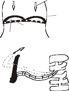
114 |
FUNDAMENTALS OF BIOMEDICAL ENGINEERING |
and sternum. The geometry of the rib cage is such that on its lifting, it increases the thoracic cavity. As thoracic cavity increases in volume, a negative pressure is created in the lungs. The negative pressure is relieved by air rushing into the lungs from the atmosphere. The lungs themselves are passive organs in the inspiration process.
Thoracic diameter increases vertically
Diaphragm contracts
& moves down
New position of diaphargm
Diaphragm
Descent of Diaphragm


 Rib cage moves up
Thoracic diameter


 increase laterally

 Rib lifted
Sternum |
Rib |
|
Vertebrae |
||
|
Antero Posterior Expansion
The expiration process is also passive and it starts with the release of the diaphragm and rib cage muscles. The volume of thorocic cavity reduces and a positive pressure is developed in the lungs which forces air out of the lungs into atmosphere. During inspiration, the pressure inside the lungs is negative (about–3 mm Hg). During expiration, the pressure inside the lungs is positive (about +3 mm Hg)
2.Deoxygenated blood is brought by the superior and inferior vena cava into the right atrium, from here it moves into the right ventricle. The blood is pumped to the lungs from the right ventricle through pulmonary artery. The blood in the lungs passes through
the pulmonary capillaries which are located in the walls of the alveolar sacs. Here oxygen is taken up by the red corpuscles which form oxyhemoglobin. Simultaneously carbon dioxide is liberated from the blood cells which is pushed out to the atmosphere by expiration. The blood pressure in the pulmonary artery is about 20 mm Hg when blood is pumped by the right ventricle and blood pressure is about 4 mm Hg when superior and inferior vena cava brings blood to the right atrium. The interchange of oxygen and carbon dioxide takes place in the capillary surface of the alveoli. The capillary surface is more than 75% surface of the alveoli (80 m2).
3.The oxygenated blood from the pulmonary capillaries is carried through the pulmonary veins to the left atrium. The blood from the left atrium moves to the left ventricle which pumps the blood into the aorta at high pressure (about 80 to 120 mm Hg) so that the blood can be circulated through all the parts of the body. The blood gives out oxygen at the cells of the tissues. The deoxygenated blood is collected by the venous systems into the superior and inferior vena cava.
4.The flow of the air in the respiratory system is usually laminar. However, during heavy breathing or when there is any obstruction during breathing, the flow of the air can become turbulent. The Reynolds number at which the flow of the air becomes turbulent is as high as 50,000. When Reynolds number is small, viscous forces dominate over inertial forces. If Reynolds number is less than one, inertial forces can be neglected. Low Reynolds number flow is the characteristic of air flows in alveolar passages of diameter less than a few hundred microns.
5.Some of the terminology used for the respiratory measurements are:
(a) Hypoventilation: Insufficient ventilation to maintain normal partial
pressure of carbon dioxide ( PCo2 )


 Valve opens