

Fundamentals of Biomedical Engineering
.pdf12 |
FUNDAMENTALS OF BIOMEDICAL ENGINEERING |
Protein |
– |
Part of cell and each cell is three- |
|
|
fourth protein. |
Pulmonary |
– |
Associated with lungs |
Pulse pressure– The difference between systolic and diastolic blood pressure.
R
Radioisotope – An isotope that is radioactive produced artificially from the basic element by the action of neutrons, protons, deutrons or alpha particles in cyclotron by chain reaction. These are used as tracer with stable element (labeled) by injecting in body to study the functioning of organs.
Radiology – The chief X-rays methods used in the examination of the chest which are fluoroscopy, radiography, tomography and bronchography.
S
Semi lunar
pulmonary valve – Outlet valve from the right ventricle into the pulmonary artery.
Sinoatrial – The pacemaker of the heart, cardiac muscle which is responsible for initiating each cycle.
Sphygmomanometer
–Instrument for measuring blood pressure (arterial).
Spirometer – Instrument for measure air which is entering and leaving the lungs.
Spleen – It is a blood forming organ in early life. It is storage organ for corpuscles and because of large number of macrophages acts as a blood filter.
Stenusis |
– Narrowing of a duct or canal. |
||
Stroke volume |
– Amount of blood pumped |
||
|
during each heartbeat. |
||
Superior vena |
|
|
|
cava |
– Main vein feeding back to the |
||
|
heart |
from |
systemic |
|
circulation above the heart. |
||
Systemic |
– Pertaining to or affecting the |
||
|
body as a whole. |
|
|
Systole |
– The contraction specially of |
||
|
ventricles during which blood |
||
|
is forced into the aorta and |
||
|
the pulmonary trunk. |
||
|
|
T |
Tachycardia |
– |
Rapid heart action. |
Tendon |
– |
A fibrous cord or band that |
|
|
connects a muscle to a bone. |
|
|
It consists of tissue fascicles |
|
|
of very densely arranged |
|
|
almost parallel collagenous |
|
|
fibres. |
Thorax |
– The part of the body between |
|
|
|
neck and abdomen. |
Thrombus |
– |
Clotting of blood within a |
|
|
blood vessel |
Tissue |
– |
Similar cells united in the |
|
|
performance of a partcular |
|
|
function. |
Trachea |
– The main trunk of the system |
|
|
|
of tubes by which air goes in |
|
|
or comes out of the lungs. |
Tricuspid valve |
– |
The valve connecting right |
|
|
atrium to right ventricle. |
|
|
V |
Ventricle |
– |
A chamber in heart which |
|
|
receives blood from atrium |
|
|
and forces it into arteries. |
Venule |
– |
A small vein. |
INTRODUCTION
HISTOLOGY
1. All organs of the body are formed of tissues. A tissue is a collection of similar type of cells, which are associated with some intercellular matrix (ground substance) governed by some laws of growth and development. These cells are adopted to perform the same function or functions. Tissues are usually classified into four main categories:
(a) Epithelial tissue (b) Connective tissue (c) Muscular tissue (d) Nervous tissue
2.Epithelium : It is a lining or covering tissue. It is a sheet of cells that cover external surface or lines of internal surface of the body. It can be simple, pseudostratified or stratified epithelium.
3.Connective tissue: It has few cells and a large amount of non living ground substance or matrix. It can be:
(a) Connective tissue proper (b) Skeletal tissue
(c) Fluid connective tissue
4.Connective tissues proper as name suggests, connect and anchor parts and give support to the body and its organs. For this reason, connective tissue is also known as supporting tissue. Connective tissue and skeletal tissue (cartilage and bone) have to perform mechanical functions.
5.The skeletal tissue includes the cartilages and the bones which form the structure of the body skeleton. The bones and cartilages have considerable rigidity. This is a feature which enables them to act as levers which is of great importance in the movement of limbs. The bones and the cartilages also provide surfaces for the attachment of muscles which provide force for the movement. Skeleton also protects the internal organs besides giving shape to the body. Cartilages
13
are four types. Hyaline cartilage is bounded by fibrous membrane which is supplied by blood vessels and through it nutritive substances diffuse into the cartilage. Cartilage grows by the addition of new layers on the outside. Hyaline cartilage occurs at the ends of the long bones. It has great resistance wear and covers the articular surfaces of nearly all synovial joints. Yellow elastic cartilage has great elasticity due to the presence of large number of yellow elastic fibers. It is found at the end of the nose and in the pinna of the ear. Calcified cartilage has its matrix impregnated with calcium salts. It is found in the pelvis and at the head of the humerus and femur bones. White fibrous cartilage has a large number of white fibers. It is found in the discs of vertebral hyaline cartilage and fibrocartilage found to calcify or even ossify in later life. Bone is a connective tissue in which the matrix is inpregnated with various salts which contribute to about two third of its weight. Bone is developed by two methods
(1) membranous (2) endochondral. In first method the bone is developed directly from a connective tissue membrane. For example, the bones of the vault of the skull are developed rapidly by the membranous method in the embryo. In the second, a cartilagious model is laid down which is replaced by bone. The long bones of the limbs are developed by endochondral ossification. Bones have fine canals which join with blood vessels and bone marrow. At birth, the marrow of all the bones of the body is red and hematopoietic (forming blood cells). The blood forming activity gradually lessens with age and red marrow is replaced by yellow marrow.
6.Fluid connective tissue: Blood is liquid connective tissue. It is red coloured fluid. It consists of liquid portion which is called plasma and of three different kinds of cells which are red blood corpuscles (erythrocytes), white blood corpuscles
14 |
FUNDAMENTALS OF BIOMEDICAL ENGINEERING |
(leucocytes) and platelets (thrombocytes). Plasma is the liquid portion of the blood of which it forms about two-third and contains about 80% of water. It is almost colourless clear fluid and contains an everchanging variety of substances in solution and suspension. Among the various substances present in the plasma are gases, absorbed food material, inorganic salts, vitamins, metabolic waste products, hormones, anti toxin and a soluble blood protein called fibrinogen. The cytoplasm of red corpuscles contains a pigment, the haemoglobin which makes these cells appear red. In bulk these cells give blood its red colour. The haemoglobin combines readily with oxygen to form an unstable compound, oxyhaemoglobin. In the tissues it breaks up releasing the oxygen. Here it combines with carbondioxide to form an unstable compound, carboxy haemoglobin, which breaks up in the lung, releasing carbondioxide for expiration. Haemoglobin, therefore, transports the gases and as such plays a vital part in respiration. The red corpuscles are formed in liver and spleen which also destroy the worn out corpuscles. As these are nucleated, they live for a pretty long time. The white blood corpuscles (leucocytes) are small, semitransparent, nucleated and amoeboid cells. These can crawl out between neighbouring endothelial cells and hence are found in every nook and corner of the body. At part of body having infections, they accumulate in very large numbers and serve to defend the body against the disease germs. They are able to eat bacteria and other germs in an amoeba like manner. This process is known as phagocytosis and hence they are known as phagocytes. Some of the WBC are killed by bacterial acids. Thus they may accumulate at the seat of infection as living or dead bacteria, leucocytes and disintegrating cells. All these form a whitish or yellow mass
which is called pus. Leucocytes are also useful in transporting waste particles and fat globules. These are produced in the bone marrow and lymphatic glands and are destroyed in the lymph organs. Thrombocytes or platelets contain a chemical which plays an important role in the clotting of blood. The various functions of blood are :
(a) Transport of gases : RBC combines with oxygen to form oxyhaemoglobin which breaks up and release oxygen in tissues. In tissues, RBC combines with carbon- di-oxide to form carboxy haemoglobin which breaks up in the lungs to release carbondioxide for expiration.
(b) Transport of food material: All the absorbed food circulated by the blood till it is taken up and used by tissue cells.
(c) Transport of substances: Many other substances such as enzymes, hormones and anti toxins are transported by the blood to the places where they are required.
(d) Defence against disease: This is effected in two ways. Firstly the white blood corpuscles feed on disease germs. Secondly blood possesses certain antioxins which unite chemically with toxins and then neutralize them.
(e) Equalization of the body temperature:
As the blood circulate throughout the body, it brings about an equalization of the body temperature by transferring heat from one part to another.
(f) Transport of metabolic wastes: The nitrogenous waste material is carried by the blood to the liver where it is converted into urea. The later is now carried by the blood to the kidney where it is removed out along with the urine.
(g) Clotting of blood: Blood has a soluble solution called fibrinogen which is converted into a mesh work of fine
INTRODUCTION
threads of insoluble fibrins. In the mesh of fibrins, various types of corpuscles get entangled to form a blood clot which is also known as coagulation. The conversion of fibrinogen into fibrin is affected by the action of an enzyme called thrombin. Free thrombin is not present in the blood but it is formed by its precursor (inactive enzyme prothromboplastin) in presence of calcium ions. Calcium ions are present in blood but free thromboplastin is absent in blood. However it is present in other solid tissues. Hence when an injury occurs, thromboplastin is released by the injured tissues. The thromboplastin then acts with calcium upon the prothrombin, changing it to thrombin. The thrombin then acts upon the fibrinogen and converts it to fibrin.
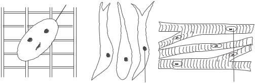
7.Muscular tissue: It consists of cells in the shape of fibres of different lengths. Intercellular elements are almost absent. The muscular tissues are of three types :
(1)Striped or voluntary
(2)Unstriped or involuntary
Nucleus
15
(3)Cardiac
The striped muscles are under the control of ‘will’ and they are wide and nontapering. In the striped muscle, fibres are united in parallel bundles which is continuous with the connective tissue sheath surrounding the tendons that unite the muscle to the skeleton. Unstriped muscles are made of elongated, spindle shaped, flattened fibres which are rarely forked at the ends. The number of unstriped muscle fibres are united together by a minute quantity of intercelluar substance into a thin and flat band and a number of such bands are bound together by connective tissues into a larger bundle. The unstriped muscles are not in the control of ‘will’ and they are found in the alimentary canal, the lungs and the blood vessels. The cardiac muscles are found only in the wall of the heart. The structure is somewhat inbetween striped and unstriped muscles. These muscles contract rhythmically and these muscles are immune to fatigue.
Nucleus
Striped Muscle |
Unstriped Muscle |
8.Nervous tissue: They consists of
(1)nerve cell
(2)nerve fibre.
Nerve cells are known as neurons.
Nucleus
Cardiac Muscle
Each neuron consists of a cell body from which arises a system of branching fibres. The number of fibres is variable. On this basis, neurons are classified into three types:

16 |
FUNDAMENTALS OF BIOMEDICAL ENGINEERING |
(1)neurons with many processes are called multipolar.
(2)neurons with two procesess arising at opposite poles are known as bipolar.
(3)neurons having two processes arising from the same pole are known as unipolar. The cytoplasm of each neuron contain a large and spherical nucleus, large number of dark staining minute particles (Nissl Granules) and numerous cytoplasmic strands known as neurofibrillae. Neurofibrillae help in the passage of the nerve impulse.
PHYSIOLOGICAL SYSTEMS OF THE BODY
1.In our body, we have mechanical, electrical, chemical, thermal, pneumatic, hydraulic and many other types of system. Each system communicates internally with other systems of the body and externally it communicates
with surroundings. We have a multi level control system with its communication network which organises these internal systems to perform many complex functions. We are able to sustain our lives due to organised operations of all these internal systems and their various subsystems. In medical terms, a study of the structure of the body and the relationship of its constituents parts to each other is known as ‘Anatomy’ while the study of function of these parts as a system is known as physiology. The major functional physiological systems of the body are:
(a) The cardiovascular system (b) The biochemical system (c) The respiratory system (d) The nervous system
(e) The excretory system (f) The locomotor system (g) The digestive system
Energy (E1) |
|
|
|
|
|
|
|
|
|
|
|
|
|
|
|
|
|
|
|
|
|
|
|
|
|
|
|
|
|
|
|
|
|
|
|
|
|
|
|
|
|
|
|
|
|
|
|
|
|
|
|
|
|
|
|
|
|
|
|
|
|
|
|
|
|
|
|
|
|
|
|
|
|
|
|
|
|
|
|
|
|
|
|
|
|
|
|
||||||
Light vision |
|
|
|
|
|
|
|
|
|
|
|
|
|
|
|
|
|
|
|
|
|
|
|
|
|
|
|
|
|
|
|
|
|
|
|
|
|
|
|
Mass (M1) |
||||||
|
|
|
|
|
|
|
|
|
|
|
|
|
|
|
|
|
|
|
|
|
|
|
|
|
|
|
|
|
|
|
|
|
|
|
|
|
||||||||||
Vibration (hearing) |
|
|
|
|
|
|
|
|
|
|
|
|
|
|
|
|
|
|
|
|
|
|
|
|
|
|
|
|
|
|
|
|
|
|
|
|
|
|
|
|||||||
Flow (smell) |
|
|
|
|
|
|
|
|
|
|
|
|
|
|
|
|
|
|
|
|
|
|
|
|
|
|
|
|
|
|
|
|
|
|
|
|
|
|
|
Food Intake |
||||||
|
|
|
|
|
|
|
|
|
|
|
|
|
|
|
|
|
|
|
|
|
|
|
|
|
|
|
|
|
|
|
|
|
|
|
|
|
|
|
|
|
|
|
|
|
|
Liquid Intake |
|
|
|
|
|
|
|
|
|
|
|
|
|
|
|
|
|
|
|
|
|
|
|
|
|
|
|
|
|
|
|
|
|
|
|
|
|
|
|
|
|
|
|
|
|||
|
|
|
|
|
|
|
|
|
|
|
|
|
|
|
|
|
|
|
|
|
|
|
|
|
|
|
|
|
|
|
|
|
|
|
|
|
|
|
|
|
|
|
|
|
|
Inspired Air |
|
|
|
|
|
|
|
|
|
|
|
|
|
|
|
|
|
|
|
|
|
|
|
|
|
|
|
|
|
|
|
|
|
|
|
|
|
|
|
|
|
|
|
|
|
|
|
Mass (M2)
 Solid Waste
 Liquid Waste


 Expired air
 Perspiration
Energy (E2)


 Body movement
 Tactile sensation
Communication with Energy and Mass Transfer with Surroundings
INTRODUCTION
2.The cardiovascular system: The cardiovascular system is a closed hydraulic system. It has heart and blood vessels. The heart works as a four chamber pump. The blood vessels are flexible and sometimes elastic tubing of varying sizes. The tubings
Blood Circuit Cardiovascular system
Right atrium  Right ventricle
  Pulm onary artery
Left |
|
Left |
|
Pulm onary |
|
Lungs |
|
|
|
|
|||||
ventricle |
atrium |
|
vein |
|
|
||
|
|
|
|
|
|
|
|
Aorta |
|
|
|
|
|
||
change their sizes to control blood pressure, for example arteries and arterioles. Certain tubings act as reservoirs as they can control their volume as per the requirements by a system of valves and variable resistance to flow by constriction and dilation of the control blood tubings. These tubings are veins and they take blood back to heart. The heart acts as two functionally isolated two stages pumps working in parallel. In first stage of each pump, the blood is taken into the reservoirs (atriums) from the system and it is pumped into second stage reservoirs (ventricles). The action of the second stage is so well coordinated that the blood is pumped into the system immediately when it is received from the first stage. The circuit of the blood is shown in the diagram. Right side of the heart collects blood from the
17
hydraulic system through veins and pumps it to the lungs for oxygenation. The left side of the heart receives blood from the lungs (oxygenation system), and pumps it into the main hydraulic system which is formed by the various organs of the body. The heart rate and stroke volume are constantly changed to control the flow of the blood in the system to meet the requirements of body parts. The blood performs all functions as elaborated in para 25 of this chapter. The blood flows in laminar manner. Superior vena cava is a large venous channel which collects blood from the upper half of the body and delivers into the right atrium. It has no valve. The inferior vena cava (larger than superior vena cava) also opens into right atrium. It returns the blood to the heart from the lower half of the body. Since the blood in the inferior vena cava has to flow against gravity at times, special one way valves are located in it to prevent gravity from pulling blood against the direction of flow. The cardiac output flow rate and volume of the fluid at various places in the body are important indicators for proper functioning of the system.
|
|
|
From veins |
|
|
|
From lungs |
|
|
|
|
|
|
||
|
|
|
|
|
|
|
|
|
|
|
|
||||
Right Atrium |
Left Atrium |
||||||
Right Ventricle |
Left Ventricle |
To Lungs |
To Aorta |
Heart Works a Pump
18 |
FUNDAMENTALSOFBIOMEDICALENGINEERING |
3.The biochemical system: There are many chemical systems in our body that produce energy for the functioning of our body. The energy is required for growth, body functions and body repairs. These chemical systems are interconnected and these can be considered as the subsystems of a very efficient chemical factory. There is a single point intake of fuel (food, water and air) for this factory which is also source for all chemical reactions which are taking place inside the body. This chemical factory also contains all monitory devices which are essential to carry out necessary control for each chemical operation. The waste disposal system is also a part of this biochemical system.
4.The respiratory system: The respiratory system is a pneumatic system which ensures exchange of gases by a biological process which is termed respiration. The body requires oxygen to combine with carbon, hydrogen and other nutrients to produce heat and energy for sustenance of life. The entire process of taking inside oxygen from surroundings, transporting it to body cells, removing the carbondioxide from the cells and pushing out the carbondioxide into surrounding is called respiration. Air enters the lungs through air passages which include the nasal cavities, pharynx, larynx, trachea, bronchi and bronchioles. The lungs are elastic bags located in a closed cavity, called the thorax. The diaphragm is a special bell shaped muscle located at the bottom of the closed cavity. When this diaphragm contracts, thorare is pulled downward, enlarging the closed cavity. The resultant increase in the volume of the closed cavity, a negative pressure (vacuum) is created which is relieved by air entering the lungs from the surroundings. When the diaphragm moves up and reduces the volume of the thorax, the used air with carbon dioxide is pushed out of the lungs. Oxygen is taken
into the blood from the incoming air in about 300 million alveoli present in the lungs. The oxygen and haemoglobins in blood form oxyhaemoglobins and carbondioxide removed from the blood is pushed out from lungs to the surroundings. An automatic control system maintains pneumatic pump operation (rate of contraction of diaphragm) at a speed that is adequate to supply oxygen and to remove carbondioxide as required by body. It is also possible to accelerate or deacelerate the operation of the pneumatic pump by manual control whenever it is required. Automatic control returns whenever manual control is not applied.
5.The nervous system: The nervous system consists of control and communication network which coordinates the functions of all parts of the body. The brain is the central information processor and it works as a computer. It has memory, power to compute, capability to make decisions and innumerous input, and output channels for communication. These channels form complicated networks with many interconnections (nodes) which take singnals from a large number of sensory devices (each sensory device detects light, sound, pressure, heat and chemicals) to the brain (computer) for analysis. Some network is again used to take the output control signals from the brain to the motor units of the muscles to carryout the desired motion or to exert force. The nerves form signal lines to carry signals (informations) generated by the nerve action potentials (sensory devices) to the brain and same signal lines are used to carry control signals generated by the brain for the motor units. In addition to the control of the brain, a large number of simple decision making devices in the form of spinal reflexes are present in the body to control independently some motor devices from certain sensory inputs. Example of this is
INTRODUCTION
the Portal system which consist of vein and capillary network.
6.The excretory system : It consists of all organs that are responsible for the removal of waste products formed by metabolism in the organisms. The kidneys are the major excretory organs in man. The left kidney is located at slightly higher level than right kidney, one on each side of the vertebral column. The kidneys have ‘bean’ shape and they are also called renes from which it is known as rent. The renal tubules act as filters to remove from the blood (1) Excess water
(2) Urea and uric acid (3) Excess mineral salts (4) Yellow pigments from the bile. The mixture of these substances forms urine. When the human kidneys fail to function, the urine accumulates in the blood resulting death of the person from toxic poisoning.
7.The locomotor system: The system provides locomotion or movement to the body. Bones and joints play an important role for this system. Statics and dynamics of the musculoskeletal system; forces and motions acting in the skeletal system; forces and movements within the body; behaviour of bones, tendons, ligaments and cartilages for stress and strains; and prosthesis design etc. will be covered in details in later chapters.
8.The digestive system: It includes all organs that help in ingestion, digestion, absorption and egestion of undigested food. It includes the alimentary canal and asociated glands like liver and pancreas etc. The liver is the largest gland in the body. It is located on the right side, just under the diaphragm. It has irregular shape and it weighs 3 to 3 ½ pounds (1/40th the weight of the body). It secretes bile juice which plays an important role in metabolism of both carbohydrate and protein. The nitrogenous waste material is carried by the blood to the liver where it is converted into urea. Bile pigments are derived from the breakdown of hemoglobin
19
from worn out red blood corpuscles. Bile pigments colour undigested food. The other important gland is pancreas which secrete digestive pancreatic juice and discharge it into intestine. This is done by exocrine part of the pancreas. The endocrine part secretes hormones like insulin. Insulin promotes glucose utilization, protein synthesis and the formation and storage of neutral lipids. Insulin is given to the persons suffering from diabetes.
9.Cell, DNA and atoms: Our body possess numerous cells (almost 10,000 trillion) of almost some few hundred varities. Each cell performs an important role to keep us fit. All activities like standing, walking, talking and playing are possible through these cells. The cells extract nutrients from food, distribute the energy and remove the waste from the body. They also fight against becteria and billion of cells die daily in this process.
10.Inside every cells is a nucleus which has 46 chromosomes (23 come from father and 23 come from mother). Chromosomes carry all instructions necessary for our growth and to maintain us. They contain long strands of chemical called DNA.
11.In microscopic level, each cell of our body is made of atoms. We have in our body about 63% by hydrogen atoms, 25.5% oxygen atoms, 9.5% carbon atoms, 1.5% nitrogen atoms and only 0.5% atoms of other atoms (Iron, Cobalt, Sodium and Potasium etc). When we die our atoms will dissemble and move off to form new uses elsewhere as atoms can not be destroyed. Some atoms may form a part of a flower or other human being or a drop of rain. It is also possible that we may be having atoms in our body which once belonged to Budha, Gandhi or Nehru.

20 |
FUNDAMENTALS OF BIOMEDICAL ENGINEERING |
12.Cloning: Cloning occurs in nature for simple organisms (bacteria and viruses) which reproduce by splitting when their DNA has replicated itself. Cloning can also be done in humans and other animals when a single fertiliged egg divides and separates to form two or more identical individuals. Gene cloning is generally done in the laboratory by means of the polymerisation chain reaction which enables to reproduce millions of
identical gene in short time. In animal cloning, the donor's DNA is introduced into egg of another animal of same species after egg's DNA has been removed. The egg is then inserted into surrogate animal's womb and pregnancy procceds as normal. Another genetic advancement is the creation of transgenic animals which can be used for the production of human compatible organs such as hearts. Pigs are being used for this purpose.
Cell with |
Cell m ultiplies |
DNA only |
producing an embryo |
Egg Donor
|
Em bryo |
|
Cell and egg are |
im planted into |
|
surrogate animal |
||
fused together |
||
|
Egg without DNA
Egg Donor
Cloning of Animal
Desired gene of |
Identification Freeing Gene Gene injection |
DNA (Hum an) |
|
Creating Transgenic Animal
INTRODUCTION |
21 |
OBJECTIVE TYPE QUESTIONS
Fill up the gaps
1.The body is made up of the head, ______
and limbs. ((a) arms (b) trunks)
2.The vertebral column is _______axis of trunk. ((a) central (b) middle)
3.The upper limbs by the sides of the trunk is
_______ position ((a) erect (b) anatomical)
4.Sagittal plane is parallel to _______ plane. ((a) median (b) lateral)
5.Horizontal plane is also known as ________
plane. ((a) median (b) transverse)
6.The adjective medial means ______ to median plane. ((a) nearer (b) farther)
7.The adjective radial and ‘ulnar’ can be used instead of ______. ((a) medial and lateral (b) lateral and medial)
8.The terms ‘anterior’ and ‘posterior’ are used to indicate _______ of the body respectively. ((a) back and front (b) front and back)
9.Nose is _______ to the ears. ((a) anterior (b) posterior)
10.The term ‘palmer’ and ‘dorsal’ surfaces of the hand. ((a) anterior and posterior (b) posterior and anterior)
11.The arm is ______ to the forearm. ((a) distal (b) proximal)
12.The mouth is _______ to the nose. ((a) superior (b) infevior)
13.If a person is lying, them he is in ________
position. ((a) supine (b) prone)
14.Flexion is the _______ of the bones and extension is _______ of the bones. ((a) unfolding, folding (b) folding, unfolding)
15.Flexion and extension of trunk takes place in the ________ plane. ((a) medial (b) lateral)
16.________ of the limb is the movement away from the midline of the body in the coronal
plane while _____ of the limb is the movement towards the body in the coronal plane. ((a) adduction, abduction (b) abduction, adduction)
17.Rotation is a term applied to the movement of a part of the body around its _______
axis. ((a) central (b) long)
18.________ is the movement of the foot so that the sole faces in medial direction while
______ is the opposite movement so that the sole faces in a lateral direction. ((a) Eversion, Inversion (b) Inversion, Eversion)
19.Blood is ______ tissue. ((a) Epithelial (b) Connective)
20.______ tissue has to perform mechanical function. ((a) skeletal (b) fluid connective)
21.All organs of the body are formed of ______. ((a) flesh (b) tissue)
22.Tissue is a collection of similar type of
_____. ((a) fibres (b) cells)
23.Epithelium is ______ tissue. ((a) covering (b) connecting)
24.Cartilages and bones are _____ tissues. ((a) supporting (b) skeletal)
25.Blood consists of liquid portion (plasma) and
______ different kinds of cells. ((a) two (b) three)
26.The haemoglobin is the pigment in ______
corpuscles which makes the blood red. ((a) erythrocytes (b) leucocytes)
27.Transport of gases (oxygen and carbon dioxide) is done by _____ of RBC (Red blood corpuscles). ((a) haemoglobin (b) platelets)
28.The haemoglobin combines with oxygen to form oxyhaemoglobin in ______. ((a)lungs (b) tissues)
29.The haemoglobin combines with carbon dioxides to form carboxy haemoglobin in
_______. ((a) lungs (b) tissues)