

Опухоли легких, плевры, тимуса и сердца ВОЗ
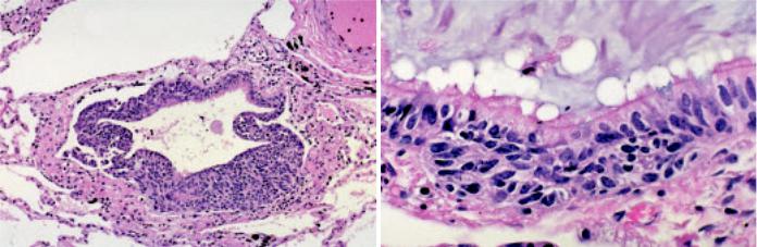
.pdfFig. 1.74 Carcinoma in situ. The bronchial mucosa is replaced with atypical squamous cells extending from the surface to the base of the epithelial. Note the severe nuclear polymorphism, hyperchromasia and enlarged nuclei.
appear as superficial or flat lesions; the remaining 25% have a nodular or polypoid appearance {967,1423}. Because nodular/polypoid lesions are elevated from the adjacent normal mucosa, lesions as small as 1-2 mm in diameter can be seen. Flat or superficially spreading lesions greater than 1-2 cm in surface diameter are generally visible as areas of focal thickening, increase in vascularity or marked irregularity of the mucosa. Flat lesions 5-10 mm in diameter usually produce non-specific thickening, redness, fine roughening, loss of luster or a slight increase in granularity which are difficult to distinguish from inflammation or squamous metaplasia {2057}. Lesions <5 mm are usually invisible on white light bronchoscopy. Bronchial dysplasia usually presents as non-specific mucosal swelling or thickening at a bronchial bifurcation.
Autofluorescence bronchoscopy
Pre-invasive lesions that have subtle or no visible findings on white-light bronchoscopy can be localized by autofluo-
rescence imaging using a violet or blue light for illumination instead of white-light and special imaging sensors attached to a fiberoptic bronchoscope for detection of the abnormal autofluorescence {1117, 1120}. Dysplastic and malignant tissues have a significant decrease in the green autofluorescence intensity relative to the red autofluorescence. These pre-inva- sive lesions are identified by their brown or brownish-red autofluorescence. Lesions as small as 0.5 mm can be localized by this method.
Cytology
Sputum cytological classification schemes for preneoplastic lesions have been published by Saccomanno {1717} and Frost {621} and consist of gradations of microscopic abnormality similar to those observed in histological sections from lower airways of smokers. Squamous metaplasia presents in sputum smears as individual cells, but mostly as flat loosely cohesive clusters. The cytologic manifestations of dysplasia occur as increasingly severe cellular
changes, ranging from mild, moderate, and severe atypia to carcinoma in situ (CIS) {1717}. There are progressive alterations including increasing variability in cellular and nuclear sizes, increasingly variable nuclear-to-cytoplasmic ratios, increasing proportions of cells with cytoplasmic eosinophilia (orangeophilia), increasing coarseness of chromatin granularity until a pyknotic-like pattern is reached in CIS, increasing irregularity in the distribution of chromatin granules, and increasing irregularities in the outlines of nuclear membranes {844,1717, 1718}. This last feature first appears in moderate atypia {1717}. According to Koprowska et al {1055}, it is this deviation from smooth nuclear outlines that is most strongly associated with the presence of carcinoma.
Localization and macroscopy
Foci of carcinoma in situ usually arise near bifurcations in the segmental bronchi, subsequently extending proximally into the adjacent lobar bronchus and distally into subsegmental branches.
Squamous dysplasia and carcinoma in situ 69
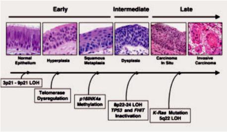
Fig. 1.75 Preinvasive lesions. Sequential molecular changes during the multistage pathogenesis of squamous cell lung carcinoma. Molecular changes occurring during lung pathogenesis may commence early
(normal or slightly abnormal epithelium), at an intermediate (dysplasia) stage, or relatively late (carcinoma in situ, invasive carcinoma).
The lesions are less frequent in the trachea. Bronchoscopically and grossly there is often no macroscopical alteration. When gross abnormalities are present, focal or multi-focal plaque-like greyish lesions resembling leukoplakia, nonspecific erythema and even nodular or polypoid lesions may be seen.
Histopathology
A variety of bronchial epithelial hyperplasias and metaplasias may occur that are not regarded as preneoplastic including goblet cell hyperplasia, basal cell (reserve cell) hyperplasia, immature squamous metaplasia, and squamous metaplasia. The term preinvasive does not imply that progression to invasion will necessarily occur. These lesions represent a continuum of cytologic and histologic changes that may show some overlap between defined categories. Squamous dysplasia does not invade the stroma. The basement membrane remains intact and is variably thickened. There may be vascular budding into the epithelium, termed angiogenic squamous dysplasia {986}. The latter lesion has also been previously reported as micropapillomatosis {724, 1407}.
Immunohistochemistry
A series of immunohistochemical changes accompany squamous dysplasia. These include increased expression of EGFR {607,1101,1710}, HER2/neu
{608}, p53 {145,211,1251}, MCM2 {1966}, Ki-67 {607,1149,1966}, cytokeratin 5/6 {54}, bcl-2 {211}, VEGF {602,1126}, maldistribution of MUC1, and loss of several proteins including FHIT {1855}, folate binding protein {609,676}, and p16 {213,1122}. A linear progression of proliferative activity, assessed with immunohistochemical staining for the proliferation marker Ki-67 (MIB-1), correlates with the extent and grade of the preneoplasia {1966}. Loss of RAR-beta expression is very frequent in the bronchial epithelium of smokers {1252,2017}. Type IV collagen staining highlights discontinuities in basement membranes that increase from basal cell hyperplasia to dysplasia, progressing to destruction in carcinoma in situ and invasive carcinoma {657}. Changes also occur in matrix metalloproteinase (MMP) and tissue inhibitor of metalloproteinase (TIMP) expression corresponding to progression in severity of dysplasia, in situ carcinoma and invasive carcinoma {657}.
Electron microscopy
There is an increase in atypical basal cells with loss of polarity. The nuclei show considerable hyperchromasia, and variations in shape with numerous invaginations. The number of nucleoli is increased, and so-called pseudoinclusion bodies may be seen within the nuclei. Some cells exhibit atypical devel-
opment via an atypical array of organelles {711,712,1407}. A special feature is seen in the basement membrane in CIS. It is subdivided by multiple tentacle-like cytoplasmic protrusions, which vary considerably in shape and size but are always directed towards and between the fibrous structures of the basement membrane {711,712,1407}.
Histogenesis
The stem cell for the squamous epithelium of the proximal airway is not certain, but it is presumed that the basal cells represent a relatively quiescent zone that is the precursor for preneoplastic epithelium. It is of interest that these cells express a different cytokeratin profile with high levels of cytokeratin 5/6 and are the only cells in the normal respiratory mucosa and express significant levels of epidermal growth factor receptor. In the earliest preinvasive lesions, this basal zone is expanded with phenotypic changes that mirror the quiescent basal zone in normal epithelium including the overexpression of EGFR, transformation from cytokeratin 5/6 negative to positive, and increased proliferative activity with high expression of Ki-67 and MCM2. It is widely supposed that low grade changes such as basal cell hyperplasia and squamous metaplasia may (with or without micropapillomatosis) progress through mild, moderate and severe dysplasia up to carcinoma in situ {392,994,2022} to invasive carcinoma. However, such a progression is rarely observed in individual subjects and the predictive power of specific grades of premalignant change for the future development of invasive carcinoma is still under investigation.
Somatic genetics
Cytogenetics and CGH
Relatively few cytogenetic studies have been performed on preneoplastic lesions because of their small size and because of the difficulty of identifying them {1449, 1854}. Classic cytogenetic studies are further limited by the necessity for shortterm cultures and the inability to identify the cell of origin of metaphase spreads. For these reasons most analyses have utilized fluorescence in situ hybridization (FISH) for detection of chromosomal or numerical changes in bronchial epithelial cells. As part of the field effect resulting from widespread smoking damage to the entire upper aerodigestive tract, cytoge-
70 Tumours of the lung - Preinvasive epithelial lesions

Table 1.11
Microscopic features of the squamous dysplasia and carcinoma in situ.
Abnormality |
Thickness |
Cell size |
Maturation/orientation |
Nuclei |
|
|
|
|
|
Mild Dysplasia |
Mildly increased |
Mildly increased |
Continuous progression of maturation from |
Mild variation of N/C ratio |
|
|
Mild anisocyto- |
base to luminal surface |
Finely granular chromatin |
|
|
sis, |
Basilar zone expanded with cellular crowd- |
Minimal angulation |
|
|
Pleomorphism |
ing in lower third |
Nucleoli inconspicuous or absent |
|
|
|
Distinct intermediate (prickle cell) zone |
Nuclei vertically oriented in lower third |
|
|
|
present |
Mitoses absent or very rare |
|
|
|
Superficial flattening of epithelial cells |
|
|
|
|
|
|
Moderate |
Moderately |
Mild increase in |
Partial progression of maturation from base |
Moderate variation of N/C ratio |
Dysplasia |
increased |
cell size; cells |
to luminal surface |
Finely granular chromatin |
|
|
often small |
Basilar zone expanded with cellular crowd- |
Angulations, grooves and lobulations pres- |
|
|
May have mod- |
ing in lower two thirds of epithelium |
ent |
|
|
erate anisocyto- |
Intermediate zone confined to upper third of |
Nucleoli inconspicuous or absent |
|
|
sis, pleomor- |
epithelium |
Nuclei vertically oriented in lower two thirds |
|
|
phism |
Superficial flattening of epithelial cells |
Mitotic figures present in lower third |
|
|
|
|
|
Severe |
Markedly |
Markedly |
Little progression of maturation from base |
N/C ratio often high and variable |
Dysplasia |
increased |
increased |
to luminal surface |
Chromatin coarse and uneven |
|
|
May have |
Basilar zone expanded with cellular crowd- |
Nuclear angulations and folding prominent |
|
|
marked anisocy- |
ing well into upper third |
Nucleoli frequently present and conspicu- |
|
|
tosis, pleomor- |
Intermediate zone greatly attenuated |
ous |
|
|
phism |
Superficial flattening of epithelial cells |
Nuclei vertically oriented in lower two thirds |
|
|
|
|
Mitotic figures present in lower two thirds |
|
|
|
|
|
Carcinoma |
May or may not |
May be markedly |
No progression of maturation from base to |
N/C ratio often high and variable |
in situ |
be increased |
increased |
luminal surface; epithelium could be invert- |
Chromatin coarse and uneven |
|
|
May have |
ed with little change in appearance |
Nuclear angulations and folding prominent |
|
|
marked anisocy- |
Basilar zone expanded with cellular crowd- |
Nucleoli may be present or inconspicuous |
|
|
tosis, pleomor- |
ing throughout epithelium |
No consistent orientation of nuclei in rela- |
|
|
phism |
Intermediate zone absent |
tion to epithelial surface |
|
|
|
Surface flattening confined to the most |
Mitotic figures present through full thick- |
|
|
|
superficial cells |
ness |
|
|
|
|
|
netic changes may be detected both in preneoplastic lesions as well as histologically normal appearing cells. Numerical changes of chromosme 7 are frequent and may predict risk for cancer development {1147,2245}. Only one study to date has performed comparative genomic hybridization on preneoplastic lesions and found that numerical alterations of chromosome 3 were the most frequent change {813}.
Molecular genetics
Precise microdissection of epithelial cells followed by molecular genetic analysis of such lesions has provided a sequence of molecular changes similar to that observed in other epithelial cancers {844,2161}. These studies have also indicated similarities and differences between the sequential changes leading
to central and peripheral tumours. The histological changes preceding squamous cell carcinomas are well documented because of accessibility of these lesions, and the developmental sequence of molecular changes is nonrandom. DNA aneuploidy is frequent in dysplastic lesions particularly in highgrade lesions. Small foci of allelic loss are common at multiple sites in the bronchial epithelium and persist long after smoking cessation {1549}.
LOH occurs at one or more chromosome 3p regions and 9p21 early in neoplastic development, commencing in histologically normal epithelium. Later changes include 8p21-23, 13q14 (RB) and 17p13 (P53) being detected frequently in histologically normal epithelium {1236,2157, 2158,2162}. In contrast, allele loss at 5q21 (APC-MCC region) mutations has
been detected at the carcinoma in situ stage, and P53 mutations appear at variable times {1900,2157,2162}. Chromosome 3p losses in normal epithelium, basal cell hyperplasia and squamous metaplasia are small and multifocal, commencing at the central (3p21) region of the chromosomal arm, while in later lesions such as carcinoma in situ, allelic loss is present along nearly all of the short arm of chromosome 3p {2157, 2158}. The clonal patches of bronchial epithelium having molecular changes (allelic loss and genetic instability) are usually small, and have been estimated to be approximately 40,000 to 360,000 cells {1549}. p16INK4a methylation has also been detected at early stages of squamous preinvasive lesions with frequency increasing during histopathologic progression from basal cell hyperpla-
Squamous dysplasia and carcinoma in situ 71
sia to squamous metaplasia to carcinoma in situ {140}. Detection of such changes in sputum samples may be of predictive value in identifying smokers at increased risk of developing lung cancer {141}. Similar changes have been detected in telomerase activation {2199}. While weak telomerase RNA expression is detected in basal layers of normal and hyperplastic epithelium, dysregulation of telomerase expression increases with tumour progression with moderate to strong expression throughout the multilayers of the epithelium in squamous metaplasia, dysplasia and carcinoma in situ.
While specific premalignant changes associated with SCLC have not been identified, extensive genetic damage occurs in the accompanying normal and hyperplastic bronchial epithelium and is characteristic of SCLC tumours {2160}. These changes are much more extensive than changes accompanying similar epithelia from lung resections of patients with squamous cell carcinoma or adenocarcinoma. These findings suggest major differences in the pathogenesis of the three major lung cancer types.
Our knowledge of the changes preceding peripheral tumours is much more limited, mainly because of the inability to
identify and have access to such lesions. However, careful examination of lung cancer resections indicates that peripheral tumours, especially adenocarcinoma, may be accompanied by specific morphologic changes known as atypical adenomatous hyperplasia (AAH). The advent of CT scans for the detection of early lung cancers has greatly increased the identification of such lesions, both in smokers with and without lung cancer {844,2078}. Inflation of the lungs prior to fixation greatly enhances the ability to detect these lesions. Multiple molecular changes have been described in these lesions {1021} including aneuploidy, ras gene mutations, COX-2 over expression, active proliferation, 3p and 9p deletions, K-ras codon 12 mutations, and disruption of the cell cycle control, but p53 gene aberrations are rare and telomerase activation is absent.
Prognostic factors
Carcinoma in situ, being a preneoplastic lesion, is classified as “Stage 0 disease.” Resection of specific lesions at this stage means 100% curability, although frequent multifocality means that other foci are liable to present elsewhere in the airways. In general, higher grades of dysplasia are more closely associated with
synchronous invasive carcinomas, although the prognostic significance of identifying dysplasia in isolation is uncertain. Currently, there are no recommendations to screen asymptomatic individuals with a history of dysplasia for development of invasive lesions {1119,1408, 1860}. There are no data to allow prediction of progression to invasive disease, depending on grade of dysplasia. It is likely that severe dysplasia/CIS carries a high risk. Progression of disease, from the early stages, probably takes many years.
Genetic predictive factors
There is a general consensus that numerous genetic and molecular abnormalities occur in very early stages of lung carcinogenesis including hyperplasia and metaplasia and even in normal appearing bronchial epithelium in smokers {1236,2162}. None of these isolated molecular abnormalities have been shown to predict progression to cancer, but their cumulative rate may be associated with the risk of cancer in the bronchial tree {926}.
.
72 Tumours of the lung - Preinvasive epithelial lesions
Atypical adenomatous hyperplasia
Definition
Atypical adenomatous hyperplasia (AAH) is a localised proliferation of mild to moderately atypical cells lining involved alveoli and, sometimes, respiratory bronchioles, resulting in focal lesions in peripheral alveolated lung, usually less than 5mm in diameter and generally in the absence of underlying interstitial inflammation and fibrosis.
Synonyms
Atypical alveolar cuboidal cell hyperplasia {1807}, alveolar epithelial hyperplasia {1434}, atypical alveolar hyperplasia {288}, atypical bronchioloalveolar cell hyperplasia {2123}, bronchioloalveolar cell adenoma {1316}.
Background
AAH is a putative precursor of peripheral pulmonary adenocarcinoma, including bronchioloalveolar carcinoma (BAC) {1807}; the ‘adenoma’ in an adenomacarcinoma sequence in the peripheral lung {1318}. Epidemiological, morphological, morphometric, cytofluorometric and genetic evidence support this hypothesis {392,994,1021,1378,2022}.
AAH is most frequently found as an incidental histologic finding in lungs already bearing primary cancer, especially adenocarcinoma. Lungs with very high num-
bers of AAH (>40) have been reported in conjunction with multiple synchronous peripheral primary adenocarcinomas or BAC {51,333,1316,1434,1928,2123}. Autopsy studies have reported AAH in 2- 4% of non-cancer bearing patients {1879,2206,2207}.
AAH has been reported in up to 19% of women and 9.3% of men with lung cancer and up to 30.2% and 18.8%, respectively, in women and men with pulmonary adenocarcinoma {333}. In Japan, this gender relationship is inconsistent {1429, 2123}. Almost all Caucasians reported with AAH have been smokers, while in Japan, an association is not clear. Data on the association of AAH with either a personal or family history of malignancy are conflicting {334,1429,1960}.
Clinical features
Signs and symptoms
There are no clinical signs or symptoms directly referable to AAH. The lesions are usually encountered as incidental findings at gross or, more often, microscopic examination of lung.
Imaging
Radiological experience of AAH is largely confined to screening studies using High Resolution CT scanning (HRCT) {979,1108}, though some have been
K. M. Kerr
A.E. Fraire
B. Pugatch
M.F. Vazquez
H. Kitamura
S. Niho
Table 1.12
AAH in lung cancer resection specimens. From references: {33,1041,1316,1387,1434,2123}
Cancer type |
Prevalence |
|
|
|
|
All primary lung cancer |
9 |
- 21% |
Adenocarcinoma |
16 |
- 35% |
Squamous cell carcinoma |
3 |
- 11% |
Large cell carcinoma |
10 |
- 25% |
Metastatic disease |
4 |
- 10% |
|
|
|
described during follow-up of patients with lung cancer {1038,1198}. In this context, small non-solid nodules, also described as localised areas of pure ground glass opacity (GGO), may be identified as areas of increased opacification with distinct borders, not completely obscuring the underlying lung parenchyma on CT scan, measuring 2- 24mm in diameter, and typically not visualized on chest radiographs. Resection of GGOs has shown a range of pathology including benign disease in up to 30%, AAH in 10-77%, BAC in up to 50% and invasive adenocarcinoma in 10-25% of cases {979,1038,1108,1431}.
Relevant diagnostic procedures
AAH may rarely be visualised radiologically and a presumptive diagnosis made. Most likely as part of an HRCT screening
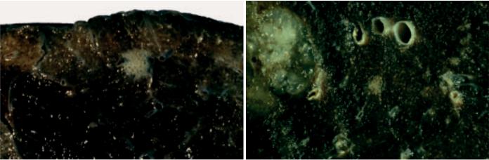
A B
Fig. 1.76 Atypical adenomatous hyperplasia. A Unusually prominent AAH lesion. Alveolar spaces are visible within the lesion. B AAH (center) detected incidentally in a lung resected for mucinous adenocarcinoma, present on the left.
Atypical adenomatous hyperplasia 73
programme for lung cancer, any detected lesion, which necessitates further investigation, can be sampled by fine needle aspiration or local resection.
Cytology
A diagnosis of AAH cannot be made on a cytology specimen. This issue is discussed further in the chapter on adenocarcinoma.
Macroscopy and localization
Most lesions are only incidentally found at microscopy but AAH may be visible on the cut surface of lung as discrete, grey to yellow foci ranging from less than 1mm to, rarely, over 10mm {408,994}. Most are less than 3mm. AAH is easier to see by flooding the lung surface with water, or after tissue fixation with Bouin’s fluid {1316}. Occasionally the alveolar spaces within the lesion create a stippled pattern of depressions. AAH lesions are more often found close to the pleura {1434} and in the upper lobes {1429}. It is likely that most occur as multiple lesions.
Histopathology
AAH is a discrete parenchymal lesion arising often in the centriacinar region, close to respiratory bronchioles. The alveoli are lined by rounded, cuboidal, low columnar or ‘peg’ cells, which have round or oval nuclei. Up to 25% of the cells show intranuclear inclusions {1434} and many have light microscopic {1434} and ultrastructural {1022} features of Clara cells and type II pneumocytes. Ciliated and mucous cells are never seen. Double nuclei are common; mitoses are extremely rare. There is some blending with normal alveolar lining cells peripherally, but most lesions are well defined. The alveolar walls may be thickened by collagen, occasional fibroblasts and lymphocytes. Lesions with these components in abundance are unusual. These interstitial changes do not extend beyond the limits of the lesion, as defined by the epithelial cell population.
Cellularity and cytological atypia vary. Many lesions show a discontinuous lining of cells with small nuclei and minimal nuclear atypia. Fewer show a more continuous single cell layer with moderate atypia. Pseudopapillae and tufts may be present. Some authors separate lesions into low and high grades: LGAAH and HGAAH {1023,1040}. This practice is not
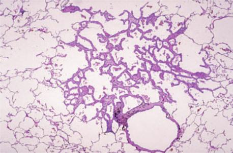
Fig. 1.77 Atypical adenomatous hyperplasia showing localised centriacinar alveolar wall thickening and increased numbers of alveolar lining cells. From Travis et al. {2024}.
universally accepted, has no known clinical significance, its reproducibility is untested, and this panel does not recommend it. The features of AAH fall short of those accepted as BAC. This issue is addressed in the discussion on BAC.
The postulated progression of disease, apparent from the increasingly atypical morphology, is supported by numerous morphometric and cytofluorometric studies {1375,1379,1438}. AAH and nonmucinous BAC probably represent a continuum of progression of pulmonary alveolar intraepithelial neoplasia.
AAH must be distinguished from reactive hyperplasia, secondary to parenchymal inflammation or fibrosis, where the alveolar lining cells are not the dominant feature and are more diffusely distributed. Generally, AAH cannot be identified in the presence of inflammatory or fibrosing disease. Distinction between more cellular and atypical AAH and BAC is difficult. BAC is generally >10mm in size, has a more pleomorphic, homogeneous columnar cell population, which is densely packed with greater cell-cell contact, overlap, mild stratification, and, usually, a less graded, more abrupt transition to adjacent alveolar lining cells. True papillae suggest papillary adenocarcinoma.
Immunohistochemistry
AAH expresses SPA, CEA {1640}, MMPs {1084}, E-cadherin, ß-catenin, CD44v6
and TTF-1. The expression of oncogene and tumour supressor gene products (TP53, C-ERB2, RB, MST1(p16), WAF1/CIP1 (p21) and FHIT) essentially reflects neoplastic progression from AAH to BAC and invasive adenocarcinomas {802,995,1021,1100}. In contrast to the data on TP53 mutations, TP53 protein accumulation seems to occur early in the proposed sequence of events {995}.
Histogenesis
The origin of AAH cells is still unknown but the differentiation phenotype derived from immunohistochemical and ultrastructural features suggests an alveolar origin. Surfactant apoprotein {1041}, and Clara cell specific 10kDd protein {1021, 1379} are expressed in almost all AAH lesions. Ultrastructurally, cytoplasmic lamellar bodies and nuclear branching microtubles, both typical of type II pneumocytes {768,1021,1316,1521}, and electron-dense Clara cell-type granules {1021,1434,1521} are found. AAH cells are likely derived from a progenitor cell with the potential for both type II pneumocyte and Clara cell differentiation.
Somatic genetics
KRAS. Mutations of the K-ras gene, particularly at codon 12, are specific for peripheral lung adenocarcinomas, as opposed to bronchogenic carcinoma, suggesting an alternative pathway of peripheral lung tumourigenesis {287,
74 Tumours of the lung - Preinvasive epithelial lesions
A B
C D
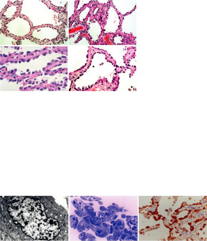
Fig. 1.78 Atypical adenomatous hyperplasia. A,B Slightly thickened alveolar walls are lined by an intermittent single layer of cuboidal cells. Occasional large cells are present. C Cuboidal pneumocytes line the alveolar walls with gaps between the adjacent cells. D Slightly thickened alveolar walls lined by an intermittent single layer of cuboidal cells, some with apical cytoplasmic snouts.
402}. K-ras codon 12 mutations are reported in 15-39% of AAH lesions, and up to 42% of concurrent adenocarcinomas. Most of the time, the K-ras mutations are different. One study found K-ras codon 12 mutations in 15% of AAH, 33% of ‘early’ BAC and 24% of ‘advanced’ BAC. {1021}, suggesting that K-ras codon 12 mutation is a very early event in the development of peripheral adenocarcinoma {1021,2126}.
TP53. Abnormalities of the P53 gene (17p), with impaired protein function, promote neoplastic transformation in affected cells. Many lung adenocarcinomas show missense mutations of the P53 gene with abnormal nuclear protein
accumulation. LOH and mutations of the P53 gene are very rare in AAH compared with adenocarcinoma; however p53 protein overexpression is frequent in AAH {1021}. P53 mutation has been demonstrated with increasing frequency in the progression from AAH, through BAC to early invasive adenocarcinoma {1836}.
LOH Allelic-specific losses at 3p and 9p loci have been detected in AAH {1044, 2187}. Some AAH lesions have shown LOH in 9q {51} and both 17q {2187} and 17p {51} LOH in the 3p and 9p loci probably occurs at a very early stage and may represent the earliest and crucial event in neoplastic transformation, with 17p events occurring later.
FHIT. The fragile histidine triad (FHIT) gene (3p) is deleted in many lung carcinomas {1856}.
p16INK4. Loss and inactivation plays an important role in the pathogenesis of lung carcinoma. However, loss of expression of p16INK4 is relatively rare in both AAH and adenocarcinoma {1021}.
TSC. A recent study on lung adenocarcinoma with concurrent multiple AAH lesions showed frequent LOH of tuberous sclerosis complex (TSC)-associated regions (TSC1 at 9q and TSC2 in 16p), suggesting that these are candidate loci for tumour suppressor genes in peripheral lung adenocarcinoma {1949}.
Aneuploidy. FISH studies of AAH have shown frequent aneuploidy of chromosome 7. The percentages of aneuploid cells and mean chromosome copy number increased from AAH to invasive adenocarcinomas, suggesting increasing polyploidy during malignant change {2245}. Some cases of AAH have been shown to be monoclonal, suggesting that it is a true preneoplastic lesion {1475}.
Prognosis and predictive factors
Assuming that AAH is always multifocal, several studies have compared postoperative survival in groups of patients with, and without AAH {333,1198,1927, 1960}. None showed any difference in outcome.
There is no indication for surgical or medical therapy in patients without cancer who are incidentally found to have AAH. In such a clinical setting, careful followup is warranted.
A B C
Fig. 1.79 Atypical adenomatous hyperplasia. A Transmission electron photomicrograph of a formalin-fixed AAH lesion showing a cuboid AAH cell having many intracytoplasmic small inclusion bodies and granules. Note scattered short microvilli on the free surface of the cell, the irregular contour of the nucleus and basal membrane. B Light photomicrograph of a thin section of a formalin-fixed, epoxy-resin-embedded AAH lesion showing a cell with an intranuclear inclusion body at the middle upper portion as well as several binucleated cells. Toluidine blue stain. C Immunostaining with a mouse monoclonal antibody against surfactant apoprotein A (PE10), showing uniformly strong positive staining of the cytoplasm of almost all the AAH cells as well as many nuclear inclusion bodies.
Atypical adenomatous hyperplasia 75
Diffuse idiopathic pulmonary neuroendocrine cell hyperplasia
J.R. Gosney
W.D. Travis
Definition
Diffuse idiopathic pulmonary neuroendocrine cell hyperplasia (DIPNECH) is a generalised proliferation of scattered single cells, small nodules (neuroendocrine bodies), or linear proliferations of pulmonary neuroendocrine cells (PNCs) that may be confined to the bronchial and bronchiolar epithelium, include local extraluminal proliferation in the form of tumourlets, or extend to the development of carcinoid tumours. It is sometimes accompanied by intraand extraluminal fibrosis of involved airways, but other pathology that might induce reactive PNC proliferation is absent.
Synonyms
The entity of DIPNECH was not fully recognised and named until 1992 {29}, but cases with its clinical and pathological features appear in the literature from the early 1950s {570}.
Clinical features
Signs and symptoms
DIPNECH may occur at any age, but presents typically in the fifth or sixth decades, and is perhaps commoner in women {29,74,1150,1317}. The history is one of a very slowly worsening dry cough and breathlessness, often over many years, sometimes misdiagnosed as mild
bronchial asthma. Physical examination usually reveals no signs, but pulmonary function tests show an obstructive or mixed obstructive/restrictive pattern of impairment with reduced diffusing capacity.
Imaging
Plain thoracic radiography is often normal, but tomographic scanning reveals a mosaic pattern of air trapping, sometimes with nodules and thickened bronchial and bronchiolar walls {1150}. Multiple nodules corresponding to tumourlets or carcinoid tumours may be present.
Macroscopy and localization
The early lesions of DIPNECH are invisible to the naked eye, but tumourlets and microcarcinoids, when present, can be just discerned as small, gray-white nodules, the latter often well-demarcated and resembling ‘miliary bodies’. Larger carcinoid tumours are firm, homogeneous, well-defined, grey or yellow-white masses. The lesions of DIPNECH usually affect one or both lungs uniformly.
Histopathology
Histopathological examination reveals widespread proliferation of PNCs {29,74, 1317}. The earliest lesions comprise
increased numbers of individual cells, small groups, or larger, nodular aggregates, confined to the bronchial or bronchiolar epithelium, the larger lesions bulging into the lumen, but not breaching the subepithelial basement membrane. The bronchiolar wall sometimes is fibrotically thickened. Bronchiolar occlusion may occur due to fibrosis and/or PNC proliferation. These are sufficient for the diagnosis of DIPNECH providing other defining criteria are met. In particular inflammatory or fibrous lesions that might cause secondary PNC hyperplasia are not seen. However, more advanced lesions are often present. These develop when the proliferating PNCs break through the basement membrane to invade locally, developing a conspicuous fibrous stroma to form small (2-5 mm) aggregates traditionally known as ‘tumourlets’. This proliferation of PNCs is sometimes accompanied by intraand extramural fibrosis of the involved airways that often obliterates them, but the surrounding lung is otherwise unremarkable. Once PNCs reach a size of 5mm or greater, they are classified as carcinoids.
Differential diagnosis
Clinically and on imaging, DIPNECH may be indistinguishable from other diffuse lung diseases characterised by cough,
A B
Fig. 1.80 Diffuse idiopathic pulmonary neuroendocrine cell hyperplasia. A The bronchiolar epithelium is almost entirely replaced by proliferating neuroendocrine cells. B Hyperplastic neuroendocrine cells form a nest at the base of the bronchiolar epithelium. From Travis et al {2024}.
76 Tumours of the lung - Preinvasive epithelial lesions
breathlessness, mixed obstructive/ restrictive pulmonary impairment and a nodular pattern of pulmonary infiltration, so that the diagnosis is usually impossible to make without recourse to biopsy. Histopathologically, DIPNECH must be distinguished from the PNC proliferation that may accompany a variety of pulmonary conditions, particularly chronic inflammatory diseases such as bronchiectasis and chronic lung abscess {717}; in the latter situation, progression of the proliferation to carcinoid tumours does not occur. DIPNECH must also be distinguished from the proliferation of PNCs not uncommonly seen adjacent to peripheral carcinoids {29,1317}.
Histogenesis
As with neuroendocrine neoplasms arising in the lungs, the origin of the proliferating PNCs that characterize DIPNECH is likely to be a yet-to-be-defined uncommitted precursor cell that is stimulated by unknown influences to differentiate along
a neuroendocrine line. However, the PNCs that proliferate in DIPNECH are found in normal lungs of adults {719, 1141}.
Somatic genetics
There are no genetic markers of DIPNECH such that it might be possible to distinguish it genetically from the limited, reactive, reversible proliferative response of PNCs that occurs after pulmonary injury. It is of interest, however, that allelic imbalance at the 11q13 region that closely approximates to the MEN1 tumour suppressor gene appears to be rare in tumourlets, but is present in the majority of carcinoid tumours {581}.
Prognosis and predictive factors
DIPNECH is a slowly progressive condition with a benign course spanning many years. Associated carcinoid tumours are indolent and atypical features have not been described. There are no predictive histologic or genetic data for DIPNECH.
Diffuse idiopathic pulmonary neuroendocrine cell hyperplasia 77
Squamous cell papilloma
D.B. Flieder
F. Thivolet-Bejui
H. Popper
Definition
A papillary tumour consisting of delicate connective tissue fronds with a squamous epithelial surface. Squamous papillomas can be solitary or multiple and can be exophytic or inverted.
ICD-O codes |
|
Squamous cell papilloma |
8052/0 |
Exophytic |
8052/0 |
Inverted |
8053/0 |
Epidemiology
Solitary squamous papillomas are very rare representing less than 0.50% of lung tumours at one large institution {1612}. Exophytic lesions far outnumber the inverted growth pattern {592}. Solitary squamous papillomas are seen predominantly in men, with a median age of 54 years {592}. Juvenile and adult laryngotracheal papillomatosis rarely involve the lower respiratory tract are always related to laryngotracheal papillomatosis {1223}.
Etiology
An association with human papilloma virus (HPV) subtypes 6 and 11 suggests a possible pathogenetic role for the virus {592}. Human papilloma virus subtypes 16,18 and 31/33/35 in squamous papillomas associated with carcinomas and in squamous cell carcinomas have been reported, suggesting that HPV infection might be related to tumoural progression {139,1611,1612}. More than half of patients are tobacco smokers, but an etiologic role has not been established {592,1937}.
Localization
Papillomas are endobronchial.
Clinical features
While up to one-third of lesions are incidental radiographic findings, patients most often present with obstructive symptoms. Computed tomography scans demonstrate a small endobronchial protuberance or nodular airway thickening. Involvement of distal air-
ways may lead to nodular opacities and/or thin-walled cavity nodules {2213}. An endobronchial biopsy may be diagnostic, but distinction from well-differenti- ated squamous cell carcinoma can be difficult, especially with superficial tissue fragments. Bronchoscopic cytologic specimens will only demonstrate the squamous nature of the lesion. Parakeratotic cells, cytologic atypia and viral cytopathic effect should not be misinterpreted as invasive carcinoma {1677}.
Macroscopy
Solitary squamous papillomas arise from the wall of either mainstem bronchi or major subdivisions and appear as cauliflower-like tan-white soft to semifirm excrescences protruding into bronchial lumens. Tumours range from 0.7-9.0 cm with a median of 1.5 cm. Distal airways may be bronchiectatic with secondary atelectasis and consolidation {965}.
Tumour spread and staging
Squamous papillomas may recur at their original site and laryngotracheal papillomatosis can spread into the lower respiratory tract. It has been suggested that electrical or laser fulguration is responsible for alveolar parenchymal seeding.
Histopathology
Squamous papillomas are composed of a loose fibrovascular core covered by stratified squamous epithelium. Exophytic lesions feature orderly squamous maturation from the basal layer to the superficial flattened and oftentimes keratinized cells. Acanthosis may be prominent. While non-keratinized epithelium may resemble transitional epithelium, the squamous nature of these cells has been demonstrated ultrastructurally and use of this term is discouraged. Over 20% of solitary squamous papillomas feature wrinkled nuclei, binucleate forms and perinuclear halos, i.e., koilocytosis related to HPV infection {592}. Scattered
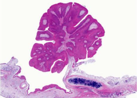
Fig. 1.81 Exophytic squamous papilloma. Mature squamous epithelial cells are growing in an exophytic papillary pattern on the surface of thin fibrovascular cores. The papilloma is attached to the underlying bronchial wall by a stalk. .
78 Tumours of the lung - Benign epithelial lesions