

Опухоли легких, плевры, тимуса и сердца ВОЗ
.pdfA
B
C
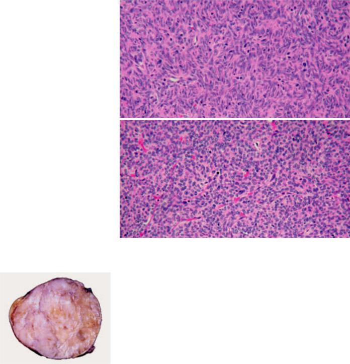
Fig. 3.02 A Cartoon of the non-lymphoid cellular components of the normal thymic cortex and medulla, including Hassall´s corpuscle (HC). Various thymic epithelial cells, dendritic cells (DC), macrophages (MA), and myoid cells (MC). Subcapsular and medullary epithelial cells delineate septa and perivascular spaces (PvS) at the corticomedullary junction. B Electron micrograph showing cortical and subcortical thymic epithelial cells (TEC), apoptotic bodies, and macrophages. C Electron micrograph of thymic medulla, depicting a medullary epithelial cell , a dendritic celland a Hassall´s corpuscle (HC).
nomas, as well as some Hodgkin, rare non-Hodgkin and NK/T-cell lymphomas.
Clinical features
Patients may exhibit symptoms due to local complications (pain, superior vena cava syndrome, respiratory insufficiency or tachycardia because of pleural or pericardial implants and effusions), as well as systemic symptoms (fever or weight loss).
In addition, thymomas can cause a large variety of autoimmune diseases (Table 3.01) which are often typical for a specific tumour type and may precede or follow thymoma resection {987}. Type A, AB and B thymomas exhibit an unrivaled frequency and spectrum of autoimmune phenomena, comprising neuromuscular, haematopoietic, dermatologic, rheumatic/vasculitic, hepatic and renal diseases. Myasthenia gravis is by far the most frequent and preferentially associated with type AB and B2, B3 thymomas, while hypogammaglobulinaemia (Good syndrome) is more typical for type A thymoma. Pure red cell aplasia is also a rare complication of type A thymomas, though recent data find a less specific association with this thymoma subtype {1096}. Thymic carcinomas are not associated with myasthenia gravis or hypogammaglobulinaemia, but occasionally with other autoimmune diseases. Cytopenias and/or hypogammaglobulinaemia can result in serious bacterial and opportunistic infections. Lymphocytosis and thrombocytosis can occur. Whether the increased incidence of second cancers in thymoma patients is related to genetic or environmental etiologies or thymoma-induced immunodeficiency is unknown {1395,1537}.
A B
Fig. 3.03 Epidemiology of mediastinal tumours A in children (< 15 years), and B in adults (45-75+ years). Thymomas are exceedingly rare in children, while they are the most common mediastinal tumours in adults.
Table 3.01
Autoimmune diseases associated with thymomas. Rare manifestations are indicated by an asterisk.
Neuromuscular Diseases
Myasthenia gravis
Neuromyotonia*, rippling muscle disease*, polymyositis/dermatomyositis*
Encephalitis* (limbic, cortical, and brainstem)
Intestinal pseudoobstruction*
Haematologic Autoimmune Diseases
Anaemia: pure red cell aplasia (in all thymoma subtypes) {168}
pernicious anaemia, haemolytic anaemia, aplastic anaemia
Other isolated cytopenias, eg eosinophils, basophils, neutrophils
Immunodeficiencies: Hypogammaglobulinaemia +/- T-cell deficiencies (Good syndrome)
Dermatologic Diseases*
Pemphigus (foliaceus, paraneoplastic)
Lichen planus
Alopecia areata
Endocrine Disorders
Addison disease, Cushing disease
Graves disease
Renal and Hepatic Diseases
Glomerulonephritis, autoimmune hepatitis
Systemic Autoimmune Diseases*
SLE, Sjögren disease, systemic sclerosis Graft-versus-Host-Disease
Histopathological classification
Thymomas
Histological classification schemes for thymomas traditionally have been descriptive (predominantly spindle, predominantly lymphocytic, predominantly epithelial, mixed lymphoepithelial) {158, 1086,1134,1172,1732}, or were based on the combined consideration of morphology (spindle, polygonal, mixed tumour cells) and lymphocyte content {1086, 1808}. Except for the spindle cell type, these classifications largely lacked prognostic significance independent of stage {1172,1253,1808}.
The histogenetic or functional classification included terms (medullary, cortical) that reflected the normal differentiation of the major functional and anatomic com-
Introduction 149

Table 3.02
Comparison of Masaoka tumour stages and corresponding TNM classification. Modified, from K. Koga et al. {1043}.
Masaoka stage |
T |
N |
M |
|
|
|
|
I |
1 |
0 |
0 |
II |
2 |
0 |
0 |
III |
3 |
0 |
0 |
IVa |
T4 |
0 |
0 |
IVb |
any T ≥ N1 or ≥ M1 |
||
|
|
|
|
partments of the thymus {1245,1404}. This classification proved to be of independent prognostic value {341,856, 1017,1540,1630} and was highly reproducible {378}. However, although some morphological and immunological studies have supported the histogenetic concept {235,859,1015,1086,1452,1510, 1886}, it has not been generally accepted {1058,1691,1917}.
In 1999, a WHO Working group suggested a non-committal terminology, preserving the distinct categories of the histogenetic classification, but using letters and numbers to designate tumour entities {1690}. The rationale for the concept to label thymomas as type A, AB, and B is derived from a growing body of morphological, functional and genetic evidence {235,859,896,1510,1631,2242}, suggesting that these thymoma subgroups form distinct entities both in morphological and clinical terms.
In recent years, this WHO classification {1690} has been well accepted and is largely retained in the current WHO classification as it provides an easy comparison of clinical, pathological and immunological studies {235,318,1196, 1511,1744,1886}.
Thymomas of type A, AB, and B exhibit organotypic (thymus-like) architectural features. These tumours have not been observed in organs other than the thymus, though they may arise from heterotopic thymic tissue in the head and neck region, anywhere in the mediastinum, pleura and lung {1691}. By contrast, the heterogeneous thymic carcinomas (called type C thymomas in the previous WHO classification) exhibit morphologies that are encountered also in organs other than the thymus.
Thymic carcinomas comprise malignant, usually invasive epithelial tumours with clear-cut atypia, largely absent organotypic features and a very diverse differ-
entiation, resembling carcinomas outside the thymus. This category includes neuroendocrine epithelial tumours.
Germ cell, lymphoid, haematopoietic and mesenchymal tumours
The classification of these tumours follows the WHO Classification of gonadal germ cell tumours {526}, tumours of haematopoietic and lymphoid tissues {919} and tumours of soft tissues and bone {590}.
Useful morphological terms
Encapsulated.
A thymoma completely surrounded by a fibrous capsule of varying thickness which is not infiltrated by tumour growth. Thymic tumours that infiltrate into, but not through, the capsule still belong in this category.
Minimally invasive.
A thymoma surrounded by a capsule which is focally infiltrated by tumour growth with invasion of the mediastinal fat. The capsular invasion needs to be complete in order for the tumour to be placed in this category. Minimally invasive thymomas are usually identifiable as such only after microscopic examination in so far as they generally appear to the surgeon indistinguishable from encapsulated thymomas at the time of excision.
Widely invasive.
A thymoma spreading by direct extension into adjacent structures such as pericardium, large vessels or lung. This type of thymoma usually appears invasive to the surgeon at the time of excision, which may be incomplete as a result.
Implants.
A thymoma in which tumour nodules separate from the main mass are found on the pericardial or pleural surface. These implants tend to be small and multiple and their microscopic appearance is usually, but not always, similar to that of the parent tumour.
Lymph node metastases.
A thymoma that involves one or more lymph nodes anatomically separate from the main mass. This excludes direct extension into the node by the tumour. The nodes most commonly involved by metastatic thymoma are mediastinal and supraclavicular. It is a rare event even in
long-standing cases. but may exceptionally be the first clinical manifestation of the tumour.
With distant metastases.
A thymoma with metastases to distant site(s), most commonly lung, liver, and skeletal system. This excludes metastases to lymph nodes and local extension into adjacent organs.
Grading of malignancy
Thymic epithelial tumours consist of several histological subtypes, i.e., thymoma types A, AB, B1, B2 and B3, and thymic carcinomas, in increasing order of malignancy {1691} Thymoma type A and AB generally behave like a benign tumour, type B1 as a low-grade malignant tumour (10-year survival rates of over 90%), type B2 has a greater degree of malignancy, and type B3 in the advanced stage shows a poor prognosis, just like thymic carcinoma and malignant tumours of other organs {1511}. Among the various subtypes of thymic carcinoma, squamous cell carcinoma, basaloid and mucoepidermoid carcinoma have a better prognosis than other histological subtypes. The malignancy grade of thymic neuroendocrine tumours (carcinoids) is intermediate between thymoma and thymic carcinoma. The rare small cell and large cell carcinomas tend to be highly malignant.
TNM Classification and stage grouping
The TNM Classification and stage-group- ing has been applied to malignant tumours of many organs {2045}, but there is currently no authorized TNM system for thymic epithelial and neuroendocrine tumours. In the TNM Supplement 2nd edition {899}, a tentative classification of malignant thymomas appeared for testing. It is mainly based on the Masaoka system and its revised versions {1255,2036,2184}. While the tentative classification applies only to malignant thymic epithelial tumours {899}, it has in this chapter been extended to include neuroendocrine tumours.
Invasion
Crucial points in defining the T-cate- gories are invasion through the capsule and invasion into neighbouring structures. Although invasive growth outside the thymus detected by the surgeon at
150 Tumours of the thymus - Introduction
the time of thoracotomy has been repeatedly reported to have significant impact on the prognosis {693,1043}, the prognostic significance of minor degrees of invasion detected by histological examination remains controversial. Many reports on thymoma have shown little or no difference in survival between Masaoka stage I and stage II thymomas {693,1043,1130,1511,1579,1645}. Furthermore, some thymomas and most thymic carcinomas are devoid of a capsule entirely or in part, which makes the definition of “encapsulation” meaningless. Therefore, the current and proposed categories “T1 (completely encapsulated)” and “T2 (with invasion of pericapsular connective tissue)” may not be biologically meaningful and may be impossible for pathologists to use. Criteria for minimal invasion need to be better defined.
Tumour size
On the other hand, tumour size has been used as an important parameter to define T-categories; critical dimensions of 11 cm and 15 cm have been reported {183,1172}. Especially in Blumberg’s report, tumour size was one of the significant parameters for survival by multivariate analysis {183}. Critical size may be quite different among thymoma and thymic carcinoma, including neuroendocrine tumours. This consideration of tumour size might also be necessary in a revised definition of the T-category.
Surgical resectability
The present T denominator includes tumours with different characteristics: one extreme is an easily resectable tumour with minimal invasion into the pericardium and a good prognosis and another extreme is a non-resectable tumour with invasive growth into multiple neighbouring organs. A further division of T3 tumours into potentially
Table 3.03
Malignant potential in terms of mortality, combining WHO histologic type and tumour stage, according to Shimosato et al. {1808}. Stage IV thymomas should be considered as tumours of high malignant potential, although metastatic type A or AB thymomas with long-term survival have been reported.
|
Histology |
Tumour Stage |
Malignant potential |
|
|
|
|
|
|
|
Type A, AB, (B1) thymoma |
I and II |
None (very low)1 |
|
|
|
III |
Low |
|
|
|
|
|
|
|
|
|
|
|
|
Type B2, B3 thymoma |
I |
Low |
|
|
|
II and III |
Moderate |
|
|
|
|
|
|
|
Thymic carcinoma: |
|
|
|
|
Low-grade squamous |
Stage I and II |
Moderate |
|
|
cell, basaloid or |
Stage III |
High |
|
|
mucoepidermoid |
|
|
|
|
carcinoma, carcinoid |
|
|
|
|
|
|
|
|
|
Other histological types |
Any stage |
High |
|
|
|
|
|
|
|
|
|
|
|
1These tumours amount to 40-50% of all thymomas {341,1511,1631}.
resectable and curable ones and nonresectable ones with a poor prognosis is desirable, especially for planning treatment.
Lymph node metastasis
This is rare in thymoma, and the basis for the definition of stage IVB (Masaoka) for thymomas with lymph node metastasis {1255}. In the tentative TNM classification, N1 (metastasis to anterior mediastinal lymph nodes) is defined as stage III. However, the prognostic equivalence between T3 and N1 has not yet been assessed. The appropriateness of the nodal grouping N1 to N3 needs to be investigated further. Depending on the tumour location in the anterior mediastinum, the lymphatic pathway by which tumour cells spread might be different. Consequently, the sentinel lymph node might be located elsewhere other than the anterior mediastinum.
Stage-Grouping
The most important issue in stage-group- ing is the definition of stages I and II. The survival curves of patients with thymomas of stages I and II are superimposed at around 100% at 5 and 10 years after surgery {693,1511}. In other reports, a minimal difference has repeatedly been reported {1043,1130,1579,1645}. If the definition of the T-category remains unchanged, the present stages I and II could be merged into a new stage I; however, no data are available on thymic carcinoma with respect to stages I and II. Stage III in the present tentative system, which is a heterogeneous group, is recommended to be divided into a potentially resectable group with a favourable prognosis and an unresectable group with a poor prognosis, respectively.
Stage grouping 151
Thymomas
A. Marx |
N.L. Harris |
Ph. Ströbel |
T.T. Kuo |
A. Zettl |
Y. Shimosato |
J.K.C. Chan |
P. Engel |
H.K. Müller-Hermelink |
|
Definitions
Thymomas (type A, AB, B thymomas) are neoplasms arising from or exhibiting differentiation towards thymic epithelial cells, regardless of the presence and relative numbers of non-neoplastic lymphocytes {1691}. Their malignant potential is either absent or low to moderate.
Thymic carcinomas are malignant epithelial tumours because of overt cytological atypia, almost invariable invasiveness and lack of “organotypic” (thymuslike) features.
Combined thymoma, combined thymoma/thymic carcinoma. These terms are used for a combination of thymoma subtypes and of thymomas with thymic carcinomas, including thymic neuroendocrine carcinomas, within one tumour mass.
Thymoma (type X) with anaplasia is the suggested diagnostic term for a very uncommon group of tumours with borderline morphological freatures between thymoma and thymic carcinoma.
Epidemiology
Thymomas and thymic carcinomas are uncommon tumours with an annual incidence of approximately 1-5 per million population. There are only very limited epidemiologic data, but cautious interpretation of data from the Danish National Board of Health suggests that the incidence has not changed significantly over the last three decades. Thymomas and thymic carcinomas occur at almost all ages (range 7-89 years) with a peak incidence between 55-65 years. They are exceedingly rare in children and adolescents {1577,1876}. There is no pronounced sex predilection {318,341,1016,1630}. Patients exhibit an increased incidence of second cancers irrespective of the histology of the thymic epithelial tumour {1537}.
Etiology
The etiology of thymomas is still largely unknown. They have been repeatedly observed in patients with MEN1 syndrome {461,1535,1644,1648,2094}.
Epstein-Barr virus appears to play an etiologic role in subsets of lymphoepithe- lioma-like, poorly differentiated squamous and undifferentiated thymic carcinomas both in Asian {343,1174,1265, 2174} and Western countries {785,894, 1234,1876}. There is no increased risk of developing thymomas in patients receiving radio-chemotherapy for mediastinal Hodgkin lymphoma {1455} or breast cancer {1498}.
Principles of thymoma classification
1.There are two major types of thymoma depending on whether the neoplastic epithelial cells and their nuclei have a spindle or oval shape, and are uniformly bland (Type A thymoma) or whether the cells have a predominantly round or polygonal appearance (Type B) {1691}.
2.Type B thymomas are further subdivided on the basis of the extent of the lymphocytic infiltrate and the degree of atypia of the neoplastic epithelial cells into three subtypes B1 (richest in lymphocytes), B2, and B3 (richest in epithelial cells).
Table 3.04
3.Thymomas combining type A with B1like or (rarely) B2-like features are designated type AB.
4.Thymic carcinomas are termed according to their differentiation (squamous cell, mucoepidermoid, etc.). In the 1999 WHO classification {1690}, the term WHO type C thymoma was the “headline designation” to stress their thymic epithelial origin. In the current classification, this term was eliminated since all non-organotypic malignant epithelial neoplasms other than germ cell tumours are designated thymic carcinomas.
5.Combined thymomas are specified by the WHO histology and approximate percentage contributed by each component of the combined thymoma.
6.Traditionally, the term “malignant thymoma” has been used for (i) thymomas with advanced stage, i.e. local invasiveness, pleural or pericardial implants or metastasis, irrespective of tumour histology or (ii) thymic epithelial tumours with marked atypia (thymic carcinomas), irrespective of tumour stage {1170,1691, 1841,1924}. The use of the term “malig-
Differential diagnosis of thymomas types A, AB, B and thymic carcinomas
Feature |
Thymomas |
Thymic carcinomas |
|
|
|
Organotypic (thymus-like) |
Almost always present (lobular |
None or abortive |
histological features |
pattern, perivascular spaces, |
|
|
immature, TdT+ / CD1a+ / |
|
|
CD99+ T-cells) |
|
|
|
|
CD5, CD70 and CD117 expres- |
No |
Frequent (~ 60%) |
sion in epithelial cells |
|
|
|
|
|
Invasion |
Variable |
Almost always |
|
|
|
Myasthenia gravis |
Variable: 10–80% |
No |
|
|
|
Other autoimmune diseases |
Common |
Rare |
|
|
|
Clinical behaviour |
Often curable by surgery; |
Often unresectable {318}; |
|
metastases are rare. |
metastases are frequent |
|
Usually long survival due to |
Often short survival due to pro- |
|
indolent clinical course |
gressive disease |
|
|
|
152 Tumours of the thymus - Thymomas
nant thymoma” as a synonym for a locally invasive thymoma irrespective of the WHO histological type is discouraged, since it may not properly reflect the excellent prognosis of type A and AB thymomas of advanced stage {318,341, 1510,1511,1630}.
Prevalence of thymoma subtypes
The predominant histological subtypes in most published series are type B2 and AB thymomas (each 20-35% of all cases), while type B1 and type A thymomas count among the rare types (5-10% in most studies) {856,1510,1967}. The percentage of thymic carcinomas has been reported to be about 10–25% {318, 341,541,1404,1510}.
In children, type A, B1 and B2 thymomas have been observed, in addition to undifferentiated and EBV-positive lymphoep- ithelioma-like thymic carcinomas {274, 1577,1876}. The morphologically heterogeneous and rare carcinomas with t(15;19) translocation typically occur in children and young adults {1081,1148, 2072}.
Genetic features
Recurrent genetic alterations have so far been reported for type A and B3 thymomas as well as for thymic squamous cell carcinomas {896,1567,2238,2242}. Type A thymomas only show few genetic alterations, with deletions of chromosome 6p reported as a recurrent genetic alteration {437,2065,2238}. Type A areas in type AB thymomas are genetically distinct from type A thymoma {699,896}. Type B3 thymomas frequently show gains of chromosome 1q and losses of chromosomes
Table 3.05
Genetic alterations reported for the different WHO histological thymoma subtypes.
WHO Type |
Chromosomal Gains |
Chromosomal Losses |
Source |
|
|
|
|
Type A |
none |
-6p |
{437,1325,2065,2238, |
|
|
|
2242} |
|
|
|
|
Type AB |
none |
-5q21-22, -6q, -12p, -16q |
{699,896,897} |
|
|
|
|
Type B3 |
+1q |
-6, -13q |
{896,897,2238,2242} |
|
|
|
|
Thymic squamous cell |
|
|
|
carcinoma |
+1q, +17q, +18 |
-3p, -6, -13q,-16q, -17p |
{896,897,1848,2238} |
|
|
|
|
6 and 13q. Type B2 thymomas are genetically related to type B3 thymomas {896}. Thymic squamous cell carcinomas frequently show gains of chromosomes 1q, 17q and 18 and losses of chromosomes 3p, 6, 16q, and 17p {2238}. The shared genetic abnormalities underline the close relationship between type B3 thymomas and thymic squamous cell carcinomas.
Prognosis and predictive factors
The most relevant prognostic factors in thymoma are tumour stage {341,1511, 1630,1808}, WHO histologic type {341, 1511} and completeness of resection {318,1419}.
Type A and AB thymomas in stages I and II virtually always follow a favourable clinical course {341,476,1511}, and even at higher stages may not be fatal due to a very slowly progressive course {1808}. They are considered benign tumours {784,1404} or neoplasms of low malignant potential. Type B1 thymomas have a
very low malignant potential; rare local recurrences or late metastases may occur {318}. Type B2 and B3 thymomas and thymic carcinomas, are clear-cut malignant tumours. B2 and B3 thymomas and well differentiated squamous, basaloid and mucoepidermoid carcinomas follow a more favourable course than poorly differentiated squamous cell carcinomas and other thymic carcinomas {1924}. The prognosis of combined thymomas may be determined by the most malignant component {1093,1912}. Paraneoplastic pure red cell aplasia, other cytopenias, or hypogammaglobulinaemia (Good syndrome) have an adverse effect {987} whereas paraneoplastic myasthenia gravis had no or a positive factor on survival {318,341}.
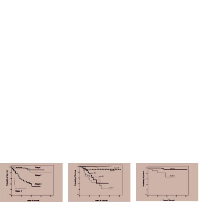
A |
|
B |
|
C |
Fig. 3.04 Kaplan-Meyer survival statistics of patients with thymic epithelial tumours. A Survival of patients with thymomas or thymic carcinomas according to stage. Masaoka tumour stage is the most important and statistically most significant independent prognostic parameter for survival in almost all clinico-pathological studies. From G. Chen et al. {341}. B Survival of patients with thymomas or thymic carcinomas according to histological type. WHO-based histology was a statistically significant prognostic parameter for survival in most clinico-pathological studies. In some studies, B3 thymomas and thymic carcinomas had a significantly worse prognosis than B2 thymomas (L. Quintanilla-Martinez et al. {1631}; M.Okumura et al. {1511}), but not in others (G. Chen et al. {341}). C WHO-based histological subtype is an independant prognostic marker in patients with thymomas and thymic carcinomas infiltrating beyond the tumour capsule into the mediastinal fat (Masaoka stage II). A no/low-risk group of tumours (type A, AB, B1 thymomas) is distinguished from a moderate/high-risk group (B2 and B3 thymomas and thymic carcinomas). From G. Chen et al. {341}.
Thymomas 153
Type A thymoma
T.T. Kuo
K. Mukai
T. Eimoto
R.H. Laeng
K. Henry
J.K.C. Chan
Definition
Type A thymoma is an organotypic thymic epithelial neoplasm composed of bland spindle/oval epithelial tumour cells with few or no lymphocytes. The tumour cells can form a variety of histologic structures.
ICD-O code |
8581/1 |
Synonyms
Spindle cell thymoma, medullary thymoma
Epidemiology
Type A thymoma is a relatively uncommon type of thymoma and accounts for 4-19% of all thymomas {541,1510,1511, 1808}. The age at manifestation ranges from 32 to 83 years, with a mean age of 61 years {1538,1540,1630}, which is higher than the mean age of 50 years of all thymoma patients {341,1095}. No consistent gender predilection has been reported {318,1511,1540,1630}.
Clinical features
Approximately 24% of type A thymomas are found in patients with myasthenia gravis {318,341,1511,1538,1540,1630}. Others are found because of local symptoms or incidentally discovered on imaging examination. Association with pure red cell aplasia may occur, but in contrast to earlier reports, pure red cell apla-
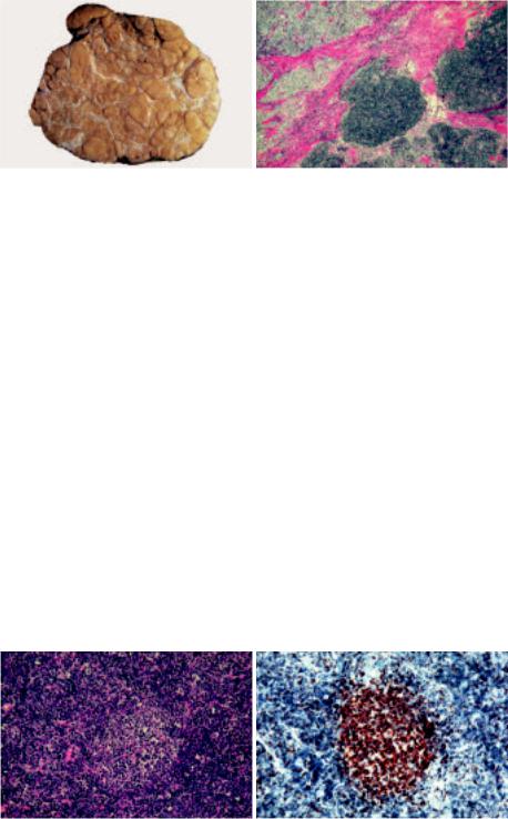
Fig. 3.05 A well encapsulated type A thymoma with vaguely lobulated appearance and thin white fibrous bands.
A
B
Fig. 3.06 Type A thymoma is composed of spindle cellls (A) or oval cells (B) with bland nuclei arranged in solid sheets without any particular pattern (B) or in a storiform patern (A).
sia may also occur in other thymoma types {1096}.
Macroscopy
Grossly, type A thymoma is usually well circumscribed and encapsulated. The cut surface is tan white and shows vague lobulation with less distinct dissecting white fibrous bands than is seen in other types. Cystic change or calcification of the capsule may be seen. Average tumour size is 10.5 cm.
Tumour spread and staging
The majority of type A thymomas (80%) occurs as Masaoka stage I in the anterior mediastinum, followed by stage II
(17%) and rarely stage III (3%) {341,541,
1095,1510,1511,1538,1540,1630,1631,1 808}. Single exceptional cases of stage IV type A thymoma have been reported {1403}.
Histopathology
Histologically, the tumour has few or no lymphocytes and shows neither distinct lobules nor dissecting fibrous bands as seen in other types of thymoma. The tumour cells are spindle and/or ovalshaped with bland nuclei, dispersed chromatin and inconspicuous nucleoli; they are arranged in solid sheets without any particular pattern or in a storiform pattern {1403,1691}. Type A thymoma
154 Tumours of the thymus - Thymomas
cells can form cysts of various size, glandular structures, glomeruloid bodies, rosettes with or without a central lumen, Masson’s haemangioma-like papillary projections in cystic spaces, or menin- gioma-like whorls {1086,1095,1403, 1538,1691,1808}. Extremely elongated fibroblast-like spindle cells may be seen focally. Vessels in the background may impart a haemangiopericytoma-like appearance {1538}. Perivascular spaces are less commonly seen than in other types of thymoma {318}. Although type A thymoma is a lymphocyte-poor tumour, spindle cell micronodules in a lymphoid stroma may be present at places {1981}. Most tumour cells are individually surrounded by reticulin fibers {1086,1691}. Cells in mitosis are seldom found, but lobular infarcts can occur. Rarely, thymic carcinoma can arise in type A thymoma. Areas of necrosis may be a clue to this phenomenon; examination of these areas reveals hyperchromatic anaplastic nuclei
and/or mitotic figures indicating the presence of carcinoma {1093}.
Immunophenotype
The tumour cells are strongly positive for AE1-defined acidic cytokeratins (CKs), and negative for AE3-defined basic CKs. Other CKs of different molecular weights show variable expression except that CK20 is negative {1086}. In general, the cystic and glandular structures express stronger CK {1808}. CD20-positive tumour cells may be detected focally {354,1403, 1538}. There is no expression of CD5 {1538}, and BCL-2, CD57 and EMA are variable and usually only focally expressed {227,1631,1808,1872, 1981}. Most tumour cells are surrounded by basement mem- brane-like deposits as demonstrated by anti-laminin and anti-type IV collagen antibodies. TP53 protein and Ki-67 show only low or no expression {1539,1872, 1980,1981}. Two antigens, metallothionein and PE-35, found in normal thymic
medullary cells are also expressed in type A thymoma cells {798,1087}. The few lymphocytes, if present, are T cells positive for CD3 and CD5. CD1a+ and CD99+ immature T cells may be present but comprise a minority of the T cells. CD20+ B cells are usually absent except in focal micronodular areas with a lymphoid stroma, if these are present.
Histogenesis
Type A thymoma has been postulated to derive from the normal thymic medullary epithelial cells {1403,1404}. Evidence in support of this postulate include their similar immunohistochemical expressions of CD20, cytokeratins, metallothionein, and PE-35 as well as the relative paucity of immature T cells {354,798, 1086,1087,1095,1452,1538,1631}.
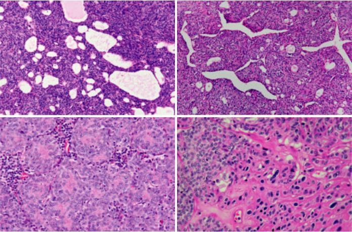
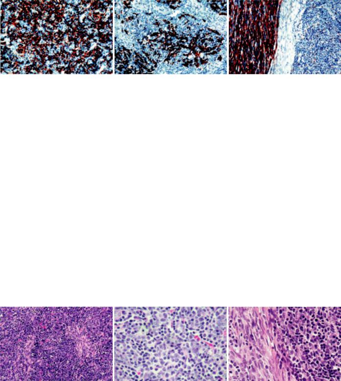
A B
C D
Fig. 3.07 Type A thymoma. A Type A thymoma cells can form cysts of various size. without a lumen. D Anaplastic malignant cells arising in type A thymoma.
B Type A tymoma cells with haemangiopericytoma-like appearence. C Rosettes
Type A thymoma 155
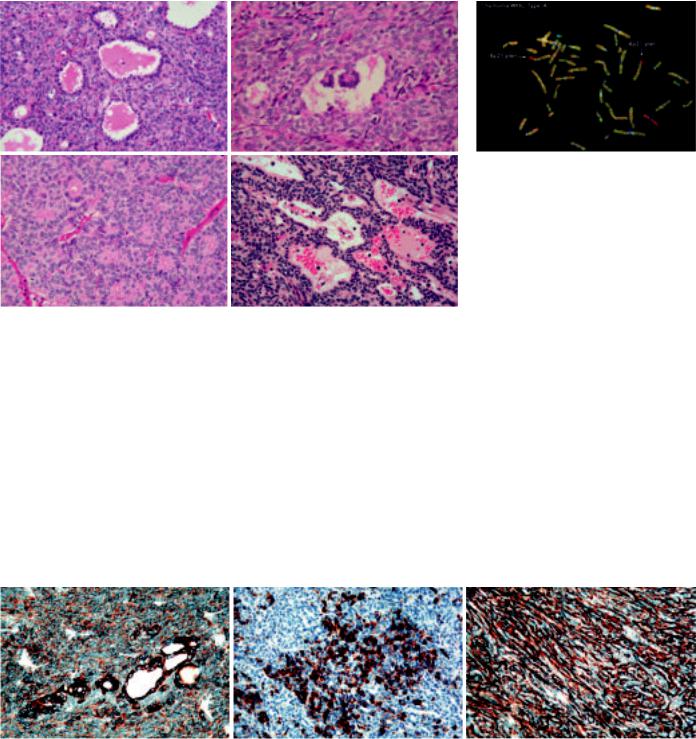
A B
C D
Fig. 3.08 Different histological patterns in type A thymoma. A Glandular structures. B Glomeruloid bodies. C Rosettes with lumens. D Perivascular spaces.
Somatic genetics
Type A thymoma has been found to have t(15;22)(p11;q11) {438} or a partial loss of the short arm of chromosome 6 {2238}. Consistent loss of heterozygosity has been found only in the region 6q23.3- 25.3, which is common to type A and B3 thymomas and squamous cell thymic carcinomas {897,2242}. Unlike type B3 thymomas and squamous cell thymic carcinoma, no aberrations in the APC, RB1, and TP53 gene loci or in regions 3p22-24.2 and 8q11.21-23 are found in
type A thymoma {2242}, which could be the genetic basis for its generally benign clinical course.
Prognosis and predictive factors
The overall survival of patients with type A thymoma has been reported to reach 100% at 5 years and 10 years {1403, 1511}, even though approximately 20% of them have stage II or stage III tumours. Generally, type A thymoma is regarded as a benign tumour without having a risk of recurrence if the tumour
Fig. 3.10 Type A thymoma. CGH hybridization of a WHO type A thymoma shows loss of chromosome 6p21-pter revealed by an excess of red hybridization signal of normal DNA as compared to the green hybridization signal of tumour DNA (highlighted by the arrows).
can be completely removed surgically {1095,1403,1404}. However, exceptional case reports of local recurrence or distant metastasis have been documented {1043,1403, 1538}. Rarely, type A thymoma can undergo malignant transformation into thymic carcinoma {1093}. The association with myasthenia gravis has been reported to have either a better or no effect on prognosis {318,1511,1630}.
A B C
Fig. 3.09 Immunophenotype of type A thymoma. A Immunopositivity for AE1-defined acidic cytokeratins. Note the stronger expression of cytokeratin by the glandular structures. B Focal epithelial expression of CD20. C Tumour cells are surrounded by abundant type IV collagen as demonstrated by immunostaining.
156 Tumours of the thymus - Thymomas
Type AB thymoma
T.T. Kuo
K. Mukai
T. Eimoto
R.H. Laeng
K. Henry
J.K.C. Chan
Definition
Type AB thymoma is an organotypical thymic epithelial neoplasm composed of a mixture of a lymphocyte-poor type A thymoma component and a more lym- phocyte-rich type B-like component. The tumour cells in the type B-like component are composed predominantly of small polygonal epithelial cells with small round, oval or spindle pale nuclei showing dispersed chromatin and inconspicuous nucleoli, and are smaller and paler than those of B1 or B2 thymomas. Lymphocytes are more numerous than in the type A component, but may be less numerous than in B1 thymomas. There is a great variation in the proportion of the two components, and while usually both components are present in most sections, either type A or type B areas can be scanty.
ICD-O code |
8582/1 |
Synonym
Mixed thymoma
Epidemiology
Type AB thymoma is either the most or the second most common type of thymoma and accounts for 15-43% of all thymomas {341,541,1095,1510,1511,1538, 1540,1631,1808}. The patients’ ages range from 29-82 years with a slightly younger mean age of 55 years than type A thymoma {1538,1540,1630}. A slight male predominance has been noted in most reports {1511,1538,1540,1630}.
Clinical features
The clinical presentation is similar to that of type A thymoma. Approximately 14% of type AB thymomas are associated with myasthenia gravis {341,541,1510,1511, 1538,1540,1630,1808}. Paraneoplastic pure red cell aplasia has also been reported {1096}. Other tumours manifest by local symptoms or can be asymptomatic and are found incidentally upon imaging examination.
A B
Fig. 3.11 A Macroscopic appearance of a type AB thymoma showing multiple nodules separated by fibrous bands. B Type AB thymoma composed of lymphocyte-associated type B nodules and diffuse lymphocytepoor type A areas.
Macroscopy
Grossly, type AB thymoma is usually encapsulated and the cut surface shows multiple tan coloured nodules of various size separated by white fibrous bands. Average tumour size is 7.7 cm.
Tumour spread and staging
The majority of type AB thymoma (71.7%) occur in the anterior mediastinum as Masaoka stage I followed by stage II (21.6%) and stage III (5.6%) {341,437,1404,1510,1511,1539,1540, 1691}. Rare cases of stage IV type AB thymoma (1.1%) have been reported {1325,1404,1510,1539,1691}.
Histopathology
Histologically, type AB thymoma shows a nodular growth pattern with diffuse areas and is composed of a variable mixture of a lymphocyte-poor type A thymoma component and a more lymphocyte-rich
type B component. All histological features of type A thymoma can be seen in the type A component. However, the type B areas are distinctive and different from either B1, B2, or B3 thymoma. The tumour cells in the type B component are composed predominantly of small polygonal epithelial cells with small round, oval or spindle pale nuclei showing dispersed chromatin and inconspicuous nucleoli {1086,1403,1691,1808}. The large, vesicular epithelial cells with nucleoli that are characteristic of B2 thymoma are only rarely seen {1086}. The type A and type B components either form discrete separate nodules or intermix together {1403,1691}. The type A component in the latter areas may form bundles of extremely elongated fibrob- last-like spindle cells. Type B areas harbour lymphocytes in variable numbers and medullary differentiation is rarely observed. In particular, Hassall corpus-
A B
Fig. 3.12 Type AB thymoma. A Medullary differention in type B component. B The lymphocytes in the focus of medullary differentiation are distinctively CD5+ T cells.
Type AB thymoma 157
A B C
Fig. 3.13 Type AB thymoma. A Type B component cells are immunopositive for cytokeratin 14. B CD20+ tumour cells in type B component. C Abundant laminin production is seen in type A component (immunostained with anti-laminin antibody).
cles are absent. There is a great variation in the proportion of both components and in particular, type A areas can be extremely scanty to almost absent {1086,1403,1691}. Unlike type A thymoma areas, type B areas show reticulin fibers around tumour nodules rather than around individual tumour cells.
Immunophenotype
The patterns of cytokeratin (CK) expression of type AB thymoma are essentially similar to those of type A thymoma, except that the epithelial cells in type B areas are usually CK14+ {1086}. CD20+ tumour cells can be seen in both type A and type B areas, and the associated lymphocytes are T cells positive for CD3 and CD5, including varying proportions of CD1a+ CD99+ immature T cells. The lymphocytes in the foci of medullary differentiation are distinctively CD5+ T cells. B cells are usually absent. The fibroblast-like elongated type A cells are strongly positive for vimentin and EMA and may show weak CK staining. There is no expression of CD5. BCL-2 and
CD57 are variably and usually weakly expressed {227,1631,1808,1872,1981}. TP53 protein and Ki-67 are extremely low or absent {1539,1872,1980,1981}. In contrast to type A thymoma areas, the type B areas show less production of laminin and type IV collagen.
Histogenesis
The cellular origin of the type A component, like in type A thymoma, has been postulated to derive from or differentiate towards thymic medullary epithelial cells {798,1087,1403,1404}. The type B component ultrastructurally resembles epithelial cells at the corticomedullary junction {1017}, but is similar to thymic subcapsular epithelial cells in expression of CK14 {1086}; thus its normal counterpart is uncertain.
Somatic genetics
Deletion of chromosome 6 with or without formation of ring chromosome 6 has been found in type AB thymoma {437, 1043,1076,2065}. In addition, complex multiple chromosomal aberrations have
been described in individual cases {699}. Loss of heterozygosity at 5q21-22 (APC), as seen in type B thymoma, has been detected in a minority of type AB thymoma {897}.
Prognosis and predictive factors
The overall survival rate of patients with type AB thymoma is 80-100% at 5 years and 10 years {1403,1511}. Although type AB thymomas may present as stage II or stage III tumours, they can be usually cured by radical surgery {1095,1403, 1404}. Therefore, they are generally regarded as clinically benign tumours {1403,1404}. Recurrence and metastasis are exceptionally rare {1043,1403,1538}. An association with myasthenia gravis has been reported to have either a better or no effect on prognosis {318,1511, 1630}.
A B C
Fig. 3.14 Type AB thymoma. A The lymphocyte-associated type B component and the lymphocyte-poor type A component intermix together. The type A component cells appear as elongated fibroblast-like spindle cells. B The type B component is composed of predominantly small polygonal cells with small round or oval pale nuclei with pale chromatin and inconspicuous nucleoli. C Type A component is poor in lymphocytes (left), while type B component is lymphocyte-rich (right).
158 Tumours of the thymus - Thymomas