

Опухоли легких, плевры, тимуса и сердца ВОЗ
.pdfSarcomatoid carcinoma
A.C.L. Chan
J.K. C. Chan
T.Eimoto
K.Mukai
Definition
Sarcomatoid carcinoma is a thymic carcinoma in which part or all of the tumour resembles soft tissue sarcoma morphologically.
ICD-O code |
8033/3 |
Synonyms
Carcinosarcoma, spindle cell thymic carcinoma
Epidemiology
Sarcomatoid carcinoma is uncommon and accounts for only up to 7% of all thymic carcinomas {1924}. It is a tumour of late adulthood, predominantly fourth to eighth decades.
Localization
The tumour is located predominantly in the anterior mediastinum, with frequent invasion of the adjacent structures.
Clinical features
The patients present with cough, dyspnoea, dysphagia, chest pain, weight loss, or superior vena cava syndrome {1478,1897,2143}. Imaging studies reveal the presence of a large anterior mediastinal mass.
Macroscopy
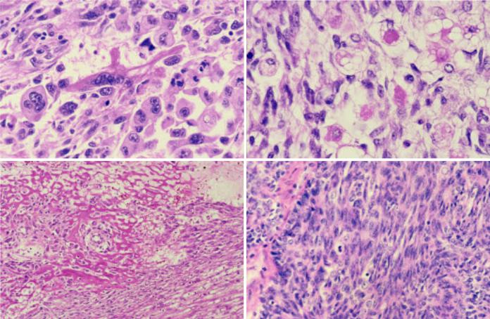
tive necrosis. It shows intimate intermingling of carcinomatous and sarcomatoid components, but the carcinomatous component can be subtle or demonstrable only by immunohistochemistry or electron microscopy in some cases. The carcinomatous component usually comprises cohesive clusters and sheets of poorly differentiated epithelial cells with significant nuclear pleomorphism, and some cases may show obvious squamous differentiation. The sarcomatoid component frequently comprises fascicles and storiform arrays of discohesive spindle tumour cells with pleomorphic nuclei, coarse chromatin, distinct nucle-
A
oli and frequent mitotic figures. Heterologous elements may be observed, most commonly rhabdomyosarcomatous and occasionally osteosarcomatous; the term ‘carcinosarcoma’ is sometimes applied for such cases {534,1478,1509,1841,1897}. In the rhabdomyosarcomatous areas, spindle cells with cross striations and large cells with abundant eosinophilic fibrillary cytoplasm are found. In the osteosarcomatous component, osteoid production by tumour cells is seen.
Immunohistochemically, the carcinomatous component expresses epithelial markers such as cytokeratin and epithe-
Grossly, the tumour is unencapsulated, often with infiltrative borders. The cut surfaces show whitish or greyish fleshy tumour with variable extent of necrosis and haemorrhage. Microcysts may be present.
Tumour spread and staging
The tumour is locally invasive, with frequent invasion of the adjacent pleura, lung and pericardium, and encroachment on the major blood vessels in the mediastinum. Metastases to mediastinal lymph nodes and parenchymal organs (especially the lungs) are common.
Histopathology
Sarcomatoid carcinoma is an infiltrative tumour often with large areas of coagula-
B
Fig. 3.48 Sarcomatoid carcinoma of the thymus. A Usually spindle cells predominate, and there are areas of geographic necrosis. B A biphasic pattern is obvious in this case. The carcinomatous component takes the form of a squamous cell carcinoma, and it gradually merges into a spindle cell (sarcomatoid) component.
Sarcomatoid carcinoma 179
A B
C D
Fig. 3.49 Sarcomatoid carcinoma of the thymus. A An elongated rhabdomyoblast with cross-striations is seen among polygonal carcinoma cells with pleomorphic nuclei. B Skeletal muscle differentiation characterized by rounded rhabdomyoblasts with vacuolated cytoplasm are interspersed among the spindle sarcomatoid cells. C Osteoid formation. D So-called spindle cell thymic carcinoma. The tumour comprises sheets and islands of compact atypical spindle cells.
lial membrane antigen. In the sarcomatoid areas, cytokeratin-positive tumour cells range from abundant to scanty or even absent {534,1093,1478,1509,1841, 1897,1916}. Variable expression of myoid markers (e.g. desmin, actin, myogenin, myoD1, myoglobin) is seen in the rhabdomyosarcomatous component {534,1478,1509,1841,1897}. The cases studied for CD5 have been negative for this marker {1093,1916}. Only rare tumours have been examined ultrastructurally, but desmosome-like junctions have been described in the spindle cell area of one case {534}.
Immunohistochemistry
In a tumour where only sarcomatoid component is identified despite extensive sampling, distinction from a sarcoma depends on the demonstration of epithelial differentiation in at least some tumour cells by immunohistochemistry
(e.g. cytokeratin, epithelial membrane antigen) or electron microscopy. Sarcomatoid carcinoma predominated by rhabdomyosarcomatous component may have been confused with mediastinal rhabdomyosarcoma in the literature. The latter sarcoma more commonly affects children and young adults. Although rhabdomyosarcoma can express cytokeratin, the positive tumour cells coexpress myoid markers, whereas at least some tumour cells in sarcomatoid carcinoma express cytokeratin only {534,1509}.
The entity reported as “spindle cell thymic carcinoma” comprises lobules and compact sheets of atypical spindle cells, and is probably an unusual form of sarcomatoid carcinoma. There is frequent transition with thymoma with spindle cell (type A) morphology. The spindle cells show epithelial characteristics and no evidence of true mesenchymal differ-
entiation on immunohistochemical evaluation {1509,1916}.
Differential diagnosis
Sarcomatoid carcinoma has to be distinguished from biphasic metaplastic thymoma, which differs in showing good circumscription of the tumour and blandlooking spindle cells, even though the interspersed squamoid epithelial islands may sometimes show nuclear pleomorphism {1919,2210}.Spindle cell carcinoid can be distinguished from sarcomatoid carcinoma by the presence of delicate fibrovascular septa, granular cytoplasm, generally less striking nuclear pleomorphism, and usually presence of a conventional carcinoid component in some foci; the diagnosis can be further confirmed by positive immunostaining for neuroendocrine markers.
The biphasic pattern of sarcomatoid carcinoma may raise the differential diag-
180 Tumours of the thymus - Thymic carcinomas
A B
C D
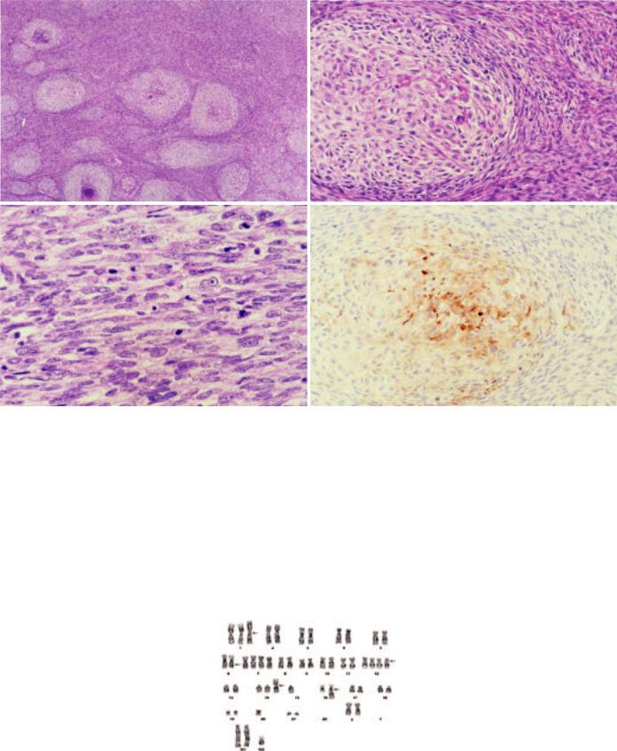
Fig. 3.50 Sarcomatoid carcinoma of the thymus. A In this example, pale-staining nodules are disposed among spindle cells. B The pale-staining nodules represent areas with subtle epithelial differentiation. This field shows some resemblance to metaplastic thymoma. C The sarcomatoid component comprises closely packed spindle cells with moderate nuclear atypia and frequent mitotic figures. D Immunostaining for EMA highlights nodular structures.
noses of synovial sarcoma and mesothelioma. Synovial sarcoma differs in showing more monotonous and uniform spindle cells and glandular differentiation in the epithelial component. The diagnosis can be further confirmed by the identification of t(X;18)(p11.2;q11.2) or SYTSSX1 or SYT-SSX2 gene fusion. Mesothelioma differs in being pleural or pericardial-based, showing papillaryglandular formation in the epithelial component, expressing mesothelial-associat- ed markers (e.g. calretinin), and showing mesothelial differentiation ultrastructurally (e.g. bushy microvilli).
Precursor lesions
Some cases show an identifiable component of thymoma, most commonly with spindle cell (type A) morphology, suggesting transformation from an underlying thymoma {1093,1897,1916}.
Histogenesis
The sarcomatoid component may arise from metaplasia of the carcinomatous component, wherein the tumour cells often gradually lose epithelial characteristics and simultaneously acquire mesenchymal or mesenchymal-like features. Alternatively, the tumour is derived from primitive cells with multidirectional differentiation.
Fig. 3.51 G-banded metaphase spread shows a complex karyotype, including der(16)t(1;16)(q12;q12.1).
Somatic genetics
Only one case has been studied by cytogenetics, with identification of a complex chromosomal abnormality including der(16)t(1;16)(q12;q12.1) {534}. Interestingly, this chromosomal translocation has also been previously reported in a case of thymic squamous cell carcinoma {1847}, suggesting a pathogenetic relationship with thymic squamous cell carcinoma in at least some cases.
Prognosis and predictive factors
Sarcomatoid carcinoma is an aggressive tumour, with most patients dying of disease within three years of diagnosis despite aggressive multi-modality therapy.
Sarcomatoid carcinoma 181
Clear cell carcinoma
M.R. Wick
A.Zettl
T.T.Kuo J.K.C. Chan
A.Marx
Definition
Clear cell carcinoma is a thymic carcinoma predominantly or exclusively composed of cells with optically clear cytoplasm. Thymomas with clear cell features are not included in this group.
ICD-O code |
8310/3 |
Synonym
Carcinoma of the thymus with clear-cell features {797}
Epidemiology
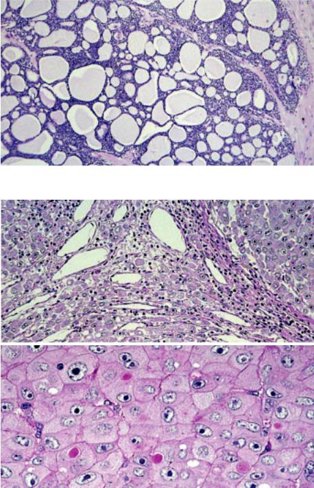
This is a very rare variant of thymic carcinoma, with only 13 “pure” cases reported to date {797,1094,1877,1924,2032, 2166}. Clear cell carcinomas constitute only 3% of all thymic carcinomas {1924}. Clear cell carcinoma has also been reported as a high-grade component in a combined thymoma/thymic carcinoma that, in addition, showed areas of spindle cell (WHO Type A) thymoma, squamous cell carcinoma and undifferentiated carcinoma {1093}. The age range of the reported cases is 33 to 84 years, and the tumour tends to prevail in men (male : female ratio 1.6) {797, 1663}.
Clinical features
Patients may show symptoms related to a mediastinal mass, e.g. chest pain or dyspnoea. Some patients are asymptomatic, with the tumour being detected by routine X-ray or during unrelated thoracotomy. There are no associated paraneoplastic autoimmune phenomena gravis.
Macroscopy
Macroscopically, the reported tumour size ranges between 4 and 12 cm (average 9 cm). The tumours may appear encapsulated and non-infiltrative, or may extensively infiltrate the surrounding tissues. The cut-surface shows solid or cystic tumour with or without haemorrhage and focal necrosis.
Histopathology
Microscopically, clear cell carcinomas of the thymus often show rather bland cel-
lular features which contrast their clinical aggressiveness. Tumour cells are rather monotonous and polyhedral, and usually display slight cellular pleomorphism with round to oval, vesicular nuclei, moderate nuclear atypia, finely dispersed chromatin, and small discernible nucleoli. They have abundant lucent, mostly clear to granular, sometimes faintly eosinophilic, cytoplasm which mostly, but not always is due to accumulation of glycogen. Clear cell carcinomas commonly show a lobulated architecture with nests, lobules or sheets of tumour cells being surrounded by a dense fibrous stroma, and lack the sinusoidal vasculature characteristic of metastatic clear cell carcinoma of the kidney. Rarely, few scattered intratumoral lymphocytes, minute foci of squamous differentiation or focal necrosis are observed. The tumour commonly exhibits an infiltrative growth, with tumour extending into the surrounding mediastinal fat and remnant thymus, even in cases which macroscopically appear well-delineated.
Special studies
Tumour cells usually show strong cytoplasmic diastase-labile PAS positivity, but PAS negative cases have also been reported {1877}. Clear cell carcinomas are keratin positive (cytokeratin 7 expression may be absent), EMA is expressed in 20% of cases studied {797}. As in other types of thymic carcinomas, a subgroup of clear cell carcinomas may
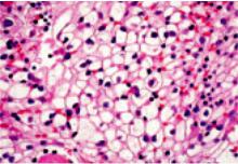
Fig. 3.52 Clear cell carcinoma. Tumour cells have abundant, optically clear cytoplasm and well defined cell membranes {1690}.
express CD5 {511,1093}. They are negative for PLAP, vimentin, CEA and S-100 {797}, and do not contain a population of immature (CD1aor CD99-positive) T-lymphocytes.
Differential diagnosis
When making the diagnosis of thymic clear cell carcinoma, metastatic clear cell epithelial malignancies, particularly renal, pulmonary and thyroid clear cell carcinoma have to be excluded. Other differential diagnoses include mediastinal diffuse large B-cell lymphoma, mediastinal seminoma, mediastinal parathyroid neoplasms, metastatic clear cell sarcoma or melanoma, glycogen-rich alveolar rhabdomyosarcoma, and clear cell paraganglioma. Furthermore, thymoma with clear cell features must be differentiated from clear cell carcinoma and from combined thymoma/thymic clear cell carcinoma {1093}. Clear cell features are common only in WHO Type B3 thymomas and they are almost always focal {797,2032}. Most tumours show a predominance of conventional B3 areas that exhibit gradual transitions to foci of bland-looking clear cells. While the conventional B3 areas harbour at least few CD1a+ and CD99+ immature T-cells, they may be absent in the clear cell areas. Significant PAS-positivity, necrosis, increased proliferative activity, desmoplastic stroma or TP53 overerexpression are typically absent in clear cell foci of WHO B3 thymomas. By contrast, the clear cell carcinoma (with squamoid features) arising in a WHO Type A thymoma (combined thymoma/thymic carcinoma) was PAS+, showed extensive necrosis, cyst formation and a desmoplastic stromal reaction {1093}.
Prognosis
Clear cell carcinomas are highly malignant, aggressive mediastinal neoplasms with frequent local recurrences and metastases. Most reported patients died of the disease. Deaths are related to metastatic disease or local infiltration of organs in recurrence {797}.
182 Tumours of the thymus - Thymic carcinomas
Papillary adenocarcinoma
Y. Matsuno
J. Rosai
Y. Shimosato
Definition
Papillary adenocarcinoma is a rare type of primary thymic carcinoma, characterized by a prominent papillary pattern of growth. Although reports of this tumour are rare, it may be the source of some metastatic papillary carcinomas with psammoma bodies in the cervical lymph nodes of patients without tumours in the thyroid gland.
ICD-O code |
8260/3 |
Synonym
Papillary carcinoma
Epidemiology
Papillary adenocarcinoma of the thymus is a rare neoplasm, and only five cases have been reported {1263}. It affects elderly individuals in their sixth to seventh decades of life. Males and females appear to be equally affected.
but prominent nucleoli. Psammoma bodies may be present. Areas of coagulation necrosis, sometimes massive, are scattered throughout the tumour. Invasion into the adhesive extrathymic tissues accompanied by a dense collagenous stroma may be seen. A small number of tumour cells show positive staining for mucin. The mitotic count may vary from 1 to 7/10 HPF among cases. Permeation of tumour cells into lymphatics such as the subpleural or intrapulmonary perivascular lymphatics may be extensive. In the majority of cases, type A thymoma is found as a component within the tumour mass; one case showed high-grade histology and a predominantly solid and sheet-like growth accompanied by welldeveloped papillary structures, highgrade atypia and high mitotic rate. In contrast to the other four cases, there was no evidence of a type A thymoma component.
phocytes are absent, but may be found in the coexisting thymoma portion.
Differential diagnosis
Differential diagnosis of this rare type of thymic carcinoma includes mediastinal thyroid neoplasm (i.e. papillary carcinoma), malignant mesothelioma, germ cell tumour, metastatic adenocarcinoma, and adenocarcinoma of foregut cyst origin {1915}.
Histogenesis
It has been suggested that papillary adenocarcinoma originates from type A thymoma as an expression of malignant transformation {1263}. This is based not only on the morphological similarities between the tubuloglandular or papillotubular structures sometimes seen in type A thymomas and those of the carcinoma, but also the occasional coexistence of a type A thymoma component within the tumour.
Clinical features
Papillary adenocarcinoma generally presents as an enlarging anterior mediastinal mass. The tumour may appear cystic. Paraneoplastic symptoms such as myasthenia gravis or pure red cell aplasia have not been described.
Macroscopy
The tumours are more or less encapsulated, and usually large (measuring 5-10 cm). The cut surface is irregularly lobulated, white and firm. Prominent cyst formation containing serohaemorrhagic fluid may be seen. Adhesion or direct invasion to the adjacent lung, pleura or pericardium is observed in most cases. Pleural implants may be found.
Histopathology
The tumour shows a tubulopapillary proliferation of uniform cuboidal to columnar cells, mainly lying in a monolayer, but occasionally showing a glomeruloid arrangement. The tumour cells have eosinophilic or clear cytoplasm. Their nuclei are round to ovoid, with coarsely condensed chromatin, and a few small
Immunophenotype
Papillary adenocarcinoma shows variable degrees of staining for LeuM1 and BerEP4. CEA and CD5 may also be positive, but CD20, thyroglobulin, pulmonary surfactant apoprotein and calretinin are negative. In addition, CD99-positive lym-
Prognosis and predictive factors
Since the number of reported cases is limited, specific information on the histopathologic prognostic factors of papillary carcinoma of the thymus is not available.
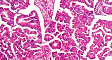
Fig. 3.53 Papillary adenocarcinoma of the thymus. Highly papillary configuration resembling papillary carcinoma of the thyroid {1690}.
Papillary adenocarcinoma 183
Non-papillary adenocarcinomas
H.K. Müller-Hermelink
A. Marx
T.T. Kuo
M. Kurrer
G. Chen
Y. Shimosato
There have been rare reports about nonpapillary adenocarcinomas in the thymus. Among them are: an adenocarcinoma with glandular differentiation arising in a thymic cyst {98}, as is also typical for papillary carcinoma {541}; an adenoid cystic carcinoma equivalent to the analogous salivary gland carcinoma {1841}; and a mucinous (colloid) carcinoma of the thymus {360}. The latter case arose in a 15 year old boy and was CD5-negative by immunohistochemistry.
An exceptional tumour in the thymus exhibiting features of a hepatoid carcinoma was observed in a 78 year-old female without an extrathymic neoplasm. The tumour had a diameter of 10 cm, was not encapsulated and virtually devoid of fibrous or inflammatory stroma. Respiratory distress was the only clinical symptom. The tumour recurred locally two years after surgery and responded to radiotherapy. Considering the female sex and protracted clinical course, lack of a yolk sac component and absence of alpha-fetoprotein in the tumour and the patient´s serum, a diagnosis of hepatoid carcinoma of thymus appears more likely than a “monophasic” variant of hepatoid yolk sac tumour of the mediastinum {606,1349,1355}.
Fig. 3.54 Thymic adenoid cystic carcinoma. This salivary gland-type thymic carcinoma shows a glandular and cribriform pattern.
A
B
Fig. 3.55 Thymic hepatoid carcinoma. A Tumour nodules composed of large polygonal tumour cells resembling activated hepatocytes. No hepatic sinuses, no portal structures and absence of a tumour stroma. B Large polygonal cells with abundant eosinophilic cytoplasm. PAS+ globules (immunorecative for alpha- 1-antitrypsin, not shown) occurred inside the cytoplasm and in between epithelial cells.
184 Tumours of the thymus - Thymic carcinomas
Carcinoma with t(15;19) translocation
Definition
Carcinoma with translocation t(15;19)(q13:p13.1) is a rare, aggressive and lethal carcinoma of unknown histogenesis arising in the mediastinum and other midline organs of young people.
Synonyms
Aggressive t(15;19)-positive carcinoma, midline lethal carcinoma
Epidemiology
Six cases of t(15;19)-positive carcinoma have been reported {439,616,985,
1081,1148,2072}. All occurred in chil- |
|
|
dren or young adults (age range: 5-34 |
A |
|
years), particularly females (F : M ratio = |
||
|
||
5:1). |
|
|
Etiology |
|
|
The etiology of t(15;19)-positive carcino- |
|
|
ma is unknown. Epstein-Barr virus does |
|
|
not play a role {985,2072} . |
|
|
Localization |
|
|
Translocation t(15;19)-positive carcino- |
|
|
ma has been reported to arise in supra- |
|
|
diaphragmatic midline organs. Three of 6 |
|
|
cases arose adjacent to the thymus in |
|
|
the mediastinum {985,1081,1148}. Other |
|
|
primary locations were epiglottis {2072}, |
|
|
sinonasal region {616}, lung {439}, and |
|
|
bladder (unpublished findings (C.A.F., |
B |
|
J.A.F.)). |
||
|
A. Marx
C.A. French
J.A. Fletcher
Clinical features
Aggressive local invasiveness is characteristic. Intracranial extension occurred in a sinonasal case. Pleural effusions and superior vena cava syndrome are common in thoracic cases. Metastases are common and may involve lymph nodes, lung, bone, skin and subcutaneous soft tissue {2027}.
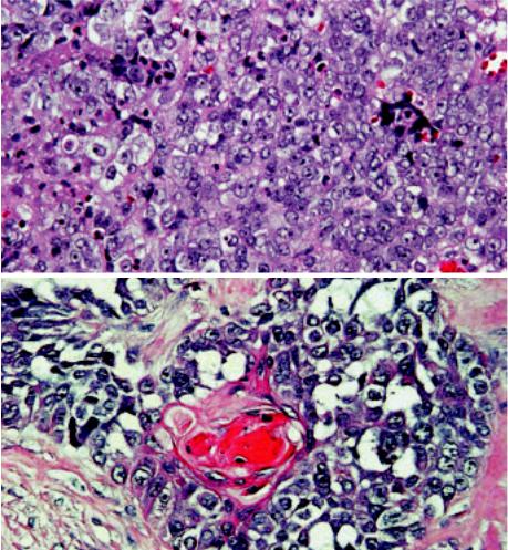
Histopathology
The presence of undifferentiated, intermediate sized, vigorously mitotic cells is characteristic. Commonly seen are sheets of undifferentiated cells forming syncytia with inter-epithelial lymphocytes, a pattern indistinguishable from
Fig. 3.56 Aggressive carcinoma with t(15;19) translocation. A The typical undifferentiated morphology of the tumour may mimic lymphoepithelioma or large cell lymphoma. B Focal squamous differentiation can be difficult to find, but is often present either histologically or ultrastructurally.
lymphoepithelioma {616}. Focal squamous differentiation is common {616, 1081,1148}, but not always seen, whereas glandular differentiation (mucoepidermoid carcinoma) {1148}) has only been reported once. Electron microscopy revealed squamous differentiation (rare desmosomes {1081,1148,2072}, tonofilaments {1148,2072}) in three cases. Care must be taken not to confuse the discohesive, undifferentiated round cells of t(15;19)-positive carcinoma with large cell lymphoma or germ cell tumour {616, 985,1081}.
Immunophenotype
The tumours consistently react, at least focally, with pan-cytokeratin markers {616,2072}. Inconsistent and usually focal positivity occurs for vimentin, EMA, and carcino-embryonic antigen (CEA) {985,2072}. CD30, CD45, PLAP, HMB45, S100, and neuroendocrine markers are negative.
Differential diagnosis
This lesion must be distinguished from large cell lymphoma, germ cell tumour, and t(15;19)-negative carcinomas, par-
Carcinoma with t(15;19) translocation 185
ticularly lymphoepithelioma-like, poorly differentiated squamous cell, mucoepidermoid, and undifferentiated carcinoma.
Histogenesis
Despite various considerations {616,985, 1081,1148,2072}, derivation of this tumour is unknown.
Somatic genetics
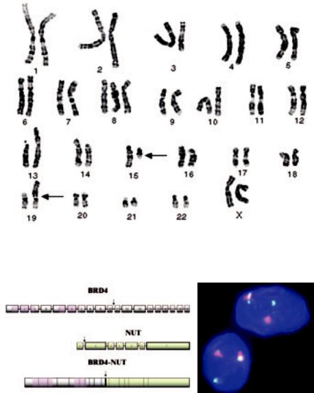
The specific t(15;19)(q13;p13.1) translocation, which generates the 6.4-kb BRD4-NUT fusion oncogenes, is often the only demonstrable cytogenetic aberration. The translocation fuses the 5´ 10 exons of the ubiquitously expressed BRD4 bromodomain gene on chromosome 19 with nearly the entire transcript of the 15q13 gene NUT (nuclear protein in testis), that is normally exclusively expressed in testis {617}. Cytogenetics, fluorescence in situ hybridization (FISH), Southern blotting and RT-PCR studies can identify the translocation {616,617}. Additional chromosomal aberrations are rare {2027}.
Prognosis and predictive factors
All cases reported so far followed an extremely aggressive clinical course (average survival 18 weeks; range 8-38 weeks) {616}.
Fig. 3.57 Aggressive carcinoma with t(15;19) translocation. Karyotype from a teenage girl with the t(15;19) carcinoma. A reciprocal translocation involving chromosomes 15q13 and 19p13.1 has occurred. The der (19) contains the functional fusion oncogene.
A B
Fig. 3.58 Aggressive carcinoma with t(15;19) translocation. A Schematic of the BRD4 and NUT genes disrupted in the t(15;19)(q13;p13.1) chromosomal translocation. Exons are represented by horizontal bars, and introns by connecting lines. All characterized breakpoints (N=2), represented by vertical arrows, occur in intron 10 of BRD4 (gray), and intron 1 of NUT (green), splitting BRD4 roughly in half, and fusing to it nearly the entire NUT transcript. Both bromodomains (pink) of BRD4 are preserved in the fusion oncogene. The oncogenic mechanism is believed, at least in part, to result from unscheduled expression of NUT (normally expressed only in testis) driven by the promoter of BRD4, which is ubiquitously expressed. B Fluorescent in situ hybridization (FISH) depicting the t(15;19)(q13;p13.1) in a paraffin section. The red-green probe doublet, which normally flanks the NUT gene on chromosome 15, is split apart by the translocation.
186 Tumours of the thymus - Thymic carcinomas
Undifferentiated carcinoma of the thymus
Definition
A thymic carcinoma growing in a solid, undifferentiated fashion but without sarcomatoid (spindle cell, pleomorphic, metaplastic) features {1924}.
ICD-O code |
8020/3 |
The diagnosis of this rare type of thymic carcinoma is one of exclusion. Defining its epithelial nature usually requires immunohistochemistry. In children and young adults, carcinoma with t(15;19) translocation should be excluded by cytogenetics or RT-PCR {617,1081}. The
most important differential diagnosis in A adults is large cell carcinoma in the lung extending or metastasizing to the mediastinum.
Small cell carcinoma without (immunohistochemically) recognizable differentiation is traditionally classified among the neuroendocrine carcinomas of the thymus.
B
A. Marx
Ph. Ströbel
J.K.C. Chan
G. Chen
H.K. Muller-Hermelink
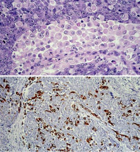
Fig. 3.59 Undifferentiated thymic carcinoma. A A solid growth pattern composed of large polygonal to round tumour cells, large nuclei and a slightly basophilic cytoplasm. No keratinization, no intercellular bridges, no glandular differentiation, no sarcomatoid features and no EBER expression by in situ hybridization. The prominent population of bland looking myoid cells with round inconspicuous nuclei and eosinophilic cytoplasm unequivocally identifies this carcinoma as a tumour of thymic differentiation. Myoid cells show no significant proliferative activity (in contrast to rhabdomyosarcoma cells). B Desmin staining demonstrates intratumorous myoid cells.
Undifferentiated carcinoma 187
Thymic neuroendocrine tumours
A. Marx
Y. Shimosato
T.T. Kuo
J.K.C. Chan
W.D. Travis
M.R. Wick
Definitions
Thymic epithelial tumours that are predominantly or exclusively composed of neuroendocrine cells are classified as neuroendocrine carcinomas (NECs) of the thymus {1691}. They have to be distinguished 1) from otherwise typical thymic carcinomas, which may contain scattered or groups of neuroendocrine cells {853,1091,1139}, and 2) from nonepithelial neurogenic tumours, particularly paragangliomas.
Neuroendocrine differentiation can be demonstrated by immunohistochemistry (positivity for chromogranin, synaptophysin, neuron-specific enolase, CD56) and/ or by ultrastructural identification of neurosecretory granules.
Neoplasms combining features of NEC and either thymoma or thymic carcinoma are included in the category of “combined thymic epithelial tumours”.
Since the seminal work of Rosai and Higa, thymic neuroendocrine tumours that are “related to carcinoid tumours” have been distinguished from thymomas {1686,1688,2144}. The epithelial neuroendocrine tumours of the thymus comprise typical and atypical carcinoids, as well as large and small cell carcinomas.
Well differentiated neuroendocrine carcinomas
In line with the nomenclature of neuroendocrine tumours occurring in other sites of the body, it is proposed that thymic carcinoids be termed well differentiated neuroendocrine carcinomas of the thymus {349,1691}. The rationale for considering all these tumours as carcinomas {1691} is the observation that even “innocent” looking and encapsulated carcinoids bear a significant risk for recurrence, metastasis and tumour-associat- ed death {628,1845,2140}. The carcinoids are further subdivided into typical and atypical carcinoids.
ICD-O codes |
|
Typical carcinoid |
8240/3 |
Atypical carcinoid |
8249/3 |
Typical and atypical carcinoids
Following the introduction of the term “atypical carcinoid” by Arrigoni et al. in 1972 for a subgroup of moderately aggressive neuroendocrine neoplasms of the lung {75}, it became clear that the vast majority of carcinoids in the thymus correspond to atypical carcinoids when the same criteria are applied as in the
lung {450,628,723,1361,1688,1808, 2062,2140}. As a group, atypical carcinoids more often show a diffuse growth pattern, advanced stage disease, and a higher degree of cytologic atypia {723, 1362,1691,1808,2062}.
Since virtually all thymic carcinoids are atypical carcinoids (see epidemiology), most studies report thymic carcinoids to have a worse prognosis compared with bronchial carcinoids {628,1361,1808, 2136}. However, varying criteria have been used for definition of atypical carcinoids of the thymus in these series {628, 1361,1845,2062}. In fact, the only clinicopathological study applying WHOdefined criteria to classify atypical thymic carcinoids {723} challenged the view that thymic atypical carcinoids are clinically more aggressive than morphologically identical carcinoids of the lung. A better prognosis of atypical thymic carcinoids as compared to pulmonary carcinoids was even suggested by a recent study {1049} (5-year and 10-year survival rates of 84% and 75% respectively, compared with 87% and 87% for pulmonary typical carcinoids and 56% and 35% for pulmonary atypical carcinoids {2028}).
A B
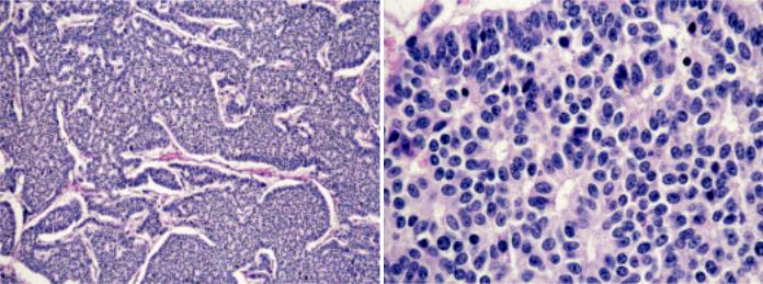
Fig. 3.60 Typical carcinoid. A Solid and trabecular growth pattern. Note absence of necrosis and mitoses. B On high magnification, rosettes, and bland cytology can be seen, but no mitoses.
188 Tumours of the thymus - Neuroendocrine tumours