

12.7 Secondary and Tertiary Structure of rna
RNA molecules (see Chapter 11) are typically single-stranded. Nevertheless, they are often rich in double-stranded regions that form when complementary sequences within the chain come together and join via intrastrand hydrogen bonding. RNA strands cannot fold to form B-DNA type double helices because their 2'-OH groups are a steric hindrance to this conformation. Instead, RNA double helices adopt a conformation similar to the A-form of DNA, having about 11 bp per turn, and the bases strongly tilted from the plane perpendicular to the helix axis (see Figure 12.13). Both tRNA and rRNA have characteristic secondary structures formed in this manner. Secondary structures are presumed to exist in mRNA species as well, although their nature is as yet little understood. (The functions of tRNA, rRNA, and mRNA are discussed in detail in Part IV: Information Transfer.)
Transfer RNA
In tRNA molecules, which contain from 73 to 94 nucleotides in a single chain, a majority of the bases are hydrogen-bonded to one another. Figure 12.34 shows the structure that typifies tRNAs. Hairpin turns bring complementary stretches of bases in the chain into contact so that double-helical regions form. Because of the arrangement of the complementary stretches along the chain, the overall pattern of H-bonding can be represented as a cloverleaf. Each cloverleaf consists of four H-bonded segments — three loops and the stem where the 3'- and 5'-ends of the molecule meet. These four segments are designated the acceptor stem, the D loop, the anticodon loop, and the T y C loop .
F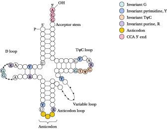 igure
12.34
•
A general diagram for the structure of tRNA. The positions of
invariant bases as well as bases that seldom vary are shown in color.
The numbering system is based on yeast tRNA Phe
.
R = 5 purine; Y = 5 pyrimidine. Dotted lines denote sites in the D
loop and variable loop regions where varying numbers of nucleotides
are found in different tRNAs.
igure
12.34
•
A general diagram for the structure of tRNA. The positions of
invariant bases as well as bases that seldom vary are shown in color.
The numbering system is based on yeast tRNA Phe
.
R = 5 purine; Y = 5 pyrimidine. Dotted lines denote sites in the D
loop and variable loop regions where varying numbers of nucleotides
are found in different tRNAs.
tRNA Secondary Structure
The acceptor stem is where the amino acid is linked to form the aminoacyl-tRNA derivative, which serves as the amino acid–donating species in protein synthesis; this is the physiological role of tRNA. The amino acid adds to the 3'-OH of the 3'-terminal A nucleotide (Figure 12.35). The 3'-end of tRNA is invariantly CCA-3'-OH. This CCA sequence plus a fourth nucleotide extends beyond the double-helical portion of the acceptor stem. The D loop is so named because this tRNA loop often contains dihydrouridine, or D, residues. In addition to dihydrouridine, tRNAs characteristically contain a number of unusual bases, including inosine, thiouridine, pseudouridine, and hypermethylated purines (see Figure 11.26). The anticodon loop consists of a double-helical segment and seven unpaired bases, three of which are the anticodon. (The anticodon is the three-nucleotide unit that recognizes and base pairs with a particular mRNA codon, a complementary three-base unit in mRNA which is the genetic information that specifies an amino acid.) Reading 3' 5', the anticodon is invariably preceded by a purine (often an alkylated one) and followed by a U.
F igure
12.35
•
Amino
acids are linked to the 3'-OH end of tRNA molecules by an ester bond
formed between the carboxyl group of the amino acid and the 3'-OH of
the terminal ribose of the tRNA.
igure
12.35
•
Amino
acids are linked to the 3'-OH end of tRNA molecules by an ester bond
formed between the carboxyl group of the amino acid and the 3'-OH of
the terminal ribose of the tRNA.
Anticodon base pairing to the codon on mRNA allows a particular tRNA species to deliver its amino acid to the protein-synthesizing apparatus. It represents the key event in translating the information in the nucleic acid sequence so that the appropriate amino acid is inserted at the right place in the amino acid sequence of the protein being synthesized. Next along the tRNA sequence in the 5' 3' direction comes a loop that varies from tRNA to tRNA in the number of residues that it has, the so-called extra or variable loop. The last loop in the tRNA, reading 5' 3', is the TC loop, which contains seven unpaired bases including the sequence , where is the symbol for pseudouridine. Ribosomes bind tRNAs through recognition of this loop. Almost all of the invariant residues common to tRNAs lie within the non-hydrogen-bonded regions of the cloverleaf structure (Figure 12.34). Figure 12.36 depicts the complete nucleotide sequence and cloverleaf structure of yeast alanine tRNA.
F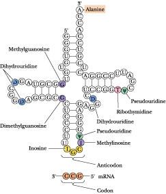 igure
12.36
•
The
complete nucleotide sequence and cloverleaf structure of yeast
alanine tRNA.
igure
12.36
•
The
complete nucleotide sequence and cloverleaf structure of yeast
alanine tRNA.
tRNA Tertiary Structure
Tertiary structure in tRNA arises from hydrogen-bonding interactions between bases in the D loop with bases in the variable and loops, as shown for yeast phenylalanine tRNA in Figure 12.37.
F igure
12.37
•
Tertiary
interactions in yeast phenylalanine tRNA. The molecule is presented
in the conventional cloverleaf secondary structure generated by
intrastrand hydrogen bonding. Solid lines connect bases that are
hydrogen-bonded when this cloverleaf pattern is folded into the
characteristic tRNA tertiary structure (see also Figure
12.36).
igure
12.37
•
Tertiary
interactions in yeast phenylalanine tRNA. The molecule is presented
in the conventional cloverleaf secondary structure generated by
intrastrand hydrogen bonding. Solid lines connect bases that are
hydrogen-bonded when this cloverleaf pattern is folded into the
characteristic tRNA tertiary structure (see also Figure
12.36).
Note that these H bonds involve the invariant nucleotides of tRNAs, thus emphasizing the importance of the tertiary structure they create to the function of tRNAs in general. These H bonds fold the D and arms together and bend the cloverleaf into the stable L-shaped tertiary form (Figure 12.38). Many of these H bonds involve base pairs that are not canonical A : T or G : C pairings (Figure 12.38). The amino acid acceptor stem is at one end of the L, separated by 7 nm or so from the anticodon at the opposite end of the L. The D and loops form the corner of the L. In the L-conformation, the bases are oriented to maximize hydrophobic stacking interactions between their flat faces. Such stacking is a second major factor contributing to L-form stabilization.
F igure
12.38•
(a) The three-dimensional structure of yeast phenylalanine tRNA
as deduced from X-ray diffraction studies of its crystals. The
tertiary folding is illustrated in the center of the diagram with the
ribose–phosphate backbone presented as a continuous ribbon; H bonds
are indicated by crossbars. Unpaired bases are shown as short,
unconnected rods. The anticodon loop is at the bottom and the -CCA
3'-OH acceptor end is at the top right. The various types of
noncanonical hydrogen-bonding interactions observed between bases
surround the central molecule. Three of these structures show
examples of unusual H-bonded interactions involving three bases;
these interactions aid in establishing tRNA tertiary structure. (b) A
space-filling model of the molecule. (After
Kim, S. H., in Schimmel, P., Söll, D., and Abelson, J. N., eds.,
1979 .
Transfer RNA: Structure, Properties, and Recognition.
New York : Cold Spring Harbor Laboratory.)
igure
12.38•
(a) The three-dimensional structure of yeast phenylalanine tRNA
as deduced from X-ray diffraction studies of its crystals. The
tertiary folding is illustrated in the center of the diagram with the
ribose–phosphate backbone presented as a continuous ribbon; H bonds
are indicated by crossbars. Unpaired bases are shown as short,
unconnected rods. The anticodon loop is at the bottom and the -CCA
3'-OH acceptor end is at the top right. The various types of
noncanonical hydrogen-bonding interactions observed between bases
surround the central molecule. Three of these structures show
examples of unusual H-bonded interactions involving three bases;
these interactions aid in establishing tRNA tertiary structure. (b) A
space-filling model of the molecule. (After
Kim, S. H., in Schimmel, P., Söll, D., and Abelson, J. N., eds.,
1979 .
Transfer RNA: Structure, Properties, and Recognition.
New York : Cold Spring Harbor Laboratory.)
Ribosomal RNA
rRNA Secondary Structure
Ribosomes, the protein-synthesizing machinery of cells, are composed of two subunits, called small and large, and ribosomal RNAs are integral components of these subunits (see Table 11.2). A large degree of intrastrand sequence complementarity is found in all rRNA strands, and all assume a highly folded pattern that allows base pairing between these complementary segments. Figure 12.39 shows the secondary structure assigned to the E. coli 16S rRNA. This structure is based on alignment of the nucleotide sequence into H-bonding segments. The reliability of these alignments is then tested through a comparative analysis of whether identical secondary structures can be predicted from nucleotide sequences of 16S-like rRNAs from other species. If so, then such structures are apparently conserved. The approach is based on the thesis that, because ribosomal RNA species (regardless of source) serve common roles in protein synthesis, it may be anticipated that they share structural features. The structure is marvelously rich in short, helical segments separated and punctuated by single-stranded loops.
F igure
12.39
•
The proposed secondary structure for E.
coli
16S rRNA, based on comparative sequence analysis in which the folding
pattern is assumed to be conserved across different species. The
molecule can be subdivided into four domains—I,
II, III,
and IV—on
the basis of contiguous stretches of the chain that are closed by
long-range base-pairing interactions. I,
the 5'-domain, includes nucleotides 27 through 556. II,
the central domain, runs from nucleotide 564 to 912. Two domains
comprise the 3'-end of the molecule. III,
the major one, comprises nucleotides 923 to 1391. IV,
the 3'-terminal domain, covers residues 1392 to 1541.
igure
12.39
•
The proposed secondary structure for E.
coli
16S rRNA, based on comparative sequence analysis in which the folding
pattern is assumed to be conserved across different species. The
molecule can be subdivided into four domains—I,
II, III,
and IV—on
the basis of contiguous stretches of the chain that are closed by
long-range base-pairing interactions. I,
the 5'-domain, includes nucleotides 27 through 556. II,
the central domain, runs from nucleotide 564 to 912. Two domains
comprise the 3'-end of the molecule. III,
the major one, comprises nucleotides 923 to 1391. IV,
the 3'-terminal domain, covers residues 1392 to 1541.
Comparison of rRNAs from Various Species
If a phylogenetic comparison is made of the 16S-like rRNAs from an archaebacterium (Halobacterium volcanii), a eubacterium (E. coli), and a eukaryote (the yeast Saccharomyces cerevisiae), a striking similarity in secondary structure emerges (Figure 12.40). Remarkably, these secondary structures are similar despite the fact that the nucleotide sequences of these rRNAs themselves exhibit a low degree of similarity. Apparently, evolution is acting at the level of rRNA secondary structure, not rRNA nucleotide sequence. Similar conserved folding patterns are seen for the 23S-like and 5S-like rRNAs that reside in the large ribosomal subunits of various species. An insightful conclusion may be drawn regarding the persistence of such strong secondary structure conservation despite the millennia that have passed since these organisms diverged: all ribosomes are constructed to a common design and all function in a similar manner.

Figure 12.40 • Phylogenetic comparison of secondary structures of 16S-like rRNAs from (a) a eubacterium (E. coli), (b) an archaebacterium (H. volcanii), (c) a eukaryote (S. cerevisiae, a yeast).
rRNA Tertiary Structure
Despite the unity in secondary structural patterns, little is known about the three-dimensional, or tertiary, structure of rRNAs. Even less is known about the quaternary interactions that occur when ribosomal proteins combine with rRNAs and when the ensuing ribonucleoprotein complexes, the small and large subunits, come together to form the complete ribosome. Furthermore, assignments of functional roles to rRNA molecules are still tentative and approximate. (We return to these topics in Chapter 33.)
Appendix to Chapter 12