

12.2 The abZs of dna Secondary Structure
D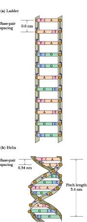 ouble-stranded
DNA molecules assume one of three secondary structures, termed A, B,
and Z. Fundamentally, double-stranded DNA is a regular two-chain
structure with hydrogen bonds formed between opposing bases on the
two chains (seeChapter
11).
Such H-bonding is possible only when the two chains are antiparallel.
The polar sugar–phosphate backbones of the two chains are on the
outside. The bases are stacked on the inside of the structure; these
heterocyclic bases, as a consequence of their π-electron clouds, are
hydrophobic on their flat sides. One purely hypothetical
conformational possibility for a two-stranded arrangement would be a
ladderlike structure (Figure 12.9) in which the base pairs are fixed
at 0.6 nm apart because this is the distance between adjacent sugars
in the DNA backbone. Because H2O
molecules would be accessible to the spaces between the hydrophobic
surfaces of the bases, this conformation is energetically
unfavorable. This ladderlike structure converts to a helix when given
a simple right-handed twist. Helical twisting brings the base-pair
rungs of the ladder closer together, stacking them 0.34 nm apart,
without affecting the sugar–sugar distance of 0.6 nm. Because this
helix repeats itself approximately every 10 bp, its pitch
is 3.4 nm. This is the major conformation of DNA in solution and it
is called B-DNA.
ouble-stranded
DNA molecules assume one of three secondary structures, termed A, B,
and Z. Fundamentally, double-stranded DNA is a regular two-chain
structure with hydrogen bonds formed between opposing bases on the
two chains (seeChapter
11).
Such H-bonding is possible only when the two chains are antiparallel.
The polar sugar–phosphate backbones of the two chains are on the
outside. The bases are stacked on the inside of the structure; these
heterocyclic bases, as a consequence of their π-electron clouds, are
hydrophobic on their flat sides. One purely hypothetical
conformational possibility for a two-stranded arrangement would be a
ladderlike structure (Figure 12.9) in which the base pairs are fixed
at 0.6 nm apart because this is the distance between adjacent sugars
in the DNA backbone. Because H2O
molecules would be accessible to the spaces between the hydrophobic
surfaces of the bases, this conformation is energetically
unfavorable. This ladderlike structure converts to a helix when given
a simple right-handed twist. Helical twisting brings the base-pair
rungs of the ladder closer together, stacking them 0.34 nm apart,
without affecting the sugar–sugar distance of 0.6 nm. Because this
helix repeats itself approximately every 10 bp, its pitch
is 3.4 nm. This is the major conformation of DNA in solution and it
is called B-DNA.
Figure 12.9 • (a) Double-stranded DNA as an imaginary ladderlike structure. (b) A simple right-handed twist converts the ladder to a helix.
Structural Equivalence of Watson–Crick Base Pairs
As indicated in Chapter 11, the base pairing in DNA is very specific: the purine adenine pairs with the pyrimidine thymine; the purine guanine pairs with the pyrimidine cytosine. Further, the A:T pair and G:C pair have virtually identical dimensions (Figure 12.10). Watson and Crick realized that units of such similarity could serve as spatially invariant substructures to build a polymer whose exterior dimensions would be uniform along its length, regardless of the sequence of bases.
F igure
12.10
•
Watson–Crick
A:T and G:C base pairs. All H bonds in both base pairs are straight,
with each H atom pointing directly at its acceptor N or O atom.
Linear H bonds are the strongest. The mandatory binding of larger
purines with smaller pyrimi-dines leads to base pairs that have
virtually identical dimensions, allowing the two sugar–phosphate
backbones to adopt identical helical conformations.
igure
12.10
•
Watson–Crick
A:T and G:C base pairs. All H bonds in both base pairs are straight,
with each H atom pointing directly at its acceptor N or O atom.
Linear H bonds are the strongest. The mandatory binding of larger
purines with smaller pyrimi-dines leads to base pairs that have
virtually identical dimensions, allowing the two sugar–phosphate
backbones to adopt identical helical conformations.
The DNA Double Helix Is a Stable Structure
Several factors account for the stability of the double-helical structure of DNA. First, both internal and external hydrogen bonds stabilize the double helix. The two strands of DNA are held together by H-bonds that form between the complementary purines and pyrimidines, two in an A:T pair and three in a G:C pair (Figure 12.10), while polar atoms in the sugar-phosphate backbone form external H bonds with surrounding water molecules. Second, the negatively charged phosphate groups are all situated on the exterior surface of the helix in such a way that they have minimal effect on one another and are free to interact electrostatically with cations in solution such as Mg2+. Third, the core of the helix consists of the base pairs, which, in addition to being H-bonded, stack together through hydrophobic interactions and van der Waals forces that contribute significantly to the overall stabilizing energy.
F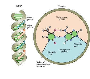 igure
12.11
•
The bases in a base pair are not directly across the helix axis from
one another along some diameter but rather are slightly displaced.
This displacement, and the relative orientation of the glycosidic
bonds linking the bases to the sugar–phosphate backbone, leads to
differently sized grooves in the cylindrical column created by the
double helix, the major groove and the minor groove, each coursing
along its length.
igure
12.11
•
The bases in a base pair are not directly across the helix axis from
one another along some diameter but rather are slightly displaced.
This displacement, and the relative orientation of the glycosidic
bonds linking the bases to the sugar–phosphate backbone, leads to
differently sized grooves in the cylindrical column created by the
double helix, the major groove and the minor groove, each coursing
along its length.
A stereochemical consequence of the way A:T and G:C base pairs form is that the sugars of the respective nucleotides have opposite orientations, and thus the sugar-phosphate backbones of the two chains run in opposite or “antiparallel” directions. Furthermore, the two glycosidic bonds holding the bases in each base pair are not directly across the helix from each other, defining a common diameter (Figure 12.11). Consequently, the sugar-phosphate backbones of the helix are not equally spaced along the helix axis, and the grooves between them are not the same size. Instead, the intertwined chains create a major groove and a minor groove (Figure 12.11). The edges of the base pairs have a specific relationship to these grooves. The “top” edges of the base pairs (“top” as defined by placing the glycosidic bond at the bottom, as in Figure 12.10) are exposed along the interior surface or “floor” of the major groove; the base-pair edges nearest to the glycosidic bond form the interior surface of the minor groove. Some proteins that bind to DNA can actually recognize specific nucleotide sequences by “reading” the pattern of H-bonding possibilities presented by the edges of the bases in these grooves. Such DNA -protein interactions provide one step toward understanding how cells regulate the expression of genetic information encoded in DNA (see Chapter 32).
Conformational Variation in Double-Helical Structures
In solution, DNA ordinarily assumes the structure we have been discussing: B-DNA. However, nucleic acids also occur naturally in other double-helical forms. The base-pairing arrangement remains the same, but the sugar -phosphate groupings that constitute the backbone are inherently flexible and can adopt different conformations. One conformational variation is propeller twist (Figure 12.12). Propeller twist allows greater overlap between successive bases along a strand of DNA and diminishes the area of contact between bases and solvent water.
F igure
12.12
•
Helical twist and propeller twist in DNA. (a) Successive base pairs
in B-DNA show a rotation with respect to each other (so-called
helical twist) of 36° or so, as viewed down the cylindrical axis of
the DNA. (b) Rotation in a different dimension—propellor
twist—allows
the hydrophobic surfaces of bases to overlap better. The view here is
edge-on to two successive bases in one DNA strand (as if the two
bases on the right-hand strand of DNA in (a) were viewed from the
right-hand margin of the page; dots represent end-on views down the
glycosidic bonds). Clockwise rotation (as shown here) has a positive
sign. (c) The two bases on the left-hand strand of DNA in (a) also
show positive propellor twist (a clockwise rotation of the two bases
in (a) as viewed from the left-hand margin of the paper). ( Adapted
from Figure 3.4 in Callandine, C. R., and Drew, H. R., 1992.
Understanding
DNA: The Molecule and How It Works.
London : Academic Press .)
igure
12.12
•
Helical twist and propeller twist in DNA. (a) Successive base pairs
in B-DNA show a rotation with respect to each other (so-called
helical twist) of 36° or so, as viewed down the cylindrical axis of
the DNA. (b) Rotation in a different dimension—propellor
twist—allows
the hydrophobic surfaces of bases to overlap better. The view here is
edge-on to two successive bases in one DNA strand (as if the two
bases on the right-hand strand of DNA in (a) were viewed from the
right-hand margin of the page; dots represent end-on views down the
glycosidic bonds). Clockwise rotation (as shown here) has a positive
sign. (c) The two bases on the left-hand strand of DNA in (a) also
show positive propellor twist (a clockwise rotation of the two bases
in (a) as viewed from the left-hand margin of the paper). ( Adapted
from Figure 3.4 in Callandine, C. R., and Drew, H. R., 1992.
Understanding
DNA: The Molecule and How It Works.
London : Academic Press .)
Alternative Form of Right-Handed DNA
An alternative form of the right-handed double helix is A-DNA. A-DNA molecules differ in a number of ways from B-DNA. The pitch, or distance required to complete one helical turn, is different. In B-DNA, it is 3.4 nm, whereas in A-DNA it is 2.46 nm. One turn in A-DNA requires 11 bp to complete. Depending on local sequence, 10 to 10.6 bp define one helical turn in B-form DNA. In A-DNA, the base pairs are no longer nearly perpendicular to the helix axis but instead are tilted 19° with respect to this axis. Successive base pairs occur every 0.23 nm along the axis, as opposed to 0.332 nm in B-DNA. The B-form of DNA is thus longer and thinner than the short, squat A-form, which has its base pairs displaced around, rather than centered on, the helix axis. Figure 12.13 shows the relevant structural characteristics of the A- and B-forms of DNA. (Z-DNA, another form of DNA to be discussed shortly, is also depicted in Figure 12.13.) A comparison of the structural properties of A-, B-, and Z-DNA is summarized in Table 12.1.


Figure 12.13 • (here and on the facing page) Comparison of the A-, B-, and Z-forms of the DNA double helix. The distance required to complete one helical turn is shorter in A-DNA than it is in B-DNA. The alternating pyrimidine–purine sequence of Z-DNA is the key to the “left-handedness” of this helix. (Robert Stodola, Fox Chase Cancer Research Center , and Irving Geis)
Although relatively dehydrated DNA fibers can be shown to adopt the A-conformation under physiological conditions, it is unclear whether DNA ever assumes this form in vivo. However, double-helical DNA:RNA hybrids probably have an A-like conformation. The 2'-OH in RNA sterically prevents double-helical regions of RNA chains from adopting the B-form helical arrangement. Importantly, double-stranded regions in RNA chains assume an A-like conformation, with their bases strongly tilted with respect to the helix axis.
|
Table 12.1 | |||
|
Comparison of the Structural Properties of A-, B-, and Z-DNA | |||
|
Double Helix Type |
A |
B |
Z |
|
Overall proportions |
Short and broad |
Longer and thinner |
Elongated and slim |
|
Rise per base pair |
2.3 Å |
3.32 Å ± 0.19 Å |
3.8 Å |
|
Helix packing diameter |
25.5 Å |
23.7 Å |
18.4 Å |
|
Helix rotation sense |
Right-handed |
Right-handed |
Left-handed |
|
Base pairs per helix repeat |
1 |
1 |
2 |
|
Base pairs per turn of helix |
~11 |
~10 |
12 |
|
Mean rotation per base pair |
33.6° |
35.9° ± 4.2° |
260°/2 |
|
Pitch per turn of helix |
24.6 Å |
33.2 Å |
45.6 Å |
|
Base-pair tilt from the perpendicular |
+19° |
-1.2° ± 4.1° |
-9° |
|
Base-pair mean propeller twist |
+18° |
116° ± 7° |
~0° |
|
Helix axis location |
Major groove |
Through base pairs |
Minor groove |
|
Major groove proportions |
Extremely narrow but very deep |
Wide and with intermediate depth |
Flattened out on helix surface |
|
Minor groove proportions |
Very broad but shallow |
Narrow and with intermediate depth |
Extremely narrow but verydeep |
|
Glycosyl bond conformation |
anti |
anti |
anti at C, syn at G |
|
Adapted from Dickerson, R. L., et al., 1982. Cold Spring Harbor Symposium on Quantitative Biology 47:14. | |||
Z-DNA: A Left-Handed Double Helix
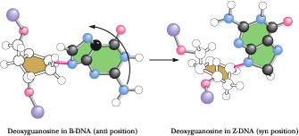
Z-DNA was first recognized by Alexander Rich and his colleagues at MIT in X-ray analysis of the synthetic deoxynucleotide dCpGpCpGpCpG, which crystallized into an antiparallel double helix of unexpected conformation. The alternating pyrimidine -purine (Py-Pu) sequence of this oligonucleotide is the key to its unusual properties. The N-glycosyl bonds of G residues in this alternating copolymer are rotated 180° with respect to their conformation in B-DNA, so that now the purine ring is in the syn rather than the anti conformation (Figure 12.14). The C residues remain in the anti form. Because the G ring is “flipped,” the C ring must also flip to maintain normal Watson–Crick base pairing. However, pyrimidine nucleosides do not readily adopt the syn conformation because it creates steric interference between the pyrimidine C-2 oxy substituent and atoms of the pentose.
 Figure
12.14 •
Comparison
of the deoxy-guanosine conformation in B- and Z-DNA. In B-DNA, the
Cl'–N-9 glycosyl bond is always in the anti position (left).
In contrast, in the left-handed Z-DNA structure, this bond rotates
(as shown) to adopt the syn conformation.
Figure
12.14 •
Comparison
of the deoxy-guanosine conformation in B- and Z-DNA. In B-DNA, the
Cl'–N-9 glycosyl bond is always in the anti position (left).
In contrast, in the left-handed Z-DNA structure, this bond rotates
(as shown) to adopt the syn conformation.
Because the cytosine ring does not rotate relative to the pentose, the whole C nucleoside (base and sugar) must flip 180° (Figure 12.15). It is topologically possible for the G to go syn and the C nucleoside to undergo rotation by 180° without breaking and re-forming the G:C hydrogen bonds. In other words, the B to Z structural transition can take place without disruption of the bonding relationships among the atoms involved.
F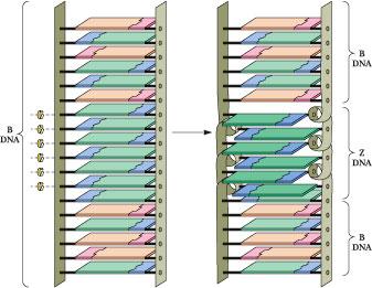 igure
12.15
•
The change in topological relationships of base pairs from B- to
Z-DNA. A six-base-pair segment of B-DNA is converted to Z-DNA through
rotation of the base pairs, as indicated by the curved arrows. The
purine rings (green) of the deoxyguanosine nucleosides rotate via an
anti to syn change in the conformation of the guanine–deoxyribose
glycosidic bond; the pyrimidine rings (blue) are rotated by flipping
the entire deoxycytidine nucleoside (base and
deoxyribose). As a consequence of these conformational changes, the
base pairs in the Z-DNA region no longer share p , p stacking
interactions with adjacent B-DNA regions.
igure
12.15
•
The change in topological relationships of base pairs from B- to
Z-DNA. A six-base-pair segment of B-DNA is converted to Z-DNA through
rotation of the base pairs, as indicated by the curved arrows. The
purine rings (green) of the deoxyguanosine nucleosides rotate via an
anti to syn change in the conformation of the guanine–deoxyribose
glycosidic bond; the pyrimidine rings (blue) are rotated by flipping
the entire deoxycytidine nucleoside (base and
deoxyribose). As a consequence of these conformational changes, the
base pairs in the Z-DNA region no longer share p , p stacking
interactions with adjacent B-DNA regions.
Because alternate nucleotides assume different conformations, the repeating unit on a given strand in the Z-helix is the dinucleotide. That is, for any number of bases, n, along one strand, n-1 dinucleotides must be considered. For example, a GpCpGpC subset of sequence along one strand is comprised of three successive dinucleotide units: GpC, CpG, and GpC. (In B-DNA, the nucleotide conformations are essentially uniform and the repeating unit is the mononucleotide.) It follows that the CpG sequence is distinct conformationally from the GpC sequence along the alternating copolymer chains in the Z-double helix. The conformational alterations going from B to Z realign the sugar-phosphate backbone along a zigzag course that has a left-handed orientation (Figure 12.13), thus the designation Z-DNA. Note that in any GpCpGp subset, the sugar-phosphates of GpC form the horizontal “zig” while the CpG backbone segment forms the vertical “zag.” The mean rotation angle circumscribed around the helix axis is -15° for a CpG step and -45° for a GpC step (giving -60° for the dinucleotide repeat). The minus sign denotes a left-handed or counterclockwise rotation about the helix axis. Z-DNA is more elongated and slimmer than B-DNA.
Cytosine Methylation and Z-DNA
The Z-form can arise in sequences that are not strictly alternating Py–Pu. For example, the hexanucleotide m5CGATm5CG, a Py-Pu-Pu-Py-Py-Pu sequence containing two 5-methylcytosines (m5C), crystallizes as Z-DNA. Indeed, the in vivo methylation of C at the 5-position is believed to favor a B to Z switch because, in B-DNA, these hydrophobic methyl groups would protrude into the aqueous environment of the major groove and destabilize its structure. In Z-DNA, the same methyl groups can form a stabilizing hydrophobic patch. It is likely that the Z-conformation naturally occurs in specific regions of cellular DNA, which otherwise is predominantly in the B-form. Furthermore, because methylation is implicated in gene regulation, the occurrence of Z-DNA may affect the expression of genetic information (see Part IV, Information Transfer).
The Double Helix in Solution
The long-range structure of B-DNA in solution is not a rigid, linear rod. Instead, DNA behaves as a dynamic, flexible molecule. Localized thermal fluctuations temporarily distort and deform DNA structure over short regions. Base and backbone ensembles of atoms undergo elastic motions on a time scale of nanoseconds. To some extent, these effects represent changes in rotational angles of the bonds comprising the polynucleotide backbone. These changes are also influenced by sequence-dependent variations in base-pair stacking. The consequence is that the helix bends gently. When these variations are summed over the great length of a DNA molecule, the net result of these bending motions is that, at any given time, the double helix assumes a roughly spherical shape, as might be expected for a long, semi-rigid rod undergoing apparently random coiling. It is also worth noting that, on close scrutiny, the surface of the double helix is not that of a totally featureless, smooth, regular “barber pole” structure. Different base sequences impart their own special signatures to the molecule by subtle influences on such factors as the groove width, the angle between the helix axis and base planes, and the mechanical rigidity. Certain regulatory proteins bind to specific DNA sequences and participate in activating or suppressing expression of the information encoded therein. These proteins bind at unique sites by virtue of their ability to recognize novel structural characteristics imposed on the DNA by the local nucleotide sequence.