

13 Laser Activated Irrigation of the Root Canal Systems. Pips (Photon-Induced Photoacoustic Streaming) |
233 |
|
|
tip was placed 22 mm away from the target area, while ultrasonic, sonic, and passive irrigation were made at the exact target area.
Jaramillo et al. [57] in an in-vitro model infected single rooted teeth with E. faecalis irrigated with three 20 s interval periods replenishing a buffered 0.5 % NaOCl solution and applied PIPS, and compared to conventional needle irrigation. Apical segments were sectioned, and then were immersed in liquid nitrogen and crushed. Serial dilutions were made and then plated. Our results showed an 83 % disinfection of the conventional group after 20 min of continuous irrigation versus 100 % disinfection on PIPS group, with a total of 1 min of irrigation with the same solution.
Alshahrani et al. [58] also found the combination of PIPS + NaOCl 6 % was more effective than water + PIPS or just irrigation with NaOCl 6 %.
According to Ordinola and Alshahrani, a better disinfection rate can be obtained with the combination of PIPS and NaOCl 6 %.
Vera et al. [59] performed root canal treatment in-vivo following standardized protocol for the cleaning and shaping of the root canals, in necrotic cases in one versus two appointments, with placement of intra-canal medication. The general and constant finding was the presence of bacteria (biofilm), infected pulp tissue, inorganic components, etc., inside the root canal lumen, isthmuses, finds, lateral canals, etc. Being aware of the fact that we will always leave all this debris behind after a root canal cleaning, shaping, and irrigation, Lloyd et al. [60] studied by means of high-resolution microcomputed tomography the effect of PIPS in the debris removal from mesial canals of lower molars, including isthmuses, fins, and lateral canals, as well as the volumetric area reached by the irrigation solution used. They compared PIPS to standard needle irrigation. Their findings were a better debris removal when PIPS was used in about 2.6 times greater than SNI group. The effect of the shockwave produced by PIPS is clearly demonstrated in this paper. These Strong Photo-acoustic shockwaves stream irrigants three dimensionally throughout the root canal
system. Its also effective debriding the isthmus area where debris tend to be trapped, allowing a better dislodgment of pulp tissue, bacteria, inorganic debris, etc., from these areas.
PIPS Protocol
According to the manufacturer, the following is the current correct protocol that should be followed when using PIPS for the irrigation of the root canal system.
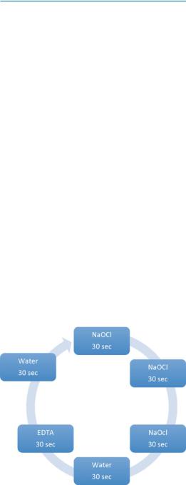
The PIPS tip is placed in the pulp chamber only (not in the root canal) and held stationary throughout the activation process. During the time of laser activation, the dental assistant applies a continuous flow of the solution from the dental irrigating syringe. It is extremely important that the pulp chamber is always kept flooded with enough irrigating solution to keep the PIPS tip submerged. The laser activation period for PIPS is in 30 s cycles. The current protocol is six 30 s cycle of laser activation, with three [3] 30 s off (rest phase) between activation when using NaOCl. Immediately after 3–30 s cycles of laseractivated irrigation with NaOCl, the canals are irrigated for an additional 30 s using PIPS with water only (Fig. 13.6). The pulp chamber is then emptied, and 17 % EDTA is used with PIPS and continuous flow for an additional 30 s. The final step in the PIPS protocol is laser activation with
Fig. 13.6 PIPS current correct protocol

234 |
D.E. Jaramillo |
|
|
an additional 30 s of water only. The canal system is now ready for obturation.
A new era of laser-activated root canal irrigation is now available with excellent results on the smear layer removal and disinfection of the root canal walls, dentinal tubules, isthmuses, lateral canals, fins, etc.
References
1. Schilder H. Cleaning and shaping of the root canal. Dent Clin North Am. 1974;18(2):269–96.
2. Bystrom A, Sundqvist G. Bacteriologic evaluation of the efficacy of mechanical root canal instrumentation in endodontic therapy. Scand J Dent Res. 1981;89(4):321–8.
3. Sjogren U, Hagglund B, Sundqvist G, Wing K. Factors affecting the long-term results of endodontic treatment. J Endod. 1990;16(10):498–504.
4. Costerton W, Veeh R, Shirtliff M, Pasmore M, Post C, Ehrlich G. The application of biofilm science to the study and control of chronic bacterial infections. J Clin Invest. 2003;112(10):1466–77.
5. Chavez de Paz LE. Redefining the persistent infection in root canals: possible role of biofilm communities. J Endod. 2007;33(6):652–62.
6. Nair PN. Pathogenesis of apical periodontitis and the cases of endodontic failures. Crit Rev Oral Biol Med. 2004;15(6):348–81.
7. Nair PNR, Henry S, Cano V, Vera J. Microbial status of the apical root canal system of human mandibular first molars with primary apical periodontitis after one visit endodontic treatment. Oral Surg Oral Med Oral Pathol Oral Radiol Endod. 2005;99:231–52.
8. Hess W. Formation of root canals in human teeth. J Am Den Assoc. 1921;8:704–34.
9. Wine FS, Healey HJ, Gerstein H, Evanson L. Canal configuration in the mesiobuccal root of the maxillary first molar and its endodontic significance. Oral Surg Oral Med Oral Pathol. 1969;28:419–25.
10.Pineda F, Kuttler Y. Mesiodistal and buccolingual roentgenographic investigation of 7,275 root canals.
Oral Surg Oral Med Oral Pathol. 1972;33:101–10. 11. Verticcu FJ. Root canal anatomy of the human per-
manent teeth. Oral Surg Oral Med Oral Pathol. 1984;58:589–99.
12.Vertucci FJ. Root canal morphology and its relationship to endodontic procedures. Endod Topics.
2005;10:3–29.
13. Arnold M, Ricucci D, Siqueira Jr JF. Infection in a complex network of apical ramifications as the cause of persistent apical periodontitis: a case report. J Endod. 2013;39(9):117–84.
14. Sjogren U, Figdor D, Spangberg L, Sundqvist G. The antimicrobial effect of calcium hydroxide
as a short-term intracanal dressing. J Endod. 1990;16(12):589–95.
15.Safavi KE, Dowdenn WE, Introcaso JH, Langeland
K.A comparison of antimicrobial effects of calcium hydroxide and iodine-potassium iodide. J Endod. 1985;11(10):454–6.
16.Weller RN, Brady JM, Bernier WE. Efficacy of ultrasonic cleaning. J Endod. 1980;6(9):740–3.
17.Van der Sluis LW, Wu MK, Wesselink PR. The efficacy of ultrasonic irrigation to remove artificially placed dentine debris from human root canals prepares using instruments of varying taper. Int Endod
J.2005;38(10):746–8.
18.Ahmad M, Pitt Ford TJ, Crum LA. Ultrasonic debridement of root canals: acoustic streaming and its
possible role. J Endod. 1987;13(10):490–9.
19. Stern RH, Sognnaes RF. Laser effect on dental hard tissues. A preliminary report. J South Calif Dent Assoc. 1965;33:17–9.
20. Meral G, Tasar F, Kocagøz S, Sener C. Factors affecting the antibacterial effects of Nd:YAG Laser in vivo. Laser Surg Med. 2003;32(3):197–202.
21. Nm S, Roth CA. Ruby laser as a microsurgical instrument. Science. 1963;141:46–7.
22. Klein E, Fine S, Ambrus J. Interaction of laser irradiation with biological system. III. Studies on biological systems in vitro. Fed Proc. 1965;14:5101–10.
23. McGuff PE, Bell EJ. The effect of laser irradiation on bacteria. Med Biol III. 1966;16:191–3.
24. Pini R, Salimbeni R, Vannini M. Laser dentistry: a new application of excimer laser in root canal therapy. Laser Surg Med. 1989;9:352–7.
25.Weichman JA, Johnson FM. Laser in endodontics. A preliminary investigation. Oral Surg Oral Med Oral Pathol. 1971;31:416–20.
26. Weichman JA, Johnsosn FM, Nitta LK. Laser use in endodontics. Part II. J Oral Surg. 1972;34:828–30.
27.Dederich D, Zachariensen K, Tulip J. Scanning electron microscopic analysis of root canal wall dentin follow Neodymium Yttrium garnet laser irradiation. J Endod. 1984;10:428.
28. Levy G. Cleaning and shaping the root canal with Nd:YAG laser beam: a comparative study. J Endod. 1992;18:123–7.
29. Kantola S. Laser induced effects on the tooth structure. IV. A study of changes in the calcium and phosphorous contents in dentine by electron probe microanalysis. Acta Odontol Scand. 1972;30:463–74.
30. Gordon W, Atabakhsh VA, Meza F, Doms A, Nissan R, Nissan R, Risoiu I, Stevens RH. The antimicrobial efficacy of the erbium, chromium:yttrium-scandium- gallium-garnet laser with emitting tips on the root canal dentin walls infected with Enterococcus faecalis. J Am Dent Assoc. 2007;138(7):992–1002.
31. Farges P, Nahas P, Bonin P. In vitro study of a Nd:YAG laser in endodontic retreatment. J Endod.
1998;24:359–63. |
|
|
|
32. Folwaczny M, |
Mehl |
A, |
Jordan C, Hickel |
R. Antibacterial |
effects |
of |
pulsed Nd:YAG laser |
13 Laser Activated Irrigation of the Root Canal Systems. Pips (Photon-Induced Photoacoustic Streaming) |
235 |
|
|
radiation at different energy settings in root canals. J Endod. 2002;28:24–9.
33. Peters OA, Schönenberger K, Laib A. Effects of four Ni-Ti preparation techniques on root canal geometry assessed by micro computed tomography. Int Endod
J. 2001;34(3):221–30.
34.Kerekes K, Tronstad L. Morphological observation on the root canals of human molars. J Endod. 1977;3(3):114–8.
35.Wu MKL, Wesselink PR. A primary observation on the preparation and obturation of oval canals. Int Endod J. 2001;34(2):137–41.
36.Tatsuta CT, Morgan LA, Baumgartner JC, Adey JD. Effect of calcium hydroxide and four irrigation regimens on instrumented and uninstrumented canal wall topography. J Endod. 1999;25(2):93–8.
37.Crane AB. A practicable root canal technique. Philadelphia: Lea & Febinger; 1920.
38. Zehnder M, Kosicki D, Luder H, Sener B, Waltimo T. Tissue – dissolving capacity and antibacterial effect of buffered and unbuffered hypochlorite solutions. Oral Surg Oral Med Oral Pathol Oral Radiol Endod. 2002;94(6):756–62.
39. Van der Sluis LW, Vogels MP, Verhaagen B, Macedo R, Wesselink PR. Study on the influence of refreshment/activation cycles and irrigants on mechanical cleaning efficiency during ultrasonic activation of the irrigant. J Endod. 2010;36(4):737–40.
40.Williams AR. Disorganization and disruption of mammalian and amoeboid cells by acoustic microstreaming. J Acoust Soc Am. 1972;52:688–93.
41.Peters OA, Bardsley S, Fong J, Pandher G, Divito E. Disinfection of root canals with photon-initiated photoacoustic streaming. J Endod. 2011;37(7):1008–12.
42.Jaramillo DE, Aprecio R, Angelov N, Divito E, McClammy TV. Efficacy of photon induced photoacoustic streaming (PIPS) on the root canals infected with Enterococcus faecalis: a pilot study. Endod Prac.
2012;7(3):28–32.
43. Divito E, Peters OA, Olivi G. Effectiveness of the erbium:YAG laser and new design radial and stripped tips in removing the smear layer after toot canal instrumentation. Laser Med Scue. 2012;27:273–80.
44.Saunders EM. In vivo findings associated with heat generation during thermomechanical compaction of gutta-percha. 1. Temperature levels at the external surface of the root. Int Endod J. 1990;23(5):263–7.
45.Sonntag KD, Kutzman B, Burkes J, Hoke J, Moshonov J. Pulpal response to cavity preparation with the Er:YAG and Mark III free electron lasers. Oral Sug Oral Med Oral Pathol Oral Radiol Endod. 1996;81:695–702.
46.Armengol V, Jean A, Marion D. Temperature rise during Er:YAG and Nd:YAP laser ablation of dentin. J Endod. 2000;26(3):138–41.
47.Wilson M. Lethal photosensitisation of oral bacteria and its potential application in the photodynamic therapy of oral infections. Photochem Photobiol Sci. 2004;3(5):412–8.
48. Lim Z, Cheng JL, Kim TW, Teo EG, Wong J, George S, Kishen A. Light-activated disinfection: an alternative endodontic disinfection strategy. Aust Dent
J. 2009;54(2):108–14.
49.George S, Kishen A. Influence of photosensitizer solvent on the mechanisms of photoactivated killing of enterococcus faecalis. Photochem Photobiol. 2008;84(3):734–40.
50. |
Boutsioukis |
C, |
Lambdrianidis |
T, |
Kastrinakis |
|
|
E. Irrigant flow within a prepared root canal using |
|||||
|
various flow rates: a computational fluid dynamic |
|||||
|
study. Int Endod J. 2009;42(2):144–55. |
|
||||
51. |
Boutsioukis |
C, |
Kastrinakis |
E, |
Lambrianidis |
|
|
T, Verhaagen |
B, |
Versluis |
M, |
van |
der Sluis |
LW. Formation and removal of apical vapor lock during syringe irrigation. A combined experimental and computational fluid dynamics approach. Int Endod J. 2014;47(2):191–201.
52. Hsieh YD, Gau CH, Kung Wu SF, Shen EC, Hsu PW, Fu E. Dynamic recording of irrigating fluid distribution in the root canals using thermal image analysis. Int Endod J. 2007;40(1):11–7.
53. Shen Y, Gao Y, Qian W, Ruse ND, Zhou X, Wu H, Haapasalo M. Three-dimensional numeric simulation of root canal irrigant flow with different irrigation needles. J Endod. 2010;36(5):884–9.
54. van der Sluis LW, Versluis M, Wu MK, Wesselink
PR. |
Passive |
ultrasonic |
irrigation |
of |
the root |
|||
canal: |
a |
review |
of the |
literature. |
Int |
Endod |
||
J. 2007;40(6):415–26. |
|
|
|
|
||||
55. Fincham |
AM, |
Jaramillo |
DE, Divito |
E, |
Peters |
|||
OA. Irrigant flow during Photo Induced Photoacoustic |
||||||||
streaming |
(PIPS) |
using |
micro |
digital |
particle |
|||
image velocimetry (μDPIV): a pilot study. IEJ. 2014;47:659–66.
56.Ordinola-Zapata R, Bramante CM, Aprecio RM, Handysides R, Jaramillo DE. Biofilm removal by 6% sodium hypochlorite activated by different irrigation
techniques. Int Endod J. 2013. doi:10.1111/iej/12202. 57. Jaramillo DE, Aguilar E, Aprecio RM, Tran K. Dentin disinfection using PIPS and conventional needle irri-
gation. LLUSD CDR, 2011. Unpublished data.
58. Alsharhrani M, Divito E, Hughes C, Nathanson D, Huang G. Enhanced removal of enterococcus faecalis biofilms in the root canal using sodium hypochlorite plus Photon Induced Photoacoustic Streaming: an in vitro study. Photomed Laser Surg. 2014;32(5): 524–30. doi:10.1089/pho2014.3714.
59. Vera J, Siqueira Jr JF, Ricucci D, Loghin S, Fernandes N, Flores B, Cruz AG. One-versus twovisit endodontic treatment of teeth with apical periodontitis: a histobacteriological study. J Endod. 2012;38(8):1040–52.
60. Lloyd A, Uhles J, Clement DJ, Garcia-Godoy F. Elimination of intracanal tissue and debris through a novel laser-activated system assessed using highresolution micro-computed tomography: a pilot study. Jendod, 2014:40(4):584–7. doi:http://dx.doi. org/10.1016/j.joen.2013.10.040.

Photodynamic Therapy for Root |
14 |
Canal Disinfection |
Anil Kishen and Annie Shrestha
Abstract
Emergence of antimicrobial-resistant microbial strains, rise of transplants, medically compromised patients, advanced cancer patients, and global spread in infection are few in the major issues related to difÞculties of managing infectious diseases. The widespread recognition of microbial bioÞlm as the contributory factor for human infection warrants the identiÞcation of a reliable and effective antimicrobial strategy to combat infectious diseases. On similar lines, treatment of infected root canals presents with a major challenge of bacterial persistence after treatment. Photodynamic therapy (PDT) is considered as one of the potential treatment modalities for the treatment of localized infections irrespective of the causative microorganism, including those that are recalcitrant to conventional antimicrobial therapies/disinfectants. The ongoing research is focused to bring about tis- sue-speciÞc innovative improvements of antimicrobial PDT by modifying photosensitizer formulation and light delivery system and increasing number of clinical trials and appropriate regulatory approvals for the usage of new photosensitizers. Cumulatively these efforts demonstrate increasing interest in the application of PDT in the coming years.
Introduction
A. Kishen, PhD, MDS, BDS (*)
Department of Endodontics, Facility of Dentistry, University of Toronto, Toronto, ON, Canada e-mail: anil.kishen@utoronto.ca
A. Shrestha, PhD, MSc, BDS
Faculty of Dentistry, Department of Endodontics, University of Toronto, Toronto, ON, Canada
Approximately 60 % of the current human infections have been associated with the presence of bacterial bioÞlms, which includes both implantrelated infections and chronic non-implant-related infections [1]. The conservative management of such infections involving topical or systemic antibiotics has been shown to be ineffective mainly due to multidrug-resistant strains and widespread systemic use of antibiotics and misuse of antibiot-
© Springer International Publishing Switzerland 2015 |
237 |
B. Basrani (ed.), Endodontic Irrigation: Chemical Disinfection of the Root Canal System, DOI 10.1007/978-3-319-16456-4_14
238 |
A. Kishen and A. Shrestha |
|
|
ics [2]. Antimicrobial resistance is constantly on rise leading to a major hindrance in the treatment of many infectious diseases [3Ð5]. Emergence of resistant microbial strains, rise of transplants, medically compromised patients, advanced cancer patients, and spread of infection due to increasing global travel between developed and developing nations are few of the major issues related to difÞculties of managing infectious diseases [5, 6]. Photodynamic therapy (PDT) is considered as one of the potential treatment modalities for the treatment of localized infections irrespective of the causative microorganism, including those that are recalcitrant to conventional antimicrobial therapies [7Ð10].
PDT involves the use of a nontoxic dye or photosensitizer (PS) in combination with visible light, which in the presence of molecular oxygen leads to the production of cytotoxic oxygen radicals such as singlet oxygen. This reactive oxygen species are responsible for the PDT cytotoxic action [11], and its production and activity depend on the PDT dose [12]. PDT was discovered by chance during the early 1900s, when a combination of nontoxic dyes and visible light resulted in the killing of cells. Oscar Raab used acridine dyes and showed that the combination of light and dyes was much more effective to kill a paramecium [13]. Application of PDT as an alternative treatment for tumors has been
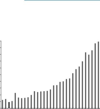
explored and tested widely. For the last two decades, series of in vitro and in vivo studies have proven the efÞcacy of PDT in the management of various infectious and noninfectious diseases. The increase in the interest toward PDT is evident as seen by the exponential increase in the number of publications in the recent years (Fig. 14.1). The introduction of photosensitizers for in vivo applications and their approval in certain countries such as Canada, the United States, the European Union, Japan, Australia, and New Zealand show increased surge in using PDT for various systemic and topical pathogenic conditions [14]. The ongoing research is focused to bring about tissue-speciÞc innovative improvements of antimicrobial PDT by modifying photosensitizer formulation and light delivery system and increasing number of clinical trials and appropriate regulatory approvals for the usage of new photosensitizers [15Ð17]. Cumulatively these efforts demonstrate increasing interest in the application of PDT in the coming years.
Mechanism of Photodynamic
Inactivation of Microbial Cells
Antimicrobial photodynamic therapy works as a combination of photosensitizer and light. Photosensitizer is a light-sensitive chemical that
Fig. 14.1 Number of publications (English language) on the PDT since 1980 till 2010 (SourceÑPubmed)
|
1,000 |
|
|
|
|
|
|
|
|
|
|
|
|
|
|
|
|
|
|
|
|
|
|
|
|
|
|
|
|
|
|
|
900 |
|
|
|
|
|
|
|
|
|
|
|
|
|
|
|
|
|
|
|
|
|
|
|
|
|
|
|
|
|
|
of publications/year |
800 |
|
|
|
|
|
|
|
|
|
|
|
|
|
|
|
|
|
|
|
|
|
|
|
|
|
|
|
|
|
|
700 |
|
|
|
|
|
|
|
|
|
|
|
|
|
|
|
|
|
|
|
|
|
|
|
|
|
|
|
|
|
|
|
600 |
|
|
|
|
|
|
|
|
|
|
|
|
|
|
|
|
|
|
|
|
|
|
|
|
|
|
|
|
|
|
|
500 |
|
|
|
|
|
|
|
|
|
|
|
|
|
|
|
|
|
|
|
|
|
|
|
|
|
|
|
|
|
|
|
400 |
|
|
|
|
|
|
|
|
|
|
|
|
|
|
|
|
|
|
|
|
|
|
|
|
|
|
|
|
|
|
|
Number |
300 |
|
|
|
|
|
|
|
|
|
|
|
|
|
|
|
|
|
|
|
|
|
|
|
|
|
|
|
|
|
|
200 |
|
|
|
|
|
|
|
|
|
|
|
|
|
|
|
|
|
|
|
|
|
|
|
|
|
|
|
|
|
|
|
|
100 |
|
|
|
|
|
|
|
|
|
|
|
|
|
|
|
|
|
|
|
|
|
|
|
|
|
|
|
|
|
|
|
0 |
|
|
|
|
|
|
|
|
|
|
|
|
|
|
|
|
|
|
|
|
|
|
|
|
|
|
|
|
|
|
|
1980 |
1981 |
1982 |
1983 |
1984 |
1985 |
1986 |
1987 |
1988 |
1989 |
1990 |
1991 |
1992 |
1993 |
1994 |
1995 |
1996 |
1997 |
1998 |
1999 |
2000 |
2001 |
2002 |
2003 |
2004 |
2005 |
2006 |
2007 |
2008 |
2009 |
2010 |
14 Photodynamic Therapy for Root Canal Disinfection |
239 |
|
|
possesses low toxicity in the absence of light. Photosensitization of the infected tissue with a photosensitizer allows uptake into the bacterial cells, and irradiation of the photosensitized tissue results in the destruction of bacteria and infected tissue. It is extremely important that the light should be at a speciÞc wavelength, which corresponds to the absorption wavelength of the photosensitizer being used. PDT can be utilized with a suitable photosensitizer and irradiation conditions to treat infections in cases where antibioticbased therapeutic strategies have failed [8]. Unlike in cancer therapy where the photosensitizer is administered intravenously, for localized infections, the photosensitizer is delivered locally by various methods such as topical application, instillation, and interstitial injection or aerosol delivery. Selectivity of photosensitizer toward microorganisms over mammalian cells and effective removal of the causative microorganisms are the key points in achieving success of PDT to manage localized infections [7].
Photosensitizers are chemicals, when excited, capable of transferring the energy absorbed to other compounds in the vicinity that, in turn, generate very reactive metastable species. The triplet-excited state of the photosensitizer releases energy to come to the ground state via two speciÞc mechanisms: type I or type II pathway [18]. Type I pathway involves production of radical ions of oxygen due to electron transfer from the photosensitizer triplet-excited state to the substrate. Radical ions such as superoxide, hydroxyl, and lipid-derived ions are the cytotoxic species responsible for type I photoreaction [19]. Type II pathway involves production of excited singlet oxygen due to energy transfer from the photosensitizer triplet-excited state to the ground-state molecular oxygen, which is responsible for the oxidation of various cellular constituents [20]. The antimicrobial effect of PDT is mainly due to type II reaction. Singlet oxygen is a strong oxidizing agent and thus highly reactive, with a lifetime of less than 0.04 μs in a biological environment and a radius of action of less than 0.02 μm [21]. The reactions of singlet oxygen with the cellular targets lead to cell death. The above two basic mechanisms account for this
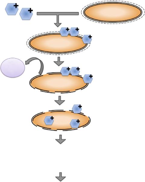
lethal damage to the bacterial cell by DNA damage and cytoplasmic membrane damage. Figure 14.2 shows the photodynamic inactivation of bacterial cells in a stepwise manner. It should be noted that the differences in microbial cell wall characteristics and bacterial growth mode should be accounted while determining the duration of photosensitization before light illumination [22, 23]. The photosensitizer with slower uptake could result only in cell wall damage and with longer incubation times; other nuclear effects such as nucleic acid strand breakage might be apparent. The choice of photosensitizer is thus critical in obtaining effective bacterial elimination.
One of the signiÞcant advantages of PDT is the targeted antibacterial effect. Choosing a photosensitizer that has high afÞnity for microbial cells and irradiating the speciÞc area of infection could result in the targeted effect of antimicrobial PDT. As the photosensitizer typically shows a higher afÞnity toward microbial cells, the host cells could be affected less during PDT. Toxicity of the photosensitizer usually occurs when high concentration/volume of photosensitizer is applied to a tissue to obtain more signiÞcant treatment response. The instant antimicrobial activity also offers added advantage as antibiotics take several days to produce comparable efÞcacy. The broad therapeutic window of PDT because of the high reactivity of ROS could effectively eliminate bacteria as well as the bacterial virulence factors such as endotoxins and proteolytic enzymes. Furthermore, due to the multiple targets of PDT on a bacterial cell, the probability of bacteria developing resistance to this treatment has been considered to be almost impossible [7, 8, 24].
Photosensitizers such as porphyrins, chlorins, and phthalocyanines, for treatment of cancer or other diseases, are chosen based upon their low dark toxicity to mammalian cells and ability to target tumor cells [8]. The photosensitizers for antibacterial PDT are chosen based on their speciÞcity to bacterial cells. Large numbers of photosensitizer potentially useful in LAD are currently in various stages of clinical trials for FDA approval. The commonly used photosensitizers
240 |
A. Kishen and A. Shrestha |
|
|
Cationic PS
Bacteria
Ca2+
Mg2+
Bacteria
Bacteria
Anionic outer wall (carboxylate groups)
Bacteria
1. Electrostatic interaction (Few minutes)
2. Increased outer wall permeability
·Displacement of Mg2+ and Ca2+ ions
·Photooxidative modification of selected proteins
3. Diffusion of PS into the cytoplasmic membrane And binding with plasma membrane
4. Photodynamic effect on multiple location In the plasma membrane
·Extension crosslinking of selective plasma membrane proteins
·Inactivation of enzymes such as NADH, succinate and lactate dehydrogenases
·Collapse of K+ and ionic balance
·DNA damage: both single/double stranded DNA breakage
5. Inhibition of cell growth and cell death
Impairment of cell functions and metabolic processes
Fig. 14.2 Schematic showing the stepwise mechanism of photodynamic inactivation of microbial cells
for antibacterial purpose are halogenated xanthenes such as rose bengal (RB) [25], phenothiazines such as methylene blue (MB) and toluidine blue (TBO) [9, 22, 26], and perylenequinones such as hypericin [27]. The factors that determine
the effectiveness of antibacterial PDT are method/ vehicle of topical application, effective time of interaction with the microbes at the site of infection, selectivity of the photosensitizer to microbes, relative non-toxicity toward host tis-

14 Photodynamic Therapy for Root Canal Disinfection |
241 |
|
|
sues at the site of infection, and ability to eliminate the microbes effectively to avoid regrowth of surviving pathogens following treatment [8].
Antimicrobial PDT on gram-positive and gram-negative bacteria induced breaks in both single and double-stranded DNA and the disappearance of the plasmid supercoiled fraction [28, 29]. In addition, the photooxidative effect caused by the phenothiazinium photosensitizer in bacteria led to the damage of multiple targets in bacterial cells such as DNA [28], membrane integrity [30], protease activity, and lipopolysaccharide (LPS) [31]. George and Kishen reported functional impairment of cell wall, extensive damage to chromosomal DNA, and degradation of membrane proteins following methylene bluemediated APDT of E. faecalis [32]. These Þndings support the hypothesis that antimicrobial PDT is a feasible alternative to antibiotics since the mode of action is markedly different from that typical of most antibiotics and chances of resistance are potentially none.
Antimicrobial PDT in Root Canal
Disinfection
The use of PDT in conjunction with conventional root canal disinfection methods resulted in signiÞcantly better bacterial elimination as compared to either of these treatments when used alone. Over the years, various efforts were made to optimize different PDT-related parameters for endodontic application. Several in vitro and in vivo studies have shown the effectiveness of PDT in eliminating root canal bioÞlms [9, 33Ð 42]. Endodontic pathogens such as E. faecalis, P. intermedia, F. nucleatum, S. intermedius, and A. actinomycetemcomitans have been shown to be killed by using photosensitizers such as methylene blue (MB), toluidine blue (TBO), and rose bengal (RB) [43Ð46].
Currently PDT is not considered a replacement for the existing root canal disinfection protocols but rather considered as a potential adjunct to improve antibioÞlm efÞcacy following current disinfection protocols during the root canal treat-
ment. Meire et al. [47] and George and Kishen [41, 43] used antimicrobial PDT to enhance the root canal disinfection. They showed that antimicrobial PDT could effectively kill bioÞlms of E. faecalis with photosensitizers such as MB and TBO along with red light. Soukos et al. conducted PDT experiments on a range of endodontic pathogens (methylene blue as photosensitizer) and reported complete elimination of all bacteria except E. faecalis (53 %) [34]. In yet another study, signiÞcant antibacterial effects on suspensions of S. intermedius, P. micros, P. intermedia, and F. nucleatum were reported by Williams et al. following PDT with TBO and red light [44]. Different in vivo studies that examined the efÞcacy of antimicrobial PDT in root canal disinfection have been summarized in Table 14.1 [26, 36Ð38]. These studies concluded that a combination of chemomechanical preparation and PDT would bring about maximum reduction in microbial loads.
Singlet oxygen is known to diffuse approximately 50 nm [18]. This emphasizes the close proximity of a photosensitizer molecule to the bacterial cell surface that allows diffusion of singlet oxygen. In a bioÞlm, only 30 % of the total mass is bacteria and remaining is the self-secreted extracellular polymeric matrix. The ability of the photosensitizer to diffuse and uniformly distribute in the bioÞlm structure is important for effective killing efÞcacy [48]. This clearly could be seen in the higher level of energy required to eliminate bacterial bioÞlms as compared to the planktonic counterparts [22, 46, 48, 49]. Bacteria existing in bioÞlms are also known to express active efßux pumps that confer their ability to transport amphiphilic chemicals and photosensitizers outside the cell [50]. This is the protective mechanism exerted by the cell to expel potentially toxic compounds. Both prokaryotic and eukaryotic cells possess various membrane proteins termed efßux pumps. Use of efßux pump inhibitors (EPI) such as verapamil would restore the antibacterial activity of a compound that is speciÞc to an efßux mechanism. Both the phenothiazinium dyes such as MB and TBO are amphipathic cations that are potential substrate for multidrug efßux pumps [51]. Use of EPI with

242 |
|
|
|
A. Kishen and A. Shrestha |
|
||||
Table 14.1 Table showing clinical studies where PDT was used for root canal disinfection |
||||
No |
Author/date |
Objective and materials |
Methodology |
Conclusion |
1 |
Bonsor |
Aimed to evaluate the antimicrobial |
Irrigation with 20 % citric |
Cleaning and shaping |
|
et al. (2006) |
efÞcacy of root canal disinfection by |
acid and 2.25 % sodium |
resulted in complete |
|
[36] |
combining conventional endodontic |
hypochlorite |
bacterial killing in 86.7 % of |
|
|
treatment with PDT |
PDT with TBO and diode |
samples |
|
|
Clinical study on 32 root canals from |
laser (12.7 mg/L−1, |
Combination of cleaning |
|
|
14 patients |
100 mW, 120 s) |
and shaping + PDT resulted |
|
|
|
Samples collected by |
in complete bacterial killing |
|
|
|
Þling |
in 96.7 % of samples |
2 |
Bonsor |
Aimed to compare the effect of a |
Procedure similar to |
Combination of 20 % citric |
|
et al. (2006) |
combination of 20 % citric acid and |
previous study |
acid and PDT resulted in |
|
[26] |
PDT with the use of 20 % citric acid |
|
complete bacterial killing in |
|
|
and 2.25 % sodium hypochlorite on |
|
91 % of samples |
|
|
bacterial load in prepared root canals |
|
20 % citric acid and 2.25 % |
|
|
64 patients were used |
|
sodium hypochlorite |
|
|
|
|
resulted in complete |
|
|
|
|
bacterial killing in 82 % of |
|
|
|
|
samples |
3 |
Garcez |
This study analyzed the antimicrobial |
Irrigation with 2.5 % |
First session produced |
|
et al. (2008) |
effect of PDT in association with |
sodium hypochlorite, 3 % |
98.5 % bacterial reduction |
|
[38] |
endodontic treatment |
hydrogen peroxide, and |
(1.83 log reduction) |
|
|
20 patients were selected |
17 % EDTA |
Second session produced |
|
|
First session of cleaning and |
PDT with |
99.9 % bacterial reduction |
|
|
shaping + PDT |
polyethylenimine (PEI) |
(1.14 log reduction) |
|
|
At the end of Þrst session, the root |
chlorin (e6) conjugate |
Second session PDT was |
|
|
canal was Þlled with Ca(OH)(2), and |
(2 min, 9.6 J, 240 s) |
observed to be more |
|
|
after 1 week, a second session of PDT |
Paper point sampling |
effective than Þrst session |
|
|
was performed |
|
|
4 |
Garcez |
Studied antimicrobial effect of PDT |
PDT used |
Endodontic therapy alone |
|
et al. (2010) |
combined with endodontic treatment |
polyethylenimine chlorin |
produced a signiÞcant |
|
[37] |
in patients with necrotic pulp infected |
(e6) as a photosensitizer |
reduction in numbers of |
|
|
with microßora resistant to a previous |
and a diode laser |
microbial species (only 3 |
|
|
antibiotic therapy |
(40 mW, 4 min, 9.6 J) |
teeth were free of bacteria) |
|
|
30 teeth from 21 patients with |
|
The combination of |
|
|
periapical lesions that had been treated |
|
endodontic therapy with |
|
|
with conventional endodontic |
|
PDT eliminated all |
|
|
treatment and antibiotic therapy were |
|
drug-resistant species and |
|
|
selected |
|
all teeth were bacteria-free |
phenothiaziniums resulted in signiÞcantly enhanced bioÞlm elimination at much lower PDT dosage [45, 52, 53]. Since efßux pumps are highly active in bacterial bioÞlms, use of EPI could potentially enhance the antibioÞlm efÞcacy of PDT inside root canals. Kishen et al. have demonstrated the enhanced ability of EPI in combination with MB photosensitizer to disinfect bioÞlm bacteria as well bioÞlm-derived bacteria [45, 52].
In addition to the limitations associated with the interaction/uptake of photosensitizer by intracanal bacterial bioÞlms, tissue-speciÞc constraining factors in the application of PDT
for endodontic disinfection also need special consideration. Some of the tissue-speciÞc constraining factors in the application of PDT for endodontic disinfection are the limited penetration of the light energy into the infected tissue, lack of optimum photosensitizer concentration within the infected tissue, low oxygen tension inside the root canals, and dentin discoloration by the photosensitizer. These issues need to be addressed before establishing PDT as a deÞnitive treatment step in root canal disinfection [33, 41].
In biological tissue, absorption of light is mainly due to the presence of free water molecules,
14 Photodynamic Therapy for Root Canal Disinfection |
243 |
|
|
proteins, pigments, and other macromolecules. The absorption coefÞcient strongly depends on the wavelength of the incoming light/laser irradiation. Scattering of light in tissue has the utmost effect on light intensity and directionality. Scattering, together with refraction, causes a widening of light beam, resulting in the loss of ßuence rate (power per unit area) and a change in directionality of the light beam. Tissue-speciÞc approach has been highlighted by George and Kishen, which improved the antimicrobial efÞcacy of PDT in root canal system. Methylene blue was dissolved in different formulations such as water, 70 % glycerol, 70 % poly ethylene glycol, and a mixture of glycerol-ethanol-water (MIX) in a ratio of 30:20:50 and analyzed for the photophysical, photochemical, and photobiological characteristics [43]. The aggregation of methylene blue molecules was signiÞcantly higher in water when compared to other formulations. In addition, the MIX-based methylene blue formulation had effective penetration into dentinal tubules and enhanced singlet oxygen generation, which in turn improved bactericidal action. A signiÞcantly higher impairment of bacterial cell wall and extensive damage to chromosomal DNA were observed when methylene blue in a MIX-based formulation was used and when compared to water [32]. The same group also showed that the incorporation of an oxidizer and oxygen carrier with photosensitizer formulation in the form of an emulsion would produce signiÞcant photooxidation capabilities, which in turn facilitated comprehensive disruption of matured endodontic bioÞlm structure [41].
Antimicrobial PDT has the potential to destroy microbial cells as well as mammalian cells. However, the selective killing of microbial cells over host cells is speciÞc to the photosensitization periods and light ßuence required for the antimicrobial effects. Soukos et al. compared the effect of PDT using a combination of toluidine blue O (TBO) and red light against S. sanguis and human gingival keratinocytes and Þbroblasts. They reported no reduction in the human cell viability, whereas the bacteria were effectively killed [54]. Soncin et al. reported the selective killing of S. aureus over human Þbroblasts and
keratinocytes (four to six fold) when subjected to PDT using cationic phthalocyanine and relatively low light ßuencies [55]. George and Kishen demonstrated a 97.7 % killing of Enterococcus faecalis compared to a 30 % human Þbroblast dysfunction following methylene blue-mediated PDT [9]. Even the newer photosensitizerconjugated chitosan nanoparticles showed favorable cell survival (Þbroblasts) as compared to highly effective antibioÞlm properties [48, 56]. All these in vitro studies suggested the targeted killing efÞcacy of antimicrobial PDT.
Conjugating photosensitizer to various agents or chemical moieties can result in improved photosensitizers for PDT. These modiÞed photosensitizers are expected to bind more effectively to the outer membrane of bacteria and upon activation of generated reactive oxygen species, which then diffused into the cells, resulting in cell death. Therefore, photo-generated oxidative species are well conÞned to the cell wall and its vicinity, which is a highly susceptible domain for photodynamic action. Soukos and coworkers formed a hypothesis that by covalently conjugating a suitable photosensitizer to a poly-l-lysine chain, a bacteria-targeted photosensitizer delivery vehicle could be constructed that would efÞciently inactivate both gram-positive and gram-negative species [57]. This was demonstrated by preparing a conjugate of chlorin (e6) and a poly-l-lysine chain (20 lysine residues), which after 1 min incubation and illumination with red light killed >99 % of the gram-positive Actinomyces viscosus and gram-negative Porphyromonas gingivalis
[58]. Conjugates of polyethylenimine and chlorin (e6) when used as a photosensitizer eliminated all the drug-resistant bacteria during retreatment in failed root canal-treated teeth [37]. This PEI-ce6 conjugate eliminated both gram-positive and gram-negative bacteria in vitro and in vivo as compared to the commonly used photosensitizer TBO [59]. Anionic photosensitizer (rose bengal) conjugated with positively charged chitosan has also been shown to be highly effective in removing bioÞlms of gram-positive, gram-negative, and multispecies bacteria [48, 60, 61] (Fig. 14.3). Shrestha et al. showed that the rose bengalconjugated chitosan presented a synergistic effect
244 |
A. Kishen and A. Shrestha |
|
|
of the antimicrobial polymer chitosan and singlet oxygen that was generated following photoactivation [48, 56]. The chitosan-conjugated rose bengal nanoparticles (CSRBnp) penetrated deep into the bioÞlm structure and photoactivation resulted in total elimination of the multispecies bioÞlms of bacteria associated with endodontic infection [61]. These modiÞed photosensitizers in nano-form were found to envelope the bacterial cells within minutes of the photosensitization
period (Fig. 14.4) [48]. Irradiation of these bacteria with closely adhered CSRBnp resulted in total killing with various stages of membrane damage as well as release of cell constituents.
Constituents of the infected root canal such as tissue remnants (pulp tissue), serum products, and dentin matrix compromised the antimicrobial efÞcacy of not only the common endodontic irrigants [62] but also the antimicrobial efÞcacy of PDT [63]. Most studies concerning the antimi-
a |
b |
|
Absorption intensity (au) |
0.5 |
|
|
|
|
0.4 |
|
|
|
|
0.3 |
|
|
|
|
0.2 |
|
|
|
|
0.1 |
|
|
|
|
0 |
|
|
|
|
475 |
500 |
525 |
550 |
575 |
|
Wavelength (nm) |
|
||
|
RB |
|
CSRBnp |
|
c |
CSRBnp |
d |
RB |
|
|
52 µM
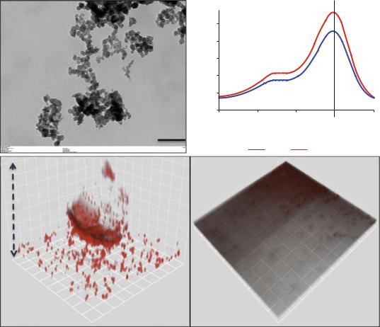
Fig. 14.3 (a) Transmission electron microscopic image of CSRBnp (scale bar = 200 nm). The CSRBnps were 60 ± 20 nm in size. (b) A typical graph showing the absorption spectrum of RB and CSRBnps. The absorption peak at 550 nm was not affected after conjugation of CSRBnps with RB. (c, d) The uptake of CSRBnps and RB into the E. faecalis bioÞlms as observed under CLSM. (eÐg) Scanning electron microscopic images of multispecies bioÞlms on dentin sections. (e) The 3-week-old bioÞlms presented as a uniformly thick matlike structure covering
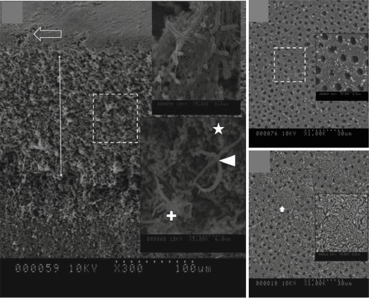
the entire dentin surface. Three speciÞc bacterial morphologies are evident in higher magniÞcation (Denoted by *, + and block white arrowhead). The surface showed an abundant polymeric matrix (open arrowhead) (magniÞed area shown by the open arrow). (f) CSRBnp treatment rendered the dentin surface clean of the bioÞlm with open dentinal tubules. (g) RB treatment showed cleaner areas of dentin along with dense bacterial aggregates (inset: magniÞed area shown by the white arrow) (Adapted with permission from Shrestha and Kishen [61])
14 Photodynamic Therapy for Root Canal Disinfection |
245 |
|
|
e f
g
Fig. 14.3 (continued)
crobial PDT of microbial pathogens use deionized water or phosphate-buffered saline to dissolve the photosensitizer. In some studies the photosensitizer was dissolved in brain-heart infusion broth wherein reduced bactericidal effect was reported. This reduction in antibacterial effect was attributed it to the presence of serum proteins in the broth [34, 64]. This effect is either due to cross-linking action or the compromised half-life of singlet oxygen in the presence of proteins.
Both coherent (lasers) and noncoherent (lamps) light sources are used for antimicrobial PDT. The choice of light source is dictated by the location, the required light dose, and the choice of photosensitizer. Laser provides monochromatic, coherent, and collimated light, offering wide range of output power. Laser light can be easily coupled into a Þber-optic cable, which can serve
as a delivery system (probe) while irradiating complex anatomy such as a root canal. Nd:YAG, KTP, HeNe, GaAlAs and diode lasers, light-emit- ting diodes (LEDs), and xenon arc lamps have been employed for APDT. The superiority of one type of light source over the other has not been clearly demonstrated [65]. Recent study evaluated the importance of using optical Þber/diffuser inside the root canal instead of laser tip at the root canal oriÞce [66] (Fig. 14.5). The rationale for using the optical Þber is mainly to allow better distribution of light energy throughout the infected root canal/root dentin. Notched optical Þber was also used to allow light distribution in 360¡ [39]. Optical Þber/diffuser allowed uniform light distribution throughout the canal length and enhanced the antimicrobial efÞcacy of PDT by reducing the bacterial bioÞlm 2 logs more than the PDT with laser tip at the canal oriÞce.
246 |
A. Kishen and A. Shrestha |
|
|
a |
c |
b |
d |
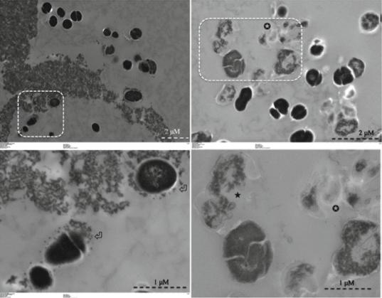
Fig. 14.4 Transmission electron microscopy images for planktonic E. faecalis after treatment with CSRBnp for 15 min (a, b). Aggregates of CSRBnp could be seen surrounding the bacterial cell. Nanoparticles were found attached to the bacterial cell surface and forming an envelope (open arrows) (b). The cells did not show any disruption of
morphology. Following PDT of the sensitized bacteria, various stages of membrane damage as well as release of cell constituents were evident (c, d). Most of the bacteria showed some kind of cell membrane disruption (black star) and release of cell constituents at higher magniÞcation (d) (Adapted with permission from Shrestha et al. [48])
There are a number of commercial PDT systems available for root canal and caries disinfection. Some of the available systems are Savedent, Denfotex PAD, and HELBO photodynamic systems that use TBO and methylene blue as photosensitizers, respectively [67]. These two systems differed in the choice of photosensitizers and their concentration, photosensitization time, Þber-optic probe design, and wavelengths of the lasers used. Although the Denfotex PAD showed signiÞcant reduction of planktonic E. faecalis [47], both these systems failed to reduce the bioÞlm bacteria grown on dentin discs. A recent sys-
tematic review by Siddiqui et al. [68] reported results of seventeen studies that used various forms of PDT to eliminate E. faecalis from infected root canals. The review clearly highlights that the discrepancies in the use of PDT for root canal disinfection are wide, resulting in highly variable Þndings from each of the studies included in the review (Tables 14.2 and 14.3). Out of the 17 studies included in the review [34, 37, 65, 70Ð83], 70 % concluded the beneÞcial effects of PDT in removing E. faecalis from root canals as compared to conventional disinfection treatments.