

Intrathoracic lymph node tuberculosis
X-RAY DIAGNOSIS
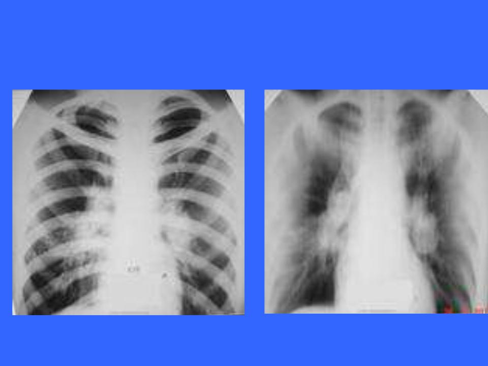
The infiltrative form of intrathoracic lymph node tuberculosis the radiograph is characterized by the presence of syndrome of lung root infiltration:
-the root shadow is enlarged in size (in width) and/or in length;
-outer contour of the root shadow is blurred; -root shadow structure is disturbed (blurred); -intensity of the root shadow - increased;
-projection of the lumen of intermediate or partially obscured or absentor.

Intrathoracic lymph node tuberculosis
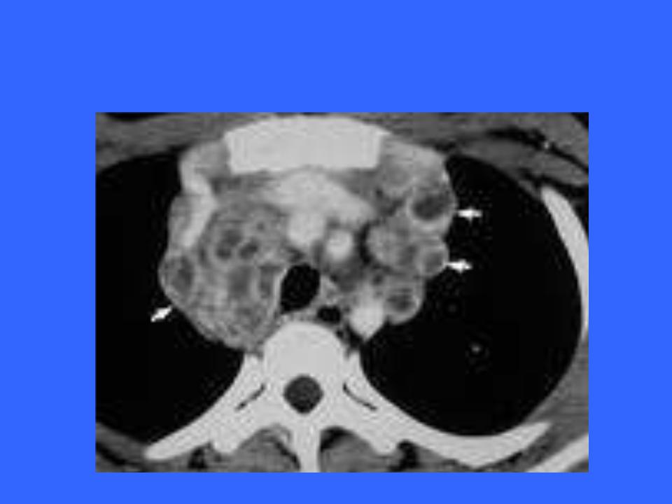
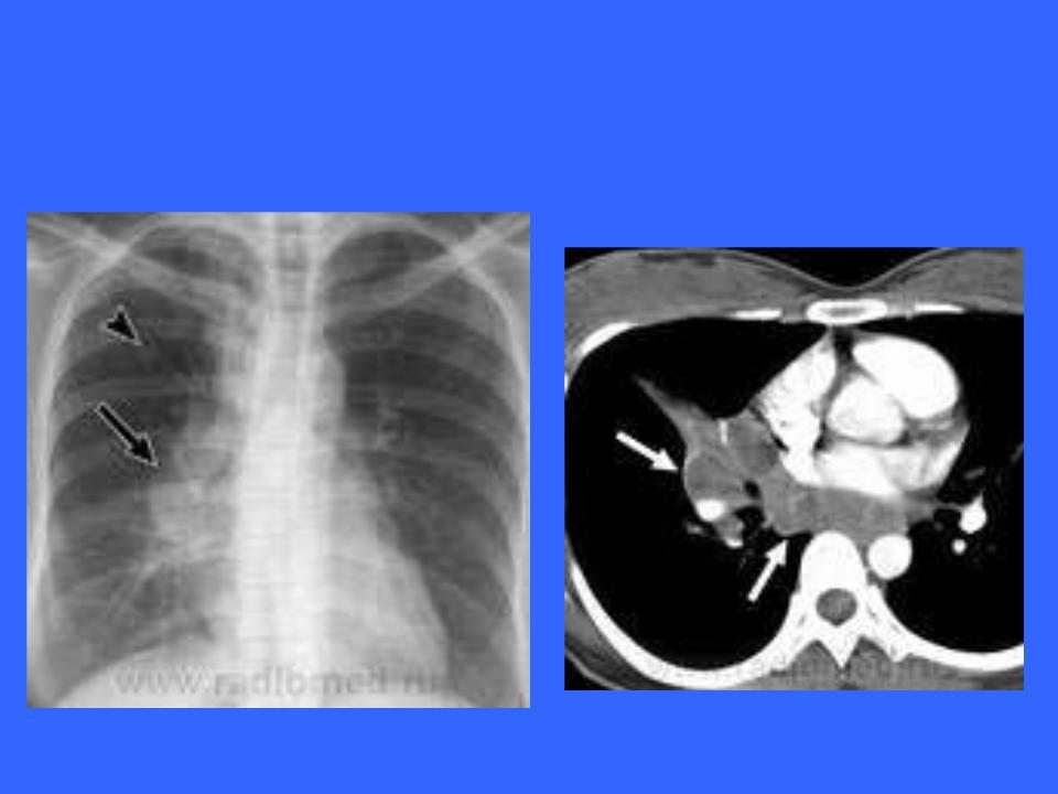
Tumor-like form of intrathoracic lymph node tuberculosis is characterized by the presence of the syndrome on the radiograph polycyclic changes of the root of the lung:
•This syndrome is characterized by all the features of the syndrome of infiltration of the lung root. The lung is infiltrated, but the outer contour of the of the shadow is clear, undulating (polycyclic);
•Increased paratracheal, tracheobronchial groups tracheobronchial groups of lymph nodes are determined in the radiograph as semicircular shadows in the area of upper mediastinum.
Intrathoracic lymph node tuberculosis
Intrathoracic lymph node tuberculosis
Intrathoracic lymph node tuberculosis

Intrathoracic lymph node tuberculosis

DIFFERENTIAL
DIAGNOSISTUBERCULES OF intrathoracic lymph nodes
• ANTERIOR MEDIASTINUM:
1.Thyroid tumors.
2.Hyperplasia of the thymus gland.
3.Teratomas and dermoid cysts.
4.Celomic cysts of the pericardium.
5.Fatty tumors of the mediastinum.
6.Aneurysm of the ascending aorta.

DIFFERENTIAL
DIAGNOSISTUBERCULES OF intrathoracic lymph nodes
• MEDIASTINUM:
1.Tuberculosis of the ILN.
2.Lymphogranulomatosis.
3.Hemodynamic abnormalities in cardiac defects.
4.Nonspecific adenopathies.
5.Lymphosarcoma.
6.Lympholeukemia.
7.Sarcoidosis.
8.Coarctation of the aorta.
9.Mediastinal cancer.
10.Aortic arch aneurysm.

|
DIFFERENTIAL |
|
DIAGNOSISTUBERCULES OF |
• |
intrathoracic lymph nodes |
POSTERIOR MEDIASTINUM: |
1.Neurogenic masses
2.Nathecal abscess.
3.Aortic aneurysm.
4.Esophageal tumors.
5.Broncho- and enteric cysts
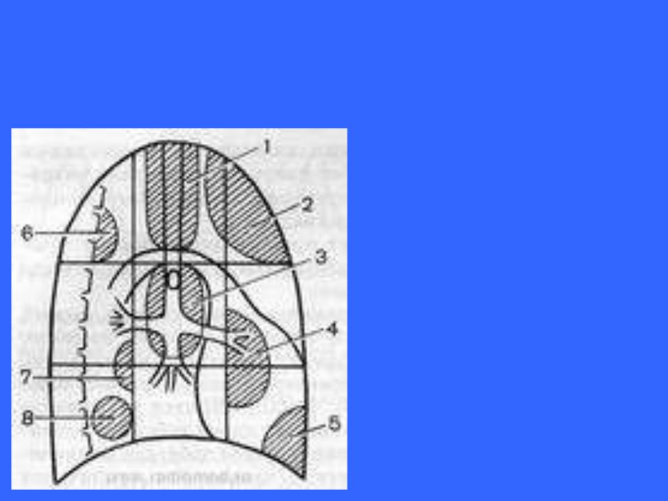
LOCALIZATION SCHEME OF THE
MOSTFREQUENT MEDIASTINAL LESIONSI CHILDHOOD AND ADOLESCENCE
1 - lymphogranulematosis;
2 - tumors of the thymusgland
3 - tumors of lymphoidtissue, tuberculosis,sarcoidosis;
4 – teratodermoid formations;
5 - coelomic cysts;
6 - neurogenic tumors;
7 - cysts of thoracic lymphatic duct;
8 - enterogenic cysts.