

4 курс / Лучевая диагностика / ЛУЧЕВАЯ_ДИАГНОСТИКА_И_ЛУЧЕВАЯ_ТЕРАПИЯ
.pdftumor is a high-grade malignancy, if the tumor is found in connective tissues, if there is extensive invasion of perineural lymphatics, or if there are positive nodes in the operative specimen.
Neoplasms of other sites
Certain lymphomas that present as subcutaneous masses or dermal lesions can be treated by electron beam therapy. In many soft tissue sarcomas, electron beam therapy can be used as total treatment or as an adjunct to photon beam treatment, with preservation of function and diminution in the number of late side effects of the treatment. Primary or recurrent carcinomas of the vulva, distal vagina, urethra, suburethral area, or other areas that recur after surgical removal and carcinomas of the cervix that recur in the vagina may be treated with electron beams (6, 9, or 12 MeV), incorporating an appropriate bolus. Intraoperative electron beam used as a boost followed by photon beam treatment represents an innovative regimen, particularly in treating gastric cancer, retroperitoneal sarcomas, head and neck cancers, and genitourinary and gynecologic cancers.
For curative cases, the typical dose for a solid epithelial tumor ranges from 60 to 80 Gy, while lymphomas are treated with 30 Gy. Preventative (adjuvant) doses are typically around 50 Gy in 2 Gy fractions (for breast, head, and neck cancers).
Brachytherapy
Skin cancer
Radiation therapy is an important management option in selected patients, offering an advantage in treating large lesions with deep tissue infiltration. Treatment margins may be as wide as necessary for facial tumors, obviating the need for extensive surgical reconstruction. Radiation may be preferred for elderly, debilitated, or medically inoperable patients as anesthesia is not necessary, and when cosmesis is not a factor, fractionation can minimize the number of treatments.
Skin cancers with perineural invasion are particularly difficult to control, and salvage after surgical recurrence is unlikely. Treatment fields encompass the nerve at risk to the base of skull with postoperative radiation to 60 Gy to the tumor bed, 50 Gy to the proximal involved nerve with negative surgical margins, and 70 Gy for microscopic or gross positive margins.
A B 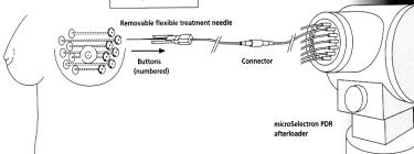
Figure 7.3 (A) The device of contact radiotherapy “MICROSELECTRON“ and (B) scheme using it for breast brachytherapy
81

Customized surface molds employing afterloading 192Ir or high-dose-rate brachytherapy sources (figure 7.3) are useful when protracted daily treatment fractionation is inconvenient or for treatment of lesions on the shin or dorsum of the hand, where conventional radiation therapy is poorly tolerated. Low-dose- rate interstitial brachytherapy can treat periorificial tumors of embryonic fusion regions of the face. Excellent disease control and cosmesis are reported with doses of 50 Gy at dose-rates 2 Gy/hr with low risk of late complications.
Esophageal cancer
In addition to external beam radiation therapy, intracavitary therapy can be used with curative or palliative intent. The advantage of brachytherapy centers on exploitation of the inverse square law and quick dose falloff, thus sparing surrounding tissues from radiation while providing focal dose escalation. The radioactive source of choice is usually iridium-192 (192Ir). High-dose-rate (HDR) techniques can deliver 100 to 400 Gy per hour, allowing treatment to be given in 5 to 10 minutes.
With brachytherapy, an afterloading catheter is introduced through the nose into the esophagus to the primary tumor site under fluoroscopic guidance. After localization films are taken and dosimetry generated, the catheter is then attached to a remote afterloader through a guide cable and the 192Ir source inserted through remote control. Dose can be shaped and modified through the use of dwell times.
For patients treated with curative intent (unifocal thoracic tumors <10 cm, no distant metastases, no airway involvement or cervical esophageal location), the American Brachytherapy Society recommends a brachytherapy dose of 10 Gy in 2 weekly fractions of 5 Gy each or 20 Gy in a single course at 1 Gy per hour. The recommended active length is the visible mucosal tumor with a 1- to 2-cm proximal and distal margins. Ideally, brachytherapy is started 2 to 3 weeks following completion of concurrent external irradiation/chemotherapy to allow mucositis resolution.
Prostate cancer
Brachytherapy for early-stage disease: preplanned transperineal implantation. With the advent of transperineal CT and ultrasound-guided permanent prostatic implantation, the accuracy of isotope source placement has dramatically improved compared with older retropubic methods.
A computerized plan is generated from the transverse ultrasound images, producing isodose distributions and the ideal location of seeds within the gland to deliver the prescription dose to the prostate. Several days to weeks later, the implantation procedure is performed. Needles are then placed under ultrasonographic guidance through a perineal template according to the coordinates determined by the preplan. Radioactive seeds are individually deposited in the needle with the aid of an applicator or with preloaded seeds on a semirigid strand containing the preplanned number of seeds.
At present, the commonly used dose for interstitial implantation is 140 Gy, prescribed to the isodose surface that completely encompasses the prostate as contoured from imaging studies.
82
Soft tissue sarcomas
Brachytherapy may be used as the sole radiation therapy mode of treatment, using doses of 50 Gy. For unresectable sarcomas, doses above 70 Gy are used, limiting the high-dose volume to the tumor plus a minimal margin.
Interstitial brachytherapy can be used to deliver all or part of the radiation dose. After surgical excision of the tumor, hollow plastic afterloading catheters are inserted using sharp metal trocars in a single plane at approximately 1-cm intervals within the tumor bed (figure 7.4). Surgical clips placed at the margin of the tumor bed permit the target volume to be delineated for planning purposes, and the catheters are secured in place.
Catheters are loaded with wired 192Ir seeds no sooner than the sixth postoperative day to reduce the risk of wound complications. After completion of the treatment, sources are removed and catheters are cut at one end for removal by pulling through the skin.
For radioimplants, a dose of 45 Gy has been shown to be adequate adjuvant treatment when used alone for high-grade lesions. Brachytherapy treatments are usually given twice daily at 5 Gy per fraction to 50 Gy.
A 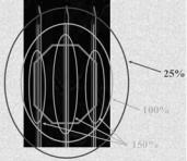 B
B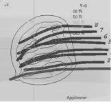
Figure 7.4 (A) Examples dose distribution from radionuclide interstitial a needles and (B) three-dimensional computer-generated image of catheters with
a radionuclide inside of the tumor
Treatment of thyroid cancer
The thyroid gland absorbs nearly all of the iodine in the blood. Because of this, radioactive iodine (also known as radioiodine or iodine-131) can be used to destroy the thyroid gland and thyroid cancer with limited effects on the rest of the body. This treatment is often used after thyroid cancer surgery to destroy any thyroid cells that may have been left behind, or to treat some types of thyroid cancer that have spread to lymph nodes and other parts of the body. An interna-
tional trial has been performed for ablation of cancer cells thyroid tissue using 3.7 GBq (100 mCi) of 131I.
Iodine-131 therapy may be combined with external radiation therapy. The dose to metastases from 131I may be calculated using single-photon emission computed tomography (SPECT) imaging.
In patients with unresectable or inoperable primary tumors, 131I does not seem to affect the rate of tumor regression appreciably or to prolong survival. A combination of 131I and external radiation therapy seems to offer the best results.
83

Side effects of radiation therapy
Radiation therapy is in itself painless. Many low-dose palliative treatments (for example, radiation therapy to bony metastases) cause minimal or no side effects, although short-term pain flare up can be experienced in the days following treatment due to oedema compressing nerves in the treated area. Higher doses (after radiology treatment of malignancy) can cause varying side effects during treatment (acute side effects), in the months or years following treatment (long-term side effects), or after re-treatment (cumulative side effects). The nature, severity, and longevity of side effects depends on the organs that receive the radiation, the treatment itself (type of radiation, dose, fractionation, concurrent chemotherapy), and the patient.
Most side effects are predictable and expected. Side effects from radiation are usually limited to the area of the patient's body that is under treatment. One of the aims of modern radiation therapy is to reduce side effects to a minimum, and to help the patient to understand and to deal with those side effects which are unavoidable.
The extent and the exact type of side effects are determined by the location of the tumor and the location of the radiation being delivered. The side effects of radiation therapy depend on the treatment dose and the part of the body that is being treated. The most common side effects may be loss of hair in the area being treated, tiredness, skin reactions (such as rash or redness) in the treated area, loss of appetite, and nausea. Radiation therapy may also cause a decrease in the number of white blood cells (cells that help protect the body against infection).
The main side effects reported are fatigue and skin irritation, like a mild to moderate sun burn. The fatigue often sets in during the middle of a course of treatment and can last for weeks after treatment ends. The irritated skin will heal, but may not be as elastic as it was before.
For instance, patients who are undergoing breast irradiation will typically experience a redness, dryness or itchiness of the breasts that usually begins two to three weeks after the treatment is commenced. It will then continue, but will eventually leave several weeks after the radiation treatment course is completed.
Another example of a side effect is diarrhea, nausea or vomiting. This is sometimes experienced by patients undergoing radiation treatment to their abdomens or bowels. In most cases, these side effects, which are called acute, take place during the radiation treatment course and will continue for a few weeks after the course is completed.
In almost all cases, these sideeffects will go away and patients willbe fine. In rare instances, some patients will experience long-term side effects or complications, becausetheradiationcausesdamagetoaninternalorganadjacenttoornearthetumorsite.
Normal tissue reactions
Normal tissues vary in their response to radiation. As with tumors, normal tissues that are dividing more rapidly may be affected and cause some of the side effects of radiation treatment. Since radiation is a local treatment, side effects are usually confined to the area being treated.
84
The early reactions are seen during the first days or weeks after irradiation (for example diarrhea or acute mucositis) and may continue for several weeks after treatments are completed. They are temporary because the cell deficit is compensated for by the repopulation of stem cells, and subsequently of differentiated cells. Late reactions due to damage to the late-reacting tissues, for instance blood vessel damage, fibrosis, telangiectasia, etc, may be seen after months.
Common side effects of radiation therapy
Hair loss in the area being treated.
Fatigue.
Skin reactions in the treated area.
Loss of appetite.
Nausea.
Acute side effects
Fatigue is a general effect of radiation but the exact cause is unknown. The tumor may cause the immune system to make substances that lead to fatigue. Fatigue may also be cause by anemia (low red blood cell count), poor nutrition, pain, medicines such as steroids or chemotherapy, depression, and stress. There is no single treatment for fatigue, but if possible, the cause of the fatigue is addressed. For example, if the fatigue is in part caused by anemia, some patients will benefit from blood transfusions or from medicines to stimulate the body to make more red blood cells.
Epithelial surfaces may sustain damage from radiation therapy. Depending on the area being treated, this may include the skin, oral mucosa, pharyngeal, bowel mucosa and ureter. The rates of onset of damage and recovery from it depend upon the turnover rate of epithelial cells. Typically the skin starts to become pink and sore several weeks into treatment. The reaction may become more severe during the treatment and for up to about one week following the end of radiation therapy, and the skin may break down. Although this moist desquamation is uncomfortable, recovery is usually quick. Skin reactions tend to be worse in areas where there are natural folds in the skin, such as underneath the female breast, behind the ear, and in the groin.
Skin
Modern radiation therapy may cause less damage to the skin than in earlier types of therapy because most of the radiation dose is delivered below the surface of the skin. During the first 2 weeks of treatment, a faint and short lasting redness may occur. Dryness and peeling of the skin, called dry desquamation, may occur in 3 to 4 weeks. The skin over the treatment area may become darker. This is because of the effect radiation has on the cells in the skin that produce pigment.
The skin may also become dry and itchy. Moisturizing the skin with aloe vera, lanolin, or vitamin E may help. After a month of treatment, some people receiving radiation may experience some extreme peeling and weeping (moist) areas. Let your medical care team know if this occurs. Later effects of radiation may include thinning of the skin. The skin may feel hard, especially if surgery
85

has also been done in the same area. Some people may have trouble with wound healing in the area that was treated.
Lung
When radiation treatments include the chest area, the lungs can be affected. One early change is a decrease in the levels of surfactant, the substance that helps keep the air passages open. This keeps the lungs from fully expanding, which may cause shortness of breath or cough. These symptoms are sometimes treated with steroids.
A possible late effect of radiation on the lungs is fibrosis (stiffening or scarring), which reduces the ability of the lungs to inflate and take in air. If a large area of the lungs is irradiated, these changes can cause shortness of breath and less tolerance for physical exercise.
Digestive tract
Radiation to the thorax and abdomen may result in swelling and inflammation of the esophagus, stomach or intestines, causing nausea, vomiting, or diarrhea. The lower bowel may be treated directly with radiation (treatment of rectal or anal cancer) or be exposed by radiation therapy to other pelvic structures (prostate, bladder, female genital tract). Typical symptoms are soreness, diarrhea, and nausea. Antacids, sometimes combined with a numbing medicine such as lidocaine, may be helpful in relieving pain from inflammation of the esophagus. Nausea and vomiting can also be treated with medicines. If it is severe, some patients may need intravenous fluids to avoid or treat dehydration. Diarrhea can be treated with medicines and by avoiding spicy.
Swelling (edema or oedema) — As part of the general inflammation that occurs, swelling of soft tissues may cause problems during radiation therapy. Surgical intervention may be considered prior to treatment with radiation. If surgery is deemed unnecessary or inappropriate, the patient may receive steroids during radiation therapy to reduce swelling.
Late side effects
Late side effects occur months to years after treatment and are generally limited to the area that has been treated. They are often due to damage of blood vessels and connective tissue cells.
Fibrosis — Tissues which have been irradiated tend to become less elastic over time due to a diffuse scarring process.
Epilation (hair loss) may occur on any hair bearing skin with doses above 1 Gy. It only occurs within the radiation field/s. Hair loss may be permanent with a single dose of 10 Gy, but if the dose is fractionated permanent hair loss may not occur until dose exceeds 45 Gy.
Dryness — The salivary glands and tear glands have a radiation tolerance of about 30 Gy in 2 Gy fractions, a dose which is exceeded by most radical head and neck cancer treatments. Dry mouth (xerostomia) and dry eyes (xerophthalmia) can become irritating long-term problems.
86
8. Radiology therapy of benign diseases
Introduction
Radiation therapy attacks reproducing cancer cells, but it can also affect reproducing cells of normal tissues. The damage to normal cells is what causes side effects. Each time radiation therapy is given it involves a balance between destroying the cancer cells and sparing the normal cells.
Radiation therapy has several applications in non-malignant conditions, such as the treatment of trigeminal neuralgia, severe thyroid eye disease, and prevention of keloid scar growth. The use of radiation therapy in non-malignant conditions is limited partly by worries about the risk of radiation-induced cancers. Radiation can sometimes play a role in the development of cancer, particularly if people are exposed to it at an early age. We are all aware of the increased frequency of cancers, especially leukemias, among Japanese survivors of World War II. Patients should bear in mind that radiation, as delivered in a radiation department, is a very careful, precise and well monitored treatment which very rarely leads to the development of cancer.
Radiation treatment of benign disease
The use of radiation in the treatment of benign diseases has a long history. Soon after the discovery of x-rays by Roentgen in 1895, the therapeutic potential of radiation was recognized. In the first half of the 20th century, radiation therapy was used empirically for a host of conditions, both benign and malignant. In many situations in which no effective therapeutic alternatives existed, radiation therapy may have been one of the few treatment options available. Even with recognition of the risks of late skin injury, carcinogenesis, leukemogenesis, and genetic damage from all ionizing radiation, radiation therapy also continues to be accepted treatment for benign diseases that do not respond to other methods of therapy.
In many cases, the use of radiation therapy for benign disorders may be able to spare the patient morbidity associated with the progression of the disease, or additional medical or surgical therapy.
The need for informed consent exists everywhere in radiation oncology, and certainly in the use of radiation therapy for benign diseases. Patients should be informed of the rationale, potential side effects, and treatment alternatives to radiation treatment before it is delivered.
Radiation carcinogenesis, although always a concern when ionizing radiation is used, appears to be a greater risk for pediatric patients and young adults than for older adults, and must be weighed in relation to the consequences of progressive disease and the side effects associated with alternative therapies. Special caution should be used before radiation is delivered to pediatric patients and young adults for benign conditions. These young patients may be at in-
87

creased risk of second malignancy induction and late atrophy, especially if the area to be treated is in proximity to the breasts or thyroid, tissues that may be sensitive to carcinogenesis. As in all aspects of radiation therapy, care should be taken to use beams of appropriate energy and to shield adjacent normal structures.
Technical considerations
1.Before institution of therapy, the quality of irradiation, total dose, overall time, underlying organs at risk, and shielding factors should be considered.
2.Meticulous radiation protection techniques, including cones and lead shields, should be used in all instances.
3.The depth of penetration of the ionizing radiation beam should be chosen in accordance with the depth of the pathologic process.
As in all radiation therapy, the choice of beam energy depends on the depth of the target volume, and every effort is made to spare normal underlying tissue in superficial lesions. Absorption data for ionizing radiation therapy are readily obtained from standard depth-dose tables.
Internal lead shields placed in oral or nasal cavities tend to increase the dose to overlying tissue for both x-ray and electron-beam treatments. This increase is caused by backscattered electrons and may be minimized by encasing the shields in plastic.
Exophthalmos
Signs and symptoms of Graves' ophthalmopathy include bilateral exophthalmos, extraocular muscle dysfunction, diplopia, blurred vision, eyelid and periorbital edema, chemosis, lid lag and retraction, and compressive optic neuropathy. The pathogenesis is believed to be an autoimmune disease in which activated T-lymphocytes invade the orbit and stimulate glycosaminoglycan production in fibroblasts, resulting in tissue edema, lymphocytic infiltration, and marked enlargement of the extraocular muscles. Because lymphocytes and fibroblasts are sensitive to radiation, retrobulbar irradiation is a logical method of treatment. Systemic high-dose steroids are customarily used, but they must be given for long periods and have many side effects. Surgical orbital decompression is used when there is rapidly progressive optic neuropathy or severe proptosis.
Megavoltage external-beam irradiation using precise planning with highresolution computed tomography (CT) scans and complete patient immobilization is recommended for optimization of dose distribution and to avoid unwanted irradiation of sensitive structures (e. g., lens, pituitary gland).
Small opposed bilateral fields are used to encompass both retrobulbar volumes with customized blocks to shield periorbital structures. Either a split-beam technique or a 5-degree posterior angulation should be used to avoid the lens.
88
The total dose is 20 Gy to the midplane, given in 10 fractions over 2 weeks. Photons of 5-MV are used in most cases.
Keloids
Some persons have a tendency to react to skin trauma with excessive production of fibrous tissue that extends beyond the wound, becomes hyalinized, and does not regress spontaneously. These keloids become unsightly masses, and they frequently cause itching and pain. They may occur in susceptible individuals after infection, burns, or (most commonly) surgical wounds.
Although suture tension and stitch infection in the wound may be contributing factors, the recurrence rate after excision is very high even in their absence, and surgical treatment alone is not recommended.
Radiation therapy is usually started within 24 hours after excision, using 120-kV x-rays or low-energy electrons. The radiation field is custom-shaped with lead, with a 0,5-cm margin around the suture lines. The ear lobes, when treated, are taped away from the face, and a direct anterior or posterior field is used with a small cone. If the lobe is more than 1-cm thick and the wound extends around it, a higher-energy beam is needed. The total dose is 10 to 15 Gy in two to five fractions in 1 to 2 weeks.
Treatment of established keloids by irradiation alone is not as successful but may be attempted if surgery is not indicated (e. g., in an elderly patient with a large symptomatic lesion or in presternal and shoulder keloids that commonly recur even after combined treatment.
Cutaneous lesions
Treatment of cavernous hemangiomas of the skin in infants by repeated doses of radium in surface applicators was commonplace many years ago. A trial of oral steroids is now the preferred treatment for skin hemangiomas requiring intervention, such as rapidly growing facial lesions causing disfigurement, and radiation therapy is reserved for lesions that threaten function or life and have failed alternative therapies.
For minor superficial hemangiomas, contact radiation therapy is most suitable. The skin dose is 5 to 10 Gy per treatment. For thicker lesions, orthovoltage irradiation is recommended, with doses of 1 to 4 Gy per treatment.
Bursitis and tendinitis
Bursitis and tendinitis most commonly affect the shoulders. These disorders are caused by degenerative and inflammatory changes in the supraspinatus and infraspinatus tendons that lead to calcium deposition, inflammation of the surface of the subdeltoid bursa, and even rupture and discharge of calcific material into the bursa. Calcification may occur without symptoms, or there may be pain, tenderness, and limitation of motion in acute, subacute, or chronic forms. It
89

is probably true, however, that irradiation is equally effective and is sometimes successful when invasive local treatments are not.
Limited fields encompass the joint only, using either opposed or occasionally a single anterior field. A daily dose of 1,5 to 2 Gy is given on 3 to 5 successive days for a total of 6 to 10 Gy. One or two additional treatments may be added after 1 or 2 weeks in chronic cases, in which results are much less satisfactory.
Rheumatoid arthritis
The patients selected had severe rheumatoid arthritis with active synovitis and significant disability treated unsuccessfully with nonsteroidal antiinflammatory agents, gold, and penicillamine. All patients would have been suitable candidates for cytotoxic therapy with agents such as azathioprine or cyclophosphamide, which, although effective in controlling advanced disease, may be accompanied by significant side effects.
In the Stanford trial, the regimen consisted of mantle field treatment with 20 Gy in 10 fractions of 2 Gy in 2 weeks. Patients were evaluated before and after therapy with respect to joint tenderness and joint swelling, duration of morning stiffness, and a global composite score. Complications of treatment included mild systemic effects, such as nausea and fatigue, and local effects, including dysphagia, xerostomia, and esophagitis.
Conclusions
Radiation therapy continues to be used in the treatment of various benign disorders because the profile of efficacy and morbidity compares well with the available treatment alternatives. There is a well-established role for radiotherapy in the management of benign neoplasms when surgical therapy is medically contraindicated or would carry undue morbidity. In addition, there are situations in which anatomically limited proliferations of lymphoid, epithelial, or mesenchymal tissue can cause significant morbidity that can be avoided or mitigated with local radiation treatment rather than systemic medical therapy or repeated surgical procedures.
90