

4 курс / Лучевая диагностика / ЛУЧЕВАЯ_ДИАГНОСТИКА_И_ЛУЧЕВАЯ_ТЕРАПИЯ
.pdfA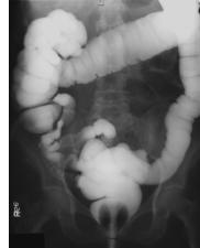 B
B 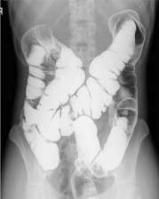 C
C 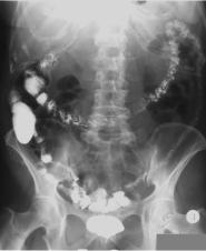
D 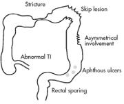 E
E 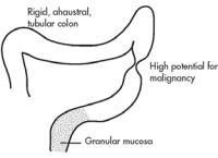
F 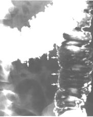
Figure 3.4 (A) Single-contrast barium enema and (B) double-contrast barium enema of the normal colon. Although a single contrast study may be easier to perform, mucosal detail is better seen on the double contrast study.
(C) Post-evacuation radiograph of abdomen and pelvic.
(D, E) Schemes variants Crohn’s disease of the large bowel. There is a stenosis of the descending and tranverse colon. The haustral pattern is lost. Numerous deep fissured ulcers can be seen and there is irregular mucosal thickening at the hepatic flexure with a cobblestone appearance. This portions of bowel is affected asymmetrically. The bowel involvement is thus asymmetrical with skip lesions.
(F) Ulcerative colitis (spot radiograph of the colon with marked multiplies of ulcers craters)
Colitis
Radiographic features of bowel inflammation (any etiology) depend mainly under of the process. Ulceration shallow: granularity of mucosa, aphthoid. Ul-
21

ceration deeper: flasklike collections of barium: collar button. Edema: displacement of barium (translucent halo around central ulcer). Spasm: localized persistent contraction/narrowing of bowel lumen.
Ulcerative colitis
Ulcerative colitis is an idiopathic chronic inflammatory disease that is characterized by superficial ulcerations, edema, and hyperemia of the colonic mucosa and submucosa. The disease usually begins in the rectum and extends proximally in a continuous pattern.
The radiographic hallmarks of ulcerative colitis are confluent, circumferencial, shallow ulcerations of the colon, granular mucosa, collar button ulcers, and "thumbprinting" (figure 3.4).
Structural abnormalities
Diverticulosis
Diverticulosis is a condition in which the mucosa and muscularis mucosae herniate through the muscularis propria of the colon wall and produce a saccular outpouching. Approximately 70 % of diverticula occur in the sigmoid colon and 25 % in the ascending colon. Plain films and barium studies reveal diverticula as gas/barium-filled sacs parallel to the lumen of the colon. Most are 5–10mm in diameter, but may range from tiny spikes to 2 cm.
Hallmarks on barium enema include distorted diverticlar sacs, evidence of abscess, and leakage of barium outside the bowel lumen (figure 3.5).
A 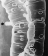 B
B 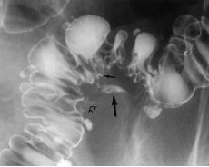 Figure 3.5 (A) Diverticulosis of the colon. Coned-down image of the splenic flexure from a left-side-down decubitus overhead from a double-contrast barium enema reveals numerous diverticula. Some barium-filled diverticula are seen in profile (thick black arrow). Most diverticula seen en face have a small pool of barium on their dependent surface resembling a meniscus (thin black arrow). Other diverticula are devoid of a barium meniscus and are depicted only as barium-coated ring shadows (open arrow). The nor-
mal haustral sacculations and interhaustral folds are preserved.
(B) Diverticulitis. Spot radiograph of the sigmoid colon from a double-contrast barium enema shows several small barium-filled tracks (open arrow) forming a flame-shaped collection (large arrow) in the pericolic space. The wall of the adjacent sigmoid colon has a spiculated contour (spicule represented by small arrow). There is an extrinsic mass impression on the inferior wall of the sigmoid colon. Compare the asymmetric inflam-
matory changes with the normal contour of the opposite colonic wall
22
Neoplasms
Polyps
Polyps are focal masses that protrude from the mucosa into the bowel lumen. In general, polyps less than 1cm in diameter have a 1 % risk of cancer, 1–2 cm have a 10 % risk, and those greater than 2cm have up to a 50 % risk. Thus, barium studies are often indicated to detect colon polyps (figure 3.6).
Radiographic finding: benign polyps are small in diameter, stable in growth, spherically shaped, have normal mucosa, long stalks, and a smooth surface. Malignant polyps on the other hand are large in diameter, sessile, irregularly shaped, may exhibit sudden growth, and have a broader base and puckered mucosa.
A 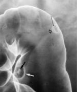 B
B  C
C 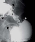 Figure 3.6 (A) Spot radiograph of the splenic flexure shows two polyps.
The small polyp resembles a hat. The top of the hat (open arrow) is the top of the polyp. The brim of the hat (thin black arrow) is barium trapped between the mucosa and the polyp as it is retracted by its stalk. This was a tubular adenoma. The second polyp has a more worrisome morphology—that of an umbilicated, sessile polyp. A small barium collection (thick black arrow) fills a central umbilication or ulceration. The edge of the polyp is coated by barium (or “etched in white”) (white arrow). This polyp was a tubulovil-
lous adenoma.
(B)Schemes variants of the colorectal cancer.
(C)Annular adenocarcinoma of the rectum. Spot radiograph from the single-contrast phase of a double-contrast barium enema shows a 5-cm-long, circumferential narrowing of the proximal rectum. The lesion has abrupt shelf-like margins (large black and white arrows) and an irregular surface (open arrows). This has been called an “apple core” lesion, but this is a misnomer because the tumor represents the part of the apple that has
been eaten away, and the lumen would be the “apple core”
Colorectal cancer
Colorectal adenocarcinoma is the most common GI malignancy and is the second most deadly cancer in the United States. Signs and symptoms depend on the location of the tumor. Approximately 50 % of these malignancies develop in the rectosigmoid area and 25 % in the cecum and ascending colon. Many likely arise from a malignant transformation of an adenomatous polyp. These tumors tend to form annular constricting lesions or bulky exophytic masses, with raised everted edges and ulcerated mucosa, ranging from 2–6 cm in diameter. They spread by direct invasion into the pericolonic fat and adjacent organs, through
23

lymphatic vessels to regional nodes, and hematogenously via systemic and portal circulation.
Apple-core constricting lesions, tumor masses, nodal involvement, and metastases may be visualized (figure 3.6).
X-ray diagnostics of some urgent abdominal conditions Intestinal obstruction
The main sign of intestinal obstruction — Kloiber's cup and arches (air-fluid levels). The small bowel obstruction — the width of horizontal fluid level is more than height of a gas bubble above it; on a background of gas the mucosal folds reminding a spring can be visible; the localization of Kloiber's cups is more often in medial parts of the abdomen. Arches — the intestinal loops, filled gas and limited by horizontal fluid levels.
A 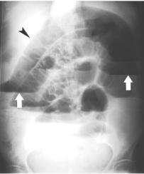 B
B 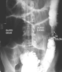
C 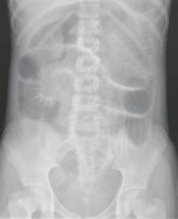 D
D 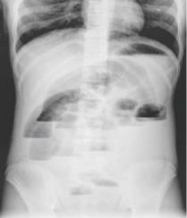
Figure 3.7 (A) Small bowel obstruction (black arrow – dilated small bowel, white arrows – air-fluid levels). (B) Large bowel obstruction.
(C) Supine and (D) upright abdominal radiographs same patient demonstrate a “stacked” appearance of multiple segments of markedly dilated small bowel with numerous air-fluid levels at high-grade distal small bowel obstruction
24
The large bowel obstruction — the width of a horizontal level is less than height of a gas bubble above it; the localization of Kloiber's cups is more often in lateral parts of the abdomen; on a background of gas colic haustres sometimes are visible (figure 3.7). For more exact determination of a level of an intestinal obstruction it is necessary to give a patient the barium sulfate per orally and to perform dynamic X-ray. The stopping of barium mass will show the localization of an obstruction. The mechanical obstruction - at a roentgenoscopy there's detected «migration» of a fluid from one loop of an intestine to another, fast motions of fluid levels. The dynamic obstruction - at a roentgenoscopy no motions of fluid levels, barium meal passes very slowly.
Free gas in an abdominal cavity
The free gas in an abdominal cavity — can be the result of perforation of hollow organs (stomach, intestine), penetrating wounds of an abdominal wall; after operations on the organs of an abdominal cavity, or owing to injection of gas in an abdominal cavity with the diagnostic purpose (diagnostic pneumoperitoneum). In a vertical position of a patient there's detected a falciform accumulation of gas under the right dome of a diaphragm (at presence of a much amount of gas — under the right and the left also) (figure 3.8), in a latero-position — under a lateral wall of an abdominal cavity.
A 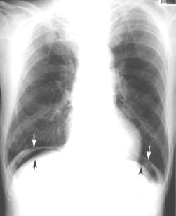 B
B 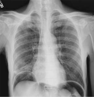
Figure 3.8 (A) Free gas of abdominal cavity (arrows).
(B) This upright chest radiograph demonstrates crescent shaped lucencies under both hemidiaphragms. This is free intraperitoneal gas, or “free air,” which rises up to the top of the peritoneal cavity when the patient is upright
Pneumoperitoneum is commonly due to recent surgery, but it may also indicate a ruptured abdominal viscus, such as a perforated duodenal ulcer.
25

4. NUCLEAR MEDICINE
Introduction
Nuclear medicine imaging (also called radionuclide, radioisotope scanning) is an excellent diagnostic tool because it shows not only the anatomy (structure) of an organ or body part, but the function of the organ as well. This additional "functional information" allows nuclear medicine to diagnose certain diseases and various medical conditions much sooner than other medical imaging examinations which provide mainly anatomic (structural) information about an organ or body part. Nuclear medicine can be valuable in the early diagnosis, treatment and prevention of numerous medical conditions and continues to grow as a powerful medical tool.
Plain films, US, CT, and MRI produce anatomic images with very high spatial resolution. A viewer can see anatomy very well, but function generally is not assessed. Nuclear medicine studies sacrifice spatial resolution, but in return offer information about organ function. Nuclear medicine studies, in general, are very sensitive, but relatively nonspecific in the detection of pathology.
Principles and methods
How it is done:
Radionuclides are injected into the body.
The nuclide concentrates in the organ that is being tested.
The imaging is done by tracing the distribution of radiopharmaceuticals within the body.
The radioactive gamma rays are emitted through the body as the natural decaying process of these isotopes takes place. The emissions of the gamma rays are captured by detectors that surround the body. This essentially means that the human is now the source of the radioactivity.
Scanning of body or organ follows.
A radionuclide can be a pure element (e. g. iodinum-131) or a biological agent labeled with radioisotopes.
All radionuclides are expelled from the body with an associated half life. Useful for:
Primarily useful to evaluate the function of the organ studied.
Nuclearimagingdetectsfunctional(vs.anatomical)propertiesofhumantissues. Advantage:
Highly sensitive. For example, early osteomyelitis may not be visible on plain films for 7-10 days, while scintigraphy will be positive at the time of presentation. Skeletal metastatic spread can frequently be detected significantly earlier by scintigraphy than by radiography.
Radionuclide imaging is safe since it does not carry the risk of allergic reaction encountered with contrast.
26
Radiation exposure is minimal.
The principal benefit of radionuclide imaging is that it is able to detect physiologic abnormalities within a given tissue.
Limitation and disadvantages
The main disadvantage of scintigraphy is its non-specificity (to take a common example, an isolated 'hot spot' on a bone scan could be due to infection, trauma, or neoplasia and correlation with clinical history and other imaging studies is of paramount importance).
Nuclear medicine applications
Radioactivity
A radioactive nucleus is one that is unstable and will spontaneously disintegrate or decay at some time. The reason for the instability is either that the nucleus is too large or because there is an imbalance between the protons and neutrons. Unstable nuclei have an excess of energy. When a nucleus disintegrates, it will emit radiation either as photons (gamma rays) or as high-velocity particles.
The particular nature and energy of emissions are characteristic of each radionuclide. The radiation generally is ionizing, meaning that it is highly energetic. Radionuclide imaging uses ionizing radiation. However, the radiation is emitted from within the patient and subsequently detected in the imaging device.
A radionuclide is an unstable atom that undergoes radioactive decay. In imaging, the most common radionuclide used is technetium, which is a gamma emitter. Pure gamma emitters (e. g. 99mTc = technetium-99m) are preferred whenever possible because beta particles give patients very much higher doses of radiation without contributing to the image. Technetium-99m is the most frequently used radionuclide in nuclear medicine today, which combines the advantages of optimum radiation properties (emission of exclusively γ-radiation with suitable energy, short half-life of 6 hours) and general availability as a generator nuclide.
Principles of nuclear medicine
In nuclear medical diagnostics, unsealed radioactive substances are used to image organ functions non-invasively. In contrast to other imaging diagnostic modalities (ultrasound, X-ray, to some extent also magnetic resonance tomography), the nuclear medical examination approach is primarily function-oriented. In this case vital processes such as blood circulation, metabolism and viability of organs or tumors can be displayed as “functional images”. Nuclear medicine provides physiological images, i.e. the metabolic activity of the organs process the radiopharmaceutical and concentrate it in the target organs for imaging.
Radiopharmaceuticals
Nuclear medical examinations are based on use of radiopharmaceuticals. Radiopharmaceuticals (radiotracers) are the possible for clinical use radionuclides (radioisotopes) and labelled medias. Nuclear medicine imaging examina-
27

tions are performed by administering various radiopharmaceuticals to the patient and subsequently recording in vivo distribution. Radiopharmaceuticals consist of two main components: (1) the main component that is distributed to various organs via a number of different mechanisms, and (2) the radionuclide that is tagged to the main component, which emits gamma rays, permitting detection of the compound in the body.
Technetium-99m is the most frequently used radionuclide in nuclear medicine today because it is widely available, easily prepared, and has a relatively short halflife (6 hours) with an acceptable whole-body radiation dose. Its parent nuclide, mo- lybdenum-99 (99Mo), can be produced in either a reactor or a cyclotron. 99Mo has a half-life of 66h and therefore 99Mo/99mTc generators can conveniently be supplied weekly to nuclear medicine departments, providing a fresh daily supply of the radionuclide. The 6-h half-life of 99mTc is sufficiently long for most imaging applications and its 140 keV gamma radiation has reasonable tissue penetration but can still be easily collimated. 99mTc can be administered in free form as the pertechnetate ion (TcO4-); however, owing to its diverse chemistry, it can also be incorporated into various pharmaceuticals for visualization of different organ systems.
The amount of radiation that is taken up and then emitted by a specific body part is linked to the metabolic activity (cellular function) of the organ or tissue. For example, cells which are dividing rapidly (like cancer tissue cells) may be seen as "hot spots" of metabolic activity on a nuclear medicine image, since they absorb more of the radionuclide.
A radionuclide that has desirable imaging properties can usually be used to make a variety of radiopharmaceuticals. Thi is done by coupling the radionuclide with variou stabl compounds that are localized b organs or disease states. Different radiopharmaceuticals are used for displaying blood circulation, blood volume, lung ventilation, gastrointestinal passage, absorption and secretion, phagocytosis, cell kinetics, antigen or receptor binding as well as metabolism and thus viability.
Common adverse, nonspecific reactions such as vasovagal reactions with syncope, hypotension and diaphoresis can occur with the intravenous administration of any drug and are not specific for radiopharmaceuticals.
Nuclear medicine can image and/or show the function a variety of organs and body parts to diagnose a number of medical conditions including:
abdomen (example given, to check for gastrointestinal bleeding);
brain (e. g., to look for tumors or aneurysms (blood vessel disease) or evaluate stroke);
blood (e. g., to test for various blood cell disorders);
breast (e. g., to image breast cancers);
hepatobiliary system (e. g., to check gallbladder and bile duct function);
heart (e. g., to look for coronary artery disease, myocardial infarction, valve disease or heart attack; to detect heart transplant rejection; check the effectiveness of bypass surgery; to select patients for angioplasty);
28
kidneys (e. g., to check renal function; to detect renal tumors; to test for renal transplant rejection);
liver/spleen (e. g., to check for cirrhosis or metastatic cancer);
lung (e. g., to check for pulmonary embolism (blood clot), check for lung transplant rejection;
lymphatic system (e. g., to detect if cancer has spread to the lymph nodes);
skeletal system (e. g., to check for metastatic cancer or to test for hidden bone trauma in sports injuries);
stomach(e.g.,tocheckforstomachfunctionandtoconfirmulcersorcancer);
thyroid and parathyroid (e. g., to check for tumor or abnormal function). Methods of nuclear medical routine diagnostics are today used most fre-
quently for examinations of the thyroid gland, skeleton, heart, kidneys, lungs and brain as well as of oncological and inflammatory diseases.
Nuclear medicine is used mainly to allow visualization of organs and regions within organs that can not be seen on conventional x-ray images. Space occupying lesions (injury or abnormality), especially tumors, may stand out on nuclear medicine images. Generally, these lesions are seen as areas of reduced radioactivity (called a "cold spot"); however, in some instances, like bone scanning, areas of increased activity (called a "hot spot") represent disease or injury (pathology).
Methods of radionuclide examinations
Radiometry — the method of nuclear examination with use of a single detector placed above the certain area of a patient's body to determine the percentage of radiotracer intake in the organ (figure 4.1).
Radiography (radionuclide, not X-ray) - the method of nuclear examination with use of a single detector placed above the certain area of a patient's body to determine the intensity and speed of radiotracer's uptake and excretion from the organ. The result is presented as a time-activity graph.
Radionuclide scanning — the old method of nuclear imaging with use of rectilinear scanners, systems which have a single pencil-like detector. By moving this detector over a body region of a patient in a meandering fashion, and printing the local gamma-ray intensity, images of the distribution of radioactivity could be obtained.
(Radionuclide) scintigraphy — the method of nuclear imaging with the use of gamma-cameras, systems which detect the radiation at a large area of a patient's body, transfer gamma photons energy to light flashes (scintillations) and electric signals with subsequent computer digitalization and image creation.
Radioimmunological examinations (RadioImmunoAssays — RIA) — the group of nuclear laboratory examinations which are used to measure minimal quantities of biological substances with the help of radioactively labelled molecules. Based on the reaction between an antibody and an antigen whose concentration has to be quantified. Radioimmunoassays are very sensitive and able to detect nanoor picomoles of substances. For this purpose uptake and kinetics of the tracers are quantitatively evaluated from measurements with external probes.
29

A 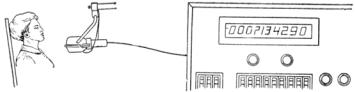
B 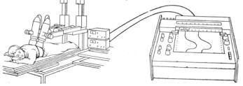
C 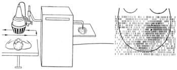
D 
Figure 4.1 Schemes of methods radionuclide examinations (nuclear medicine).
(A)Radiometry. (B) Radiography (radionuclide, not X-ray radiography).
(C)Radionuclide scanning. (D) (Radionuclide) scintigraphy
Techniques of image acquisition and processing
A major strength of nuclear medicine imaging is its ability to show and quantify physiological function. This capability is supported by a range of acquisition protocols and image processing software. This section introduces the main types of study.
Static image is the basic image; other acquisitions consist of a series of such images. This determines the number of pixels there will be in the image. Each gamma ray that is accepted by the camera is saved as a count in the pixel corresponding to its location. A typical image may contain between 100 000 and 1 million counts, and take up to several minutes to acquire.
Dynamic imaging consists of a sequence of basic images or frames acquired over time. The frame rate is chosen according to the rate of change of the
30