

6 курс / Кардиология / Практическая_электрокардиография_Марриотта_Galen_S_W_,_David_G_S
.pdfBarney Marriott was one of those bigger-than-life icons who populated the 20th century. To those who knew him at all, he was simply Barney. Born on the eve of St. Barnabas’ day in 1917 in Hamilton, Bermuda, he was never referred to as Henry J.L. Marriott. Those who did were likely destined to remain strangers . . .
but not for long. He was never a stranger to me. I have had the wonderful and rare privilege of spanning the charmed lives and careers of both authors of this book. Galen Wagner, my mentor, friend, and colleague for the past nearly 40 years, has asked me to pen a reminiscence of Barney because, for the last 25 years of Barney’s life, he and I were buddies. Therein lies a tale.
Following his early formative years in Bermuda, this “onion,” as Bermudans call themselves, went to Oxford as a Rhodes scholar. He enrolled at Brasenose College. The principal of Brasenose was a German named Sonnenschein (later changed to Stallybrass), about whom Barney painted me a picture of respect, awe, and perhaps a little disdain. Traveling to London during the war (not The War), he matriculated at St. Mary’s as a medical student, then as a registrar. During our many luncheon outings together, Barney would regale me to stories of St. Mary’s. Not uncommonly, the Germans would launch their V-1 missiles called “buzz bombs” (because of their ramjet engines) to rain terror on the English populous, especially London. Barney would laugh in his usually reserved guffaw as he told me that the medical students had been fascinated by these weapons. The V-1 missiles emitted a characteristic high-pitched “clack-clack-clack” as they approached the city, then silence as the missiles entered their final path to their target. Barney said that the clacking drew the students to the wide open windows of the anatomy lab on the top floor of St. Mary’s, except for Barney, who, not quite ready to meet his maker, had dived under the cadaver dissection table seeking some sort of premortem protection provided by his postmortem colleague. Happily for all concerned, there were no acute casualties in the St. Mary’s Medical School anatomy lab during those wartime adventures.
In another tale of St. Mary’s, Sir Alexander Fleming had performed his initial studies into the isolation and first clinical use of penicillin in that institution. By the time of Barney’s registrar years, the original “penicillin lab” had become a registrar’s on-call room. Barney was the registrar on the Penicillin Service, where he and his attending made fateful decisions about who was to receive the new lifesaving antibiotic and who was not. Dr. Marriott’s attending of that era was George
Pickering, later knighted and a much later successor to Osler as Regius Professor of Medicine at the Radcliffe Infirmary at Oxford.
Following the war, Barney came to the United States. After a fellowship year in allergy at Johns Hopkins Children’s Center, Barney moved across town to the University of Maryland. As a young faculty member there and director of the Arthritis Clinic, Dr. Marriott was drafted into the role of teaching and supervising ECGs, a job he embraced with a fervor that was infectious and illuminating. By the late 1950s, Barney had grown tired of Baltimore and its cold, wet winters. He accepted a position at Tampa General Hospital in 1961 as director of Medical Education, where he remained for several years.
In 1965, Dr. Marriott was approached by Frank LaCamera of the Rogers Heart Foundation to relocate across the bay to St. Petersburg, where he began his series of seminars on ECG interpretation. Many greats of cardiology nationally and internationally were invited to speak at these seminars. Regardless, it was Barney who set the curriculum and the informality that characterized his personal approach to teaching. Those landmark courses put Barney and his talents in front of literally tens of thousands of doctors and nurses around the world for the next 40 years. All the while, he published over 17 books, mostly on electrocardiography. His scholarly writing was not limited to books. His list of published scientific papers is prodigious. The New England Journal of Medicine alone published papers spanning over 50 years of his vibrant productivity. Barney’s love of language is apparent in one of his least well-recognized contributions. For many years, Dr. Marriott was the author of the Medical Etymology section of Stedman’s Medical Dictionary. He reveled in and revered English and its many quirky words and grammatical rules.
In addition to his visiting professorships at Emory and the University of Florida, the University of South Florida (USF) in Tampa was fortunate to have Barney on its volunteer clinical faculty beginning in the 1980s. Monthly or quarterly, Barney would bring a mountain of carousel slide trays to our evening conferences. It was the glorious, now bygone era of big pharma. The fellows and faculty alike would be repeatedly skewered by Barney’s rapier-like witticisms as he led and pushed us to be better ECG readers. His acumen and sharpness for his task and his boundless enthusiasm were hallmarks of the conferences. Aphorisms such as “Every good arrhythmia has at least three possible interpretations” poured forth like the sangria that fueled raucous audience participation. Barney’s old friends from around the United States and the world would drop by to be toasted and roasted by the master. David Friedberg, an immigrant to the United States from South Africa, was one of the first I encountered. Later, Bill Nelson joined our faculty at USF and became a suitable stage partner and foil for Barney. One particularly memorable evening, Leo Schamroth himself, from South Africa, joined Barney, David, and me for an evening at Bill Nelson’s home, where we argued about concealed conduction and AV block late into the night.
As the decades in the Tampa Bay region wore on, Barney and his companion, Jonni Cooper, RN, spent more time at their place in Riverview, Florida, where he had a large library and workspace for his many books and teaching projects. Chief among those books was his personal favorite, Practical Electrocardiography, a bestseller up to today. It remained a single-author volume through the eight edi-
tions he wrote. He graciously facilitated Galen Wagner’s evolution of print and electronic formats through the subsequent editions. In those first eight editions, beginning in 1954, Barney loved to write with his uniquely conversational style, unlike just about any textbook that you might find in a medical bookstore. Practical Electrocardiography was and remains, however, a very special, now multiformat text suitable for students of all ages and skills at ECG interpretation.
Barney and I continued our monthly lunches as he and Bill Nelson and I put together his last book, Concepts and Cautions in Electrocardiography. Barney’s health held on until his terminal bout with lung cancer; we increased the frequency of those meetings as his health declined. To the very end, he remained gracious, charming, curious, and firmly attached to his ECGs. Every week, tracings continued to come to him from former students around the globe. On my Thursdays with Barney, my task was to bring the Guinness so that we could chat, look at ECGs together, lift a few pints, and reminisce a bit. He reminded me, as his life ebbed away, that being bitter and holding grudges was “a useless waste of time.” It was a lesson for all of us. His legacy remains much more than the eponymic moniker for this volume. Pour me another Guinness. Cheers, Barney.
Douglas D. Schocken, MD
Durham, North Carolina
July 2013

Barney Marriott created Practical Electrocardiography in 1954 and nurtured it through eight editions. After assisting him with the 8th edition, Galen Wagner enthusiastically accepted the challenge of writing the subsequent editions. The 9th edition had extensive revisions to the text, the 10th edition had almost completely new illustrations, and the 11th edition had further text and figure updates and also an accompanying DVD with interactive animations. For this 12th edition, David Strauss joined Galen as coauthor. Galen and David have been working together on electrocardiographic teaching and research challenges for the past 9 years.
One of the strengths of Marriott’s Practical Electrocardiography through its more than 50-year history has been its lucid foundation for understanding the basis for ECG interpretation. Again, in this revision, we have attempted to retain the best of the Marriott tradition—emphasis on the concepts required for everyday ECG interpretation and the simplicities, rather than complexities, of the ECG recordings. Tobin Lim coauthored many of the 11th edition chapters and served as the primary developer of the digital content associated with that edition.
Tobin Lim’s input continues into this 12th edition, and David Strauss has led even further into the electronic-based interactive learning experiences. More than 30 of the figures that evolved through previous editions have now been converted through the creative expertise of Mark Flanders into animated movies accessed via QR codes embedded in the book. David has also collaborated with electrocardiographic educators who are especially skilled in e-based education to add interactive video content to many of the 12th edition chapters. These include Raymond Bond and Dewar Finlay in Chapter 2, Charles (Bill) Olson in the new Chapter 4, and Peter van Dam in Chapter 9.
The chapters are in the same order as in the 11th edition; however, two new chapters have been added. In Chapter 4, Bill Olson, Harvey Estes, Vivian Kamphuis, and Esben Carlsen contribute to the introduction of “The Three-Dimensional Electrocardiogram”; and in Chapter 8, Albert Sun presents “Inherited Arrhythmia Disorders.” Each of the now 24 chapters is divided (as indicated in the table of contents) into discrete, compact “learning units.” Each learning unit begins on a new page to provide blank space for the reader’s notes. The purpose of the learning units is to make this book easier to use by allowing the reader to be selective regarding the material to be considered at a particular time. Because the modern student of electrocardiography is primarily oriented to a visual perspective, we have typically begun each page with an illustration.
The four chapters in Section I (Basic Concepts) provide an introductory orientation to electrocardiography. In Chapter 1 (“Cardiac Electrical Activity”), we include a basic perspective for those with no previous experience in reading ECGs. The reader is asked to consider, “What can this book do for me?” and “What can I expect from myself after I have completed this book?” Also in Chapter 1, the magnetic resonance images of the normal heart in the thorax provide orientation to the relationship between the cardiac structures and the body surface ECG recording sites. Animated video has been added to many of the illustrations to enhance understanding of the basic electrophysiologic principles of electrocardiography. Jacob Simlund provided a new perspective on QT interval correction in Chapter 3.
In the nine chapters of Section II (Abnormal Wave Morphology), the standard 12-lead ECG recordings have been modified from their typical format. Single cardiac cycles are included for each of the standard leads to show how the morphology of the ECG waveforms characteristically appears in each of these 12 different views of the cardiac electrical activity. Ljuba Bacharova added her enthusiasm of studying left-ventricular hypertrophy to Chapter 5 (“Chamber Enlargement”). There have been extensive revisions of the four chapters on myocardial ischemia and infarction (Chapters 9 to 12) because of the many recent advances in understanding their electrocardiographic manifestations. A broad spectrum of health care providers are being challenged to learn the ECG interpretive skills required for rapid prehospital diagnosis and management of patients with acute coronary syndrome.
The Marriott legacy is particularly strong in Section III (Abnormal Rhythms). Barney Marriott and Galen Wagner worked extensively in the preparation for the 9th edition to retain his methodical and innovative approach while including the more recent concepts. In the 10th edition, Galen organized perspectives from clinical electrophysiologists into a practical classification of the various tachyarrhythmias. In the 11th and 12th editions, in-depth electrophysiologic principles were added to enhance understanding of the basic pathophysiology. Ten-second rhythm strips from three simultaneously recorded ECG leads are typically used for the illustrations. Chapter 23 (“Artificial Cardiac Pacemakers”) has been extensively revised by Wesley (Ken) Haisty because of the current availability of a wide variety of sophisticated devices.
Marcel Gilbert, an electrophysiologist at Laval University in Quebec, provided the ECG illustrations for all of the chapters on tachyarrhythmias and contributed to rewriting Chapter 18 (“Reentrant Junctional Tachyarrhythmias”) and Chapter 19 (“Reentrant Ventricular Tachyarrhythmias”).
Ken Haisty, an electrophysiologist at Wake Forest University in Winston-Salem, and Tobin Lim share authorship with Galen Wagner of Chapter 23 (“Artificial Cardiac Pacemakers”). It had become clear that advances in pacing had made the chapter in the 11th edition obsolete.
We coordinated our communication with LWW personnel, which included editorial support from Julie Goolsby (Acquisitions Editor) and Leanne Vandetty (Product Development Editor), digital media support from Freddie Patane (Art Director, Media) and Mark Flanders (Creative Media Director, BioMedia Communications), production support from Marian Bellus (Production Project Manager) and Russ
Hall (Executive Director, Absolute Service, Inc.), and marketing support from Stephanie Manzo (Marketing Manager).
Our goal for the 12th edition is to continue to preserve the “spirit of Barney Marriott” through the many changes in words and images. He had been a tough but most helpful critic as Galen justified the maintenance of the title Marriott’s Practical Electrocardiography. Barney passed away during the time of production of the 11th edition, so this is the first edition without his own unique input. However, his long-time Tampa colleague Douglas Schocken provides his warm personal tribute to Barney in the foreword to this 12th edition, and “Dr. Marriott’s Systematic Approach to the Diagnosis of Arrhythmias” remains the final chapter.
Galen S. Wagner and David G. Strauss
Durham, North Carolina, and Washington, District of Columbia

ГЛАВА 1 ЭЛЕКТРИЧЕСКАЯ АКТИВНОСТЬ СЕРДЦА
Galen S. Wagner, Tobin H. Lim, David G. Strauss
КНИГА: ПРАКТИЧЕСКАЯ ЭЛЕКТРОКАРДИОГРАФИЯ MARRIOTT, 12-ИЗДАНИЕ
Что эта книга может сделать для меня?
Этот 12-ое издание Практической Электрокардиографии Мариотта специально предназначено, чтобы предоставить Вам практический подход к чтению электрокардиограмм. Никакой предыдущий текст или опыт не требуются. Вы должны решить, как Вам учится лучше всего, прежде, чем обратиться
кэтой книге.
Все медицинские термины определены в словаре в конце каждой главы.
Каждое отдельное «практическое понятие» представлено в «изучаемом блоке». Каждый изучаемый блок начинается на новой странице с заголовка, который подчеркнут зеленой линией. Изучаемые блоки перечислены в оглавлении для легкой справки. Эта книга будет более полезной, если Вы сделаете свои собственные аннотации; пробелы обеспечен с этой целью.
Иллюстрации полностью объединены с текстом, избавив от необходимости обширных рисунков. Розовый фон используется для примеров кардиограмм, чтобы предоставить контраст записи, которая появляется в черно-белом изображении. Поскольку чтение кардиограммы - визуальный опыт, большинство иллюстраций книги - типичные примеры различных клинических ситуаций, для которых записаны кардиограммы. Ссылка на эти примеры должна предоставить Вам помощь в точной интерпретации ЭКГ, с которыми Вы сталкиваетесь в своей собственном клинической практике.
Что я могу ожидать от себя, когда я «закончу» эту книгу?
Эта книга не предназначена, чтобы её «закончить». Скорее она предназначено как рекомендация в вопросах электрокардиографии, с которыми Вы сталкиваетесь. Через Ваш опыт с этой книгой Вы должны научиться идентифицировать «нормальную» ЭКГ и точно диагностировать множество обычных отклонений кардиограммы. У Вас должно также придти понимание практических аспектов основ патофизиологии каждого из этих отклонений ЭКГ.
ЭЛЕКТРОКАРДИОГРАММА
Что такое электрокардиограмма?
Электрокардиограмма - регистрация электрической активности клеток сердца, которая достигает поверхности тела. Эта электрическая активность инициирует мышечное сокращение сердца, которое перекачивает кровь к телу. Каждый регистрирующий электрод ЭКГ создает точку, из которой он «видит» электрическую активность сердца на поверхности тела. Наблюдение из 12 позиций, обеспеченное обычной клинической ЭКГ, позволяет видеть эту электрическую активность, как если бы Вы видели сердце с различных точек зрения.
Что фактически измеряет электрокардиограмма?
ЭКГ записывает напряжение на своей вертикальной оси во времени по своей горизонтальной оси. Измерения вдоль горизонтальной оси указывают на частоту сердечного ритма, его регулярность и временные интервалы во время
электрической активации, которая перемещается от одной части сердца к другой. Измерения вдоль вертикальной оси указывают на напряжение, измеренное на поверхности тела. Это напряжение представляет «сумму» электрической активности всех клеток миокарда. Некоторые отклонения могут диагностироваться по одиночной ЭКГ, а другие становятся очевидными только при динамической регистрации в течение длительного времени.
Какие проблемы со здоровьем могут быть диагностированы при электрокардиографии?
Много сердечных отклонений могут диагностироваться интерпретацией ЭКГ, включая увеличение сердечной мышцы, блокады проведения, недостаточного коронарного кровотока и смерти сердечной мышцы из-за тромбоза коронарных сосудов. ЭКГ может даже определить, какая из коронарных артерий сердца содержит эту преграду, когда это все еще только угрожает поразить область сердечной мышцы. ЭКГ является также первичным методом идентификации проблем с сердечным ритмом и его регулярностью. В дополнение к ее ценности для понимания кардиальных проблем, ЭКГ может использоваться в диагностике заболеваний других органов и систем. Например, ЭКГ может показать аномальные уровни ионов в крови, такие как калий и кальций, и аномальную функцию желёз, например, щитовидной железы. Она также может обнаружить потенциально опасные уровни определенныхле карств.
Было бы полезно записать мою собственную электрокардиограмму?
В процессе изучения электрокардиографии может быть полезно зарегистрировать Вашу собственную кардиограмму. Вот список возможных причин этого:
• Вы будете в состоянии понять значение размещения отведений ЭКГ и их ориентацию, потому что Вы ощутите электроды, размещенные на Ваше тело.
•Вы можете принести свою кардиограмму с собой, если заподозрите какието изменения.
•Вы можете сравнить ЭКГ с чьей-либо, чтобы увидеть варианты нормы.
•Вы можете сравнить ЭКГ в разное время Вашей жизни, чтобы увидеть, как она изменяется.
•Вы можете сделать глубокий вдох, чтобы увидеть, как получающееся небольшое движение Вашего сердца затрагивает Вашу ЭКГ.
•Вы можете переместить электроды в неправильные позиции, чтобы увидеть, как это искажает регистрацию.
АНАТОМИЧЕСКАЯ ОРИЕНТАЦИЯ СЕРДЦА
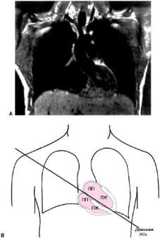
Позиция сердца в пределах тела определяет«вид» электрической активности сердца, которая может наблюдаться из любой точки на поверхности тела. Фронтальное изображение сердца с использованием МРТ в пределах грудной клетки показано на рисунке1.1A. Предсердия расположены в верхней части или основании сердца, а желудочки конусом к дну или верхушке. Длинная ось сердца, которое простирается от основания до верхушки, наклонена влево в ее апикальном конце на схематическом рисунке этой фронтальной плоскости (см. Рис. 1.1B).
Рисунок 1.1. А. Фронтальное изображение сердца при МРТ. В. Камеры сердца.
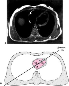
Однако правые предсердие/желудочек и левые предсердие/желудочек непосредственно не выровнены с правыми и левыми сторонами тела, как показано на поперечном изображении сердца с использованием МРТ в пределах грудной клетки (Рис. 1.2A). Схематический рисунок показывает, как правые камеры сердца расположены спереди левых камер, так что в итоге межпредсердные и межжелудочковые перегородки создают диагональ в этой поперечной плоскости (см. Рис. 1.2B).1,2
Рисунок 1.2. А. Фронтальное изображение сердца при МРТ (вид снизу). В. Камеры сердца.
КАРДИАЛЬНЫЙ ЦИКЛ
Механическая насосная функция сердца производится клетками миокарда (кардиомиоцитами), которые содержат сокращающиеся белки. Время и синхронность сокращения этих клеток контролируется несокращающимися клетками водителей ритма и проводящей системы. Импульсы, генерируемые в этих специализированных клетка, создают ритмичные повторяющиеся события, названные кардиальными циклами. Каждый цикл включает электрическую и механическую активацию клеток миокарда(систола) и восстановление (диастола). Термины, которые относятся к этим компонентам кардиального цикла, перечислены в таблице 1.1. Поскольку электрические события инициируют механические, имеется краткая задержка между началом элек-