

Цитология (Э.К.Гасымов)
.pdf161
распада. Гемоглобин содержит в своем составе железо, имеет ярко-красный цвет и участвует в переносе кислорода. Окончившие свой жизненный цикл (примерно 120 дней) эритроциты, поглощаются и перевариваются макрофагами. В это время из молекул гемоглобина образуются содержащий железо пигмент гемосидерин и не содержащий железо –
билирубин.
Гемосидерин состоит в основном из белка ферритина и имеет желто-каштановый цвет. При электронной микроскопии гемосидерин состоит из электронно-плотных гранул размером 8-9 нм. Данные гранулы расположены по отдельности или в виде скоплений неопределенной формы. Наиболее часто сидерин встречается в цитоплазме макрофагов, локализованных в селезенке, печени, легких, красном костном мозгу, в местах кровотечений и кровяных сгустков.
Впервые кристаллы пигмента желтого цвета на месте давнего кровотечения, обнаружил известный немецкий патолог Р.Вирхов (1848). Он также указал, что они образуются в результате расщепления гемоглобина. Автор произвел химический анализ данных кристаллов именуемых гематоидином и обнаружил, что они имеют идентичный состав с пигментом билирубина, окрашивающим желчь в желтовато-каштановый цвет. Пигмент билирубина, не имеющий в своем составе железо, легко растворяется в биологических
жидкостях |
в сравнении с гемосидерином. Поэтому билирубин не скапливается |
в |
цитоплазме |
поглотивших эритроциты макрофагов в виде гранул (как гемосидерин), |
а |
поступает в кровь при помощи диффузии. Далее, из крови он захватывается клетками печени и включается в состав желчи.
Наряду с гемоглобином, широко распространенным эндогенным пигментом является меланин. Меланин, имеющий темно каштановый или черный цвет, синтезируется в цитоплазме меланоцитов из аминокислоты тирозина при участии фермента тирозиназы. Меланоциты встречаются в эпидермисе кожи, в сетчатой и радужной оболочках глаза, в черном веществе среднего мозга. Меланин играет важную роль в защите организма от ультрафиолетового излучения и в восприятии лучей света.
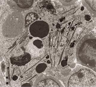
В определенных группах клеток встречаются скопления белков кристаллической формы. По своему строению они встречаются либо в виде отдельных кристаллов неопределенной формы и размеров, либо в виде кристаллиновой (кристалоподобной) сердцевины.
У человека кристалоподобные включения встречаются в основном в составе половых желез (семенник и яичники). Примером могут служить кристаллы Рейнке (Reinke) в клетках Лейдига, Чаркот-Ботчера (Charkot-Bottcher) в клетках Сертоли и Любарша (Lubarsh) в сперматогониях. Следует отметить, что кристаллы Рейнке встречаются также в клетках, расположенных во входной части яичников. Данные кристаллы окружены мембраной и располагаются в цитоплазме соответствующих клеток в свободном виде (рис. 4.29). Хоть и отмечено, что по составу они представлены белками, их функция еще не выяснена.
162
Рис.4.29. Электронно-микроскопическое строение кристалообразных включений в макрофаге.
Включения с кристаллиновой сердцевиной, в отличие от кристаллов, расположены в мембране не свободно, а в виде окруженных мембраной гранул. Примером являются кристаллиновая сердцевина в составе специфических гранул эозинофилов.
Кристаллиновая сердцевина имеет структуру кубовидной сети, повторяющейся через каждые 4 нм, и состоит из основных щелочных белков (major basic protein), имеющих молекулярную массу 9,2кД. Данный белок участвует в уничтожении гельминтов шистосомы, паразитирующих в организме человека.
Литература:
Bornens M, Paintrand M, Berges J, Marty MC, Karsenti E. Structural and chemical characterization of isolated centrosomes. Cell Motil. Cytoskeleton, 1987; 8(3):238-49.
Chang P, Gidding ThH, Mark W, Srearns T. Tubulin is required for centiole duplication and microtubule organization. Nature Cell Biology, 2002;5:71-76.
Chen D, Purohit A, Halilovic E, Doxsey SJ, Newton AC. Centrosomal anchoring of protein kinase C betaII by pericentrin controls microtubule organization, spindle function, and cytokinesis. J. Biol. Chem., 2004; 279(6):4829-4839.
Dammermann A, Merdes A. Assembly of centrosomal proteins and microtubule organization depends on PCM-1. The Journal of Cell Biology, 2002; 159(2):255266.
Dammermann A, Muller-Reichert T, Pelletier L, Habermann B et al. Centriole assembly requires both centriolar and pericentriolar material proteins. Dev. Cell, 2004; 7(6):815-29.
Delattre M, Gonczy P. The arithmetic of centrosome biogenesis. Journal of Cell Science, 2004; 117:1619-1630.
Delgehyr N, Sillibourne J, Bornens M. Microtubule nucleation and anchoring at the centrosome are independent processes linked by ninein function. Journal of Cell Science, 2005; 118:1565-1575.
Dictenberg JB, Wendy Z, Sparks CA, Young A et al. Pericentrin and tubulin form a protein complex and are organized into a novel lattice at the centrosome. J. Cell Biol., 1998; 141(1):163-174.
Faragher AJ, Fry A.M. Nek2A kinase stimulates centrosome disjunction and is required for formation of bipolar mitotic spindles. MBC Online, 2003; 14(7):28762889.
Fry AM. The Nek2 protein kinase: a novel regulator of centrosome structure.
163
Oncogene, 2002; 21(40):6184-6194.
Manandhar G, Schatten H, Sutovsky P. Centrosome reduction during gametogenesis and its significance. Biology of Reproduction, 2005; 72:2-13.
Meraldi P, Nigg EA. Centrosome cohesion is regulated by a balance of kinase and phosphatase activities. Journal of Cell Science, 2001; 114:3749-3757.
Ou YY, Mack GJ, Zhang M, Rattner J.B. CEP110 and ninein are located in a specific domain of the centrosome associated with centrosome maturation. Journal of Cell Science, 2002; 115:1825-1835.
Ou YY, Zhang M, Chi S, Matyas JR, Rattner JB. Higher order structure of the PCM adjacent to the centriole. Cell Motil. Cytoskeleton, 2003; 55(2):125-33.
Piehl M, Tulu US, Wadsworth P, Cassimeris L. Centrosome maturation: measurement of microtubule nucleation throughout the cell cycle by using GFPtagged EB1. PNAS, 2004; 101(6):1584-1588.
Pihan GA, Wallace J, Zhou Y, Doxsey SJ. Centrosome abnormalities and chromosome instability occur together in pre-invasive carcinomas. Cancer Research, 2003; 63:1398-1404.
Selvapandiyan A, Debrabant A, Duncan R, Muller J et al. centrin gene disruption impairs stagespecific basal body duplication and cell cycle progression in leishmania. J. Biol. Chem., 2004; 279(24):25703-25710.
Sonn S, Khang I, Kim K, Rhee K. Suppression of Nek2A in mouse early embryos confirms its requirement for chromosome segregation. Journal of Cell Science, 2004; 117:5557-5566.
Stearns T. Centrosome duplication. a centriolar pas de deux. Cell, 2001; 105(4):417-420.
Terra SL, English ChN, Hergert P, McEwen BF et al. The de novo centriole assembly pathway in
HeLa cells : cell cycle progression and centriole assembly/maturation. J. Cell Biol., 2005; 168(5):713-22. Mitoxondri
Andersson SG, Karlberg O, Canback B,
Kurland CG. On the origin of mitochondria: a genomics perspective. Philos. Trans. R. Soc. Lond. B. Biol. Sci., 2003; 358(1429):165-
77.
Frey TG, Mannella CA. The internal structure of mitochondria. Trends Biochem. Sci., 2000; 25:319-
324.
Frey TG, Renken CW, Perkins GA. Insight into mitochondrial structure and function from electron tomography. Biochim. Biophys. Acta, 2002; 1555:196-203.
Gray MW, Burger G, Lang BF. Mitochondrial evolution. Science, 1999; 283: 1476-1481.
Griparic L, van der Bliek AM. The many shapes of mitochondrial membranes. Traffic, 2001; 2:235-
44.
Ko CH, Shen SC, Hsu CS, Chen YC. Mitochondrial-dependent, reactive oxygen speciesindependent apoptosis by myricetin: roles of protein kinase C, cytochrome c, and caspase cascade. Biochem. Pharmacol., 2005; 69(6):913-27.
Lea PJ, Hollenberg MJ. Mitochondrial structure revealed by high-resolution scanning electron microscopy. Am. J. Anat., 1989; 184:245-57.
Mannella CA, Marko M, Buttle K. Reconsidering mitochondrial structure: new views of an old organelle. Trends Biochem. Sci., 1997; 22:37-8.
Perkins G, Ellisman MH, Fox DA. Three-dimensional analysis of mouse rod and cone mitochondrial cristae architecture: Bioenergetic and functional implications. Molecular Vision, 2003; 9:60-73.
Perkins GA, Song JY, Tarsa L, Deerinck TJ, Ellisman MH, Frey TG. Electron tomography of mitochondria from brown adipocytes reveals crista junctions. J. Bioenerg. Biomembr., 1998; 30:431-42.
Prince FP. Lamellar and tubular associations of the mitochondrial cristae: unique forms of the cristae present in steroid-producing cells. Mitochondrion, 2002; 1:381-9.
Reichert AS, Neupert W. Contact sites between the outer and inner membrane of mitochondria-role in protein transport. Biochim. Biophys. Acta, 2002; 1592:41-9. Renken C, Siragusa G, Perkins G, Washington L, Nulton J, Salamon P, Frey TG. A thermodynamic model describing the nature of the crista junction: a structural motif in the mitochondrion. J. Struct. Biol., 2002; 138:137-44.
Semak I, Naumova M, Korik E, Terekhovich V, Wortsman J, Slominski A. A novel metabolic pathway of melatonin: oxidation by cytochrome c. Biochemistry, 20055; 44(26):9300-9307.
Theissen U, Hoffmeister M, Grieshaber M, Martin W. Single eubacterial origin of eukaryotic sulfide: quinone oxidoreductase, a mitochondrial enzyme conserved from the early evolution of eukaryotes during anoxic and sulfidic times. Mol. Biol. Evol., 2003; 20(9):1564-74.
Vijayasarathy C, Damle S, Lenka N, Avadhani NG. Tissue variant effects of heme inhibitors on the mouse cytochrome c oxidase gene expression and catalytic activity of the enzyme complex. Eur. J.
Biochem., 1999; 266(1):191-200. Ribosom
164
Blobel G, Dobberstein B. Transfer of proteins across membranes. I. presence of proteolytically processed and unprocessed nascent immunoglobulin light chains on membrane-bound ribosomes of murine myeloma. Journal of Cell Biology, 1975; 67:835-851.
Dong G, Nowakowski J, Hoffman DW. Structure of small protein B: the protein component of the tmRNA-SmpB system for ribosome rescue. EMBO J., 2002; 21:18451854.
Gutmann S, Haebel PW, Metzinger L, Sutter M, Felden B, Ban N. Crystal structure of the transferRNA domain of transfer-messenger RNA in complex with SmpB. Nature, 2003; 424:699-703.
Ivanova N, Pavlov MY, Bouakaz E, Ehrenberg M, Schiavone LH Mapping the interaction of SmpB with ribosomes by footprinting of ribosomal RNA. Nucleic Acids Res., 2005; 33(11):3529-39.
Lafontaine DL, Tollervery D. The function and synthesis of ribosomes. Nat. Rev. Mol. Cell Biol., 2001; 2:514-520.
Larsson SL, Nygard O. Proposed secondary structure of eukaryote specific expansion segment 15 in 28S rRNA from mice, rats, and rabbits Biochemistry, 2001; 40:32223231.
Martoglio B, Dobberstein B. Signal sequences: more than just greasy peptides. Trends Cell Biol., 1998; 8:410-415.
Moazed D, Noller HF. Intermediate states in the movement of transfer RNA in the ribosome Nature, 1989; 342:142-148.
Noller HF, Hoffarth V, Zimmak L. Unusual resistance of peptidyl transferase to protein extraction procedures. Science, 1992; 256:1416-1419.
Pavlov MY, Freistroffer DV, MacDougall J, Buckingham RH, Ehrenberg M. Fast recycling of Escherichia coli ribosomes requires both ribosome recycling factor (RRF) and release factor RF3. EMBO J., 1997; 16:4134-4141.
Phelps SS, Jerinic O, Joseph S. Universally conserved interactions between the ribosome and the anticodon stem-loop of A site tRNA important for translocation. Mol. Cell., 2002; 10:799-807.
Ramakrishnan V. Ribosome structure and the mechanism of translation. Cell, 2002; 108, 557-572. Ramkrishnan V, White SW. Ribosomal structures: Insights into the architecture, machinery and
evolution of the ribosome. Trends Biochem. Sci., 1998; 23:208-212.
Stroud RM, Walter P. Signal sequence recognition and protein targeting. Curr. Opin. Struct. Biol., 1999; 9:754-749.
Valle M, Gillet R, Kaur S, Henne A, Ramakrishnan V, Frank J. Visualizing tmRNA entry into a stalled ribosome. Science, 2003; 300:127-130.
Yusupov MM, Yusupova GZ, Baucom
Lieberman K et al. Crystal structure of the ribosome at 5.5 A resolution. Science, 2001;
292:883-896.
Endoplazmatik şəbəkə
Ahearn GA, Mandal PK, Mandal A.
Mechanisms of heavy-metal sequestration and detoxification in crustaceans: a review. J. Comp. Physiol., 2004; 174(6):439-52.
Alder NN, Shen Y, Brodsky JL, Hendershot LM, Johnson AE. The molecular mechanisms underlying BiP-mediated gating of the Sec61 translocon of the endoplasmic reticulum. J. Cell Biol., 2005; 31; 168(3):389-99.
Domenech RJ, Sanchez G, Donoso P, Parra V, Macho P. Effect of tachycardia on myocardial sarcoplasmic reticulum and Ca2+ dynamics: a mechanism for preconditioning? J. Mol. Cell Cardiol., 2003; 35(12):1429-37.
Johnson AE, van Waes MA. The translocon: A dynamic gateway at the ER mambrane. Annu. Rev. Cell Dev. Biol., 1999; 15:799-842.
Llach A, Huang J, Sederat F, Tort L, Tibbits G, Hove-Madsen L. Effect of betaadrenergic stimulation on the relationship between membrane potential, intracellular [Ca2+] and sarcoplasmic reticulum Ca2+ uptake in rainbow trout atrial myocytes. J. Exp. Biol., 2004; 207(Pt 8):1369-77.
Schafer A, Wolf DH. Endoplasmic reticulum-associated protein quality control and degradation: genome-wide screen for ERAD components. Methods Mol. Biol., 2005; 301:289-92.
Waters MG, Pfeffer SR. Membrane tethering in intracellular transport. Curr. Opin. Cell Biol., 1999; 11:453-459.
Holci kompleksi və sitoplazmada qovuqcuqlarm nəqli - COP
BaHHH B.B. Kyma BefleT "üyTB ronBHXH"? (K 10 0-neTHro OTKPHTHH KOMnneKca ronbflXH). OTjqen Mop^onorHH (3aB. - npo^. B.B. BaHHH) PoccHäcKoro rocyäapCTBeHHoro MeflHöHHCKoro yHHBepcHTeTa, 1999, MocKBa, YflK
576.31(091), CTp. 90-97.
Allan BB, Balch WE. Protein sorting by directed maturation of Golgi compartments. Science, 1999; 285:63-66.
165
Farquhar MG, Palade GE. The Golgi apparatus: 100 years of progress and controversy. Trends in Cell Biology, 1998; 8:2-10.
Joglekar AP, Xu D, Rigotti D, Fairman R, Hay JC. The SNARE motif contributes to rbet1 intracellular targeting and dynamics independently of SNARE interactions. .J. Biol. Chem., 2003; 278(16):14121-14133.
Kamena F, Spang A. Tip20p prohibits back-fusion of COPII vesicles with the endoplasmic reticulum. Science, 2004; 304(5668):286-289.
Kunau WH, Agne B, Girzalsky W. The diversity of organelle protein transport mechanisms. Trends in Cell Biology, 2001; 11(9):358-361.
Lederkremer GZ, Cheng Y, Petre BM, Vogan E et al. Structure of the Sec23p/24p and Sec13p/31p complexes of COPII. PNAS, 2001; 98(19):10704-10709.
Love HD, Lin Ch-Ch, Short CS, Ostermann J. isolation of functional Golgiderived vesicles with a possible role in retrograde transport. J. Cell Biol., 1998; 140(3):541-551.
Matsuoka K, Schekman R, Orci L, Heuser JE. Surface structure of the COPIIcoated vesicle. PNAS, 2001; 98(24):1370513709.
Mironov AA, Polishchuk RS, Luini A. Visualizing membrane traffic in vivo by combined video fluorescence and 3D electron microscopy. Trends in Cell Biology, 2000; 10:349-351.
Orci L, Ravazzola M, Mack GJ, Barlowe Ch, Otte S. Mammalian Erv46 localizes to the endoplasmic reticulum-Golgi intermediate compartment and to cis-Golgi cisternae. PNAS, 2003; 100(8):586-4591.
Orci L, Ravazzola M, Volchuk A, Engel Th et al. Anterograde flow of cargo across the Golgi stack potentially mediated via bidirectional "percolating" COPI vesicles. PNAS, 2000; 97(19):10400-10405.
Parlati F, Varlamov O, Paz K, McNew JA et al. Distinct SNARE complexes mediating membrane fusion in Golgi transport based on combinatorial specificity. Proc. Natl. Acad. Sci USA, 2002; 99(8):5424-9.
Peppelenbosch MP, DeSmedt M, ten
Hove T, van Deventer SJH, Grooten J. Lipopolysaccharide regulates macrophage fluid phase pinocytosis via CD14-dependent and CD14-independent pathways. Blood, 1999; 93(11):4011-4018.
Rohn WM, Rouille Y, Waguri S, Hoflack B. Bi-directional trafficking between the trans-Golgi network and the endosomal/lysosomal system. Journal of Cell Sciense, 2000; 113:2093-2101.
Xu D, Hay JC. Reconstitution of COPII vesicle fusion to generate a pre-Golgi intermediate
compartment. JCB, 2004; 167(6):997-1003. Endosom, lizosom və peroksisomlar
Bucci C, Thomson P, Nicoziani P, McCarthy J, van Deurs B. Rab7: a key to lysosome biogenesis. Molecular Biology of the Cell, 2000; 11:467-480.
Edinger AL, Thompson CB. Defective autophagy leads to cancer. Cancer Cell, 2003; 4(6):422-4. Fineschi B, Miller J. Endosomal proteases and antigen processing. Trends Biochem. Sci., 1997;
22:377-382.
Fouraux MA, Deneka M, Ivan V, van der Heijden A et al. Rabip4' is an effector of rab5 and rab4 and regulates transport through early endosomes. Mol. Biol. Cell., 2004; 15(2):611-24.
Haraguchi CM, Mabuchi T, Yokota S. Localization of a mitochondrial type of NADP-dependent isocitrate dehydrogenase in kidney and heart of rat: an immunocytochemical and biochemical study. J. Histochem. Cytochem., 2003; 51(2):215-26.
Heiland I, Erdmann R. Biogenesis of peroxisomes. Topogenesis of the peroxisomal membrane and matrix proteins. FEBS J., 2005; 272(10):2362-72.
Hislop JN, Marley A, Zastrow M. Role of mammalian vacuolar protein-sorting proteins in endocytic trafficking of a non-ubiquitinated G protein-coupled receptor to lysosomes. J. Biol. Chem., 2004; 279(21):2252222531.
Kjeken R, Egeberg M, Habermann A, Kuehnel M et al. Fusion between phagosomes, early and late endosomes: a role for actin in fusion between late, but not early endocytic organelles. Mol. Biol. Cell., 2004; 15(1):345-58.
Klionsky DJ. The molecular machinery of autophagy: unanswered questions. J. Cell Sci., 2005; 118(1):7-18.
Lefkir Y, Malbouyres M, Gotthardt D, Ozinsky A et al. Involvement of the AP-1 adaptor complex in early steps of phagocytosis and macropinocytosis. Mol. Biol. Cell., 2004; 15(2):861-9.
Mayran N, Parton RG, Gruenberg J. Annexin II regulates multivesicular endosome biogenesis in the degradation pathway of animal cells. The EMBO Journal, 2003; 22(13):3242-3253.
Mullins C, Bonifacino JS. The molecular machinery for lysosome biogenesis. Bioessays, 2001; 23:333-343.
Nardacci R, Sartori C, Stefanini S. Selective autophagy of clofibrate-induced rat liver peroxisomes. Cytochemistry and immunocytochemistry on tissue specimens and on fractions obtained by Nycodenz density gradient centrifugation. Cell Mol. Biol. (Noisy-le-grand), 2000; 46(7):1277-90.
166
Petiot A, Faure J, Stenmark H, Gruenberg J. PI3P signaling regulates receptor sorting but not transport in the endosomal pathway. The Journal of Cell Biology, 2003; 162(6):971-979.
Raiborg C, Bache KG, Mehlum A, Stang E, Stenmark H. Hrs recruits clathrin to early endosomes. The EMBO Journal, 2001; 20(17):5008-5021.
Rohn WM, Rouille Y, Waguri S, Hoflack
Bi-directional trafficking between the trans-Golgi network and the endosomal/lysosomal system. Journal of Cell Sciense, 2000; 113:2093-2101.
Sachse M, Ramm G, Strous G, Klumperman J. Endosomes: multipurpose designs for integrating housekeeping and specialized tasks. Histochem. Cell Biol., 2002; 117:91-104.
Sachse M, Urbe S, Oorschot V, Strous GJ, Klumperman J. Bilayered clathrin coats on endosomal vacuoles are involved in protein sorting toward lysosomes. Mol. Biol. Cell, 2002; 13(4):1313-28.
Slagsvold T, Stenmark H. The structure of an endosomal protein sorter. Dev. Cell, 2004; 7(4):457-8. Tschantz WR, Digits JA, Pyun HJ, Coates RM, Casey PJ. Lysosomal prenylcysteine lyase is a FAD-
dependent thioether oxidase. J. Biol. Chem., 2001; 276(4):2321-4.
Ungewickell A, Ward ME, Ungewickell E, Majerus PW. The inositol polyphosphate 5-phosphatase Ocrl associates with endosomes that are partially coated with clathrin. Cell Biology, 2004; 101(37):1350113506.
Van Diggelen OP, Schindler D, Willemsen R, Boer M et al. alpha-N-acetylgalactosaminidase deficiency, a new lysosomal storage disorder. J. Inherit. Metab. Dis., 1988; 11(4):349-57.
Vonderheit A, Helenius A. Rab7 Associates with Early Endosomes to Mediate Sorting and Transport of Semliki Forest Virus to Late Endosomes. PLoS Biol., 2005; 37:e233.
Wilcke M, Alexson SE. Differential induction of peroxisomal populations in subcellular fractions of rat liver. Biochim. Biophys. Acta, 2001; 1544(1-2):358-69.
Yamamoto K, Volkl A, Hashimoto T, Fahimi HD. Catalase in guinea pig hepatocytes is localized in cytoplasm, nuclear matrix and peroxisomes. Eur. J. Cell Biol., 1988; 46(1):129-35.
Yokota S, Oda T, Fahimi HD. The role of 15-lipoxygenase in disruption of the peroxisomal membrane and in programmed degradation of peroxisomes in normal rat liver. J. Histochem. Cytochem., 2001; 49(5):613-22.
Yoshihara T, Hamamoto T, Munakata R, Tajiri R, Ohsumi M, Yokota S. Localization of cytosolic NADP-dependent isocitrate dehydrogenase in the peroxisomes of rat liver cells: biochemical and
immunocytochemical studies. J. Histochem. Cytochem., 2001; 49(9):1123-31. Proteasom və ubikvinasiya
Ciechanover A. Proteolysis: from the lysosome to ubiquitin and the proteasome. Nat. Rev. Mol. Cell Biol., 2005; 6(1):79-87.
Hershko A, Ciechanover A, Rose IA. Resolution of the ATP-dependent proteolytic system from reticulocytes: A component that interacts with ATP. Proc. Natl. Acad. Sci. USA, 1979; 76:3107-3110.
Hershko A, Heller H, Elias S, Ciechanover A. Components of ubiguitinprotein ligase system. J. Biol. Chem., 1983; 258:8206-8214.
Hoang T, Fenne IS, Cook C, Borud B et al. cAMP-dependent protein kinase regulates ubiquitin- proteasome-mediated degradation and subcellular localization of the nuclear receptor coactivator GRIP1. J. Biol. Chem., 2004; 279(47):49120-30.
Maki CG, Huibregtse JM, Howley PM. In vivo ubiquitination and proteasome-mediated degradation of p53. Cancer Res., 1996; 56:2649-2654.
Nohl H, Staniek K, Kozlov AV, Gille L. The biomolecule ubiquinone exerts a variety of biological functions. Biofactors, 2003; 18(1-4):23-31.
Pickart CM. Mechanisms underlying Ubiquitination. Annu. Rev. Biochem., 2001; 70:503-534. Sasajima H, Nakagawa K, Yokosawa H. Antiproliferative proteins of the BTG/Tob family are
degraded by the ubiquitin-proteasome system. Eur. J. Biochem., 2002; 269(14):3596-604. Ubiguitin-mediated proteolysis.
Advanced information on the Nobel Prize in Chemistry, 6 October 2004. www.kva.se. Varshavsky A. The ubiquitin system. Trends Biochem. Sci., 1997; 22:383-387.
Voges D, Zwickl P, Baumeister W. The 26S proteasome: A molecular machine designed for controlled proteolysis. Annu. Rev. Biochem., 1999; 68:1015-1068.
Wilkinson KD, Urban MK, Haas AL. Ubiquitin is the ATP dependent proteolysis factor I of rabbit reticulocytes. J. Biol. Chem., 1980; 255:7529 7532
167
ЯДРО
Ядро является одним из важнейших компонентов всех клеток человеческого организма (за исключением эритроцитов, некоторых стареющих и закончивших свою жизнедеятельность клеток). Удаление ядра из цитоплазмы становится причиной полной остановки синтеза белка и гибели клетки.
Ядро, являясь самой большой органеллой клетки, содержит в себе компактно упакованные структуры с кислыми (ДНК и РНК) и щелочными (гистоны) свойствами, что дает возможность с легкостью увидеть его в световой микроскоп в гистологических препаратах, окрашенных с использованием различных красителей. Не случайно, что еще в 1825-1827 годах ученым Я.Пуркинье в различного рода клетках было выявлено ядро. В 1831 году Р.Броун отметил ядро, как одну из обязательных частей растительной клетки. Несмотря на то, что ядро обнаружено более чем 170 лет назад, гистотопография его структурных элементов, характер их строения, а также молекулярные основы процессов взаимодействия цитоплазмы при синтезе белков с нуклеоплазмой, глубоко были изучены только за последние годы.
Свойства |
избирательной |
проницаемости ядерной |
оболочки, |
отделяющей |
|
нуклеоплазму |
от цитоплазмы, делает ядро |
важным участком не только для расположения |
|||
генетического материала, но |
также и для |
выполнения |
таких важных процессов, как |
||
репликация молекул ДНК, транскрипция и созревание иРНК (processing). Так, при помощи комплекса ядерной поры ядро контролирует поступление в нуклеоплазму из цитоплазмы факторов транскрипции, а также участвует в синтезе иРНК из первичных транскриптов с помощью специальных сплайсеосом, находящихся в ядре и пр. Все это свидетельствует о том, что ядро играет роль интегративного центра управления синтезом белков и тем самым деятельностью клетки в целом.
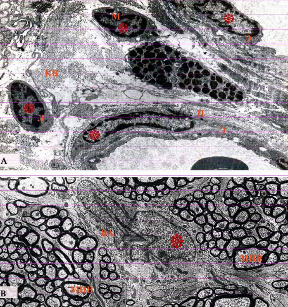
Ядро, поперечный срез которого составляет 5-10 мкм, находится в основном в центре клетки, имеет различные (круглую, овальную, грушевидную, бобовидную, многодольчатую, палочковидную, полиморфную с неровными краями и т.д.) формы (рис. 5.1 и 5.2). У млекопитающих большинство клеток являются одноядерными, реже встречаются двухили многоядерные к примеру соответственно - гепатоциты и костеразрушающие клетки (остеокласты).
168
Рисунок 5.1. Электронно-микроскопический рисунок клеток с различными формами ядер.
А - Взаимная гистотопография клеток и неклеточных структур, располагающихся в стенке кровеносных сосудов и вокругсосудистых областях. Ультратонкий срез проходит через перицит (П), лимфоцит (Л), макрофаг (М) центральную (ядросодержащую) часть эндотелиальных клеток (Э) и периферическую часть тучных клеток (Т).
В - в центре поперечного среза зрительного нерва видно ядро неопределенной формы волокнистого астроцита (электронограмма к.м.н. Оруджева М.Т. и проф. Гасымова Э.К.)
МНВ - миелиновое нервное волокно; ВА - волокнистый астроцит; КВ - коллагеновые волокна; ядра отмечены звездочками.
Структурными компонентами ядра являются ядерная оболочка, нуклеоплазма, хроматин и ядрышко.
169
ЯДЕРНАЯ ОБОЛОЧКА
Ядерная оболочка, располагаясь между нуклеоплазмой и цитоплазмой, обладает избирательной проницаемостью (в основном для макромолекул), тем самым принимает участие в процессе синтеза белков и в формировании хроматина.
На гистологических препаратах, окрашенных гематоксилин-эозином, светооптически за ядерную оболочку принимается обращенная к цитоплазме синеокрашенная, относительно более толстая в форме очерчивающей линии, структура ядра. Однако на гистологических препаратах, окрашенных после предварительного действия на срезы фермента нуклеазы, было выявлено, что ядерная оболочка имеет исключительно тончайшую толщину. Электронно-микроскопические исследования показали, что в действительности толщина оболочки, разделяющая цитоплазму и нуклеоплазму, составляет всего лишь 40-60 нм и
поэтому светооптически увидеть ее как отдельную структуру технически |
просто |
невозможно. |
|
Ультраструктурно ядерная оболочка состоит из наружной и внутренней |
ядерных |
оболочек, перинуклеарного пространства и комплекса ядерных пор (рис.5.2). |
|
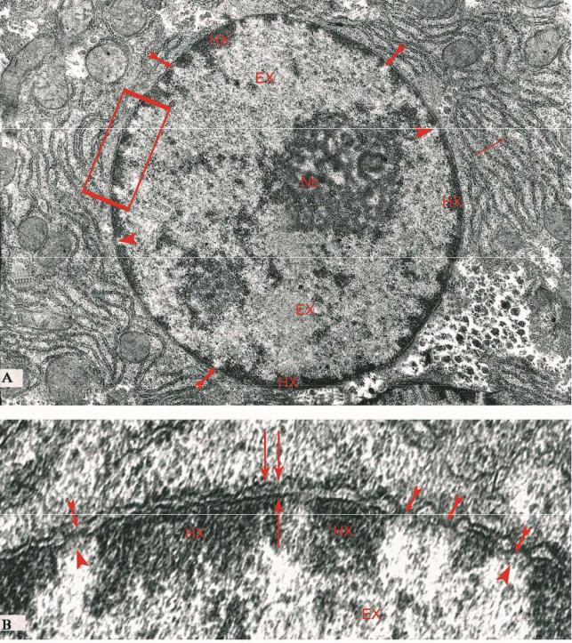
Наружная и внутренняя ядерные оболочки вокруг ядерных пор непрерывно переходят друг в друга, в остальных местах между ними имеются промежутки шириной в 25-30нм в форме щелевидных вокругядерных (перинуклеарных) пространств (рис.5.2.В). По своему строению ядерные мембраны (состоящие из двухслойного фосфолипидного слоя толщиной 6-7 нм), напоминают оболочки в других участках клетки. Но между этими оболочками есть и отличия, особенно в составе внутренней ядерной оболочки, где были обнаружены мембранные белки, свойственные только ядру.
170
Рисунок 5.2. Электронно-микроскопический рисунок ядра и его составных элементов.
А - ясно виден, |
находящийся |
в области ядерной оболочки и в нуклеоплазме |
гетерохроматин (HX), эухроматин (EX), ядерные поры ( ), каналы ядерных пор (▲) и топографическое положения ядрышка (Nc).
В - увеличенный фрагмент А ; видны наружная и внутренняя оболочки ядра (↑↑), перинуклеарное пространство ( ↑) и ядерная пора