

Полезные материалы за все 6 курсов / Учебники, методички, pdf / INBDEBooster Oral Pathology
.pdfINBDE ORAL PATHOLOGY NOTES |
31 |
•Multiple Myeloma - cancer affecting plasma cells, also known as plasma cell myeloma
Must know: this can be observed radiographically via punched out radiolucencies usually in the skull (think: Mixed Martial (MM) artists Punch the skull)
-Another characteristic sign of multiple myeloma is amyloidosis, the accumulation of amyloid proteins, that develop from antibody light chains
Treatment: chemotherapy with poor prognosis
•Non-Hodgkin’s Lymphoma (NHL) - also a cancer affecting the lymphatic system, NHL differs from Hodgkin’s Lymphoma in that Reed-Sternberg cells are not present
Must know: NHL is a neoplasm of B or T cells, and Burkitt’s Lymphoma is a type of B cell NHL characterized by THe LiP Bone:
-Tooth mobility
-Halted root development
-Lip paresthesia
-Pain and swelling
-Bone marrow involvement
Treatment: chemotherapy/radiotherapy
Figure 5.04 NHL histology
Figure 5.03 MM Radiolucencies
6 Odontogenic Cysts and Tumors
We are now ready to discuss conditions directly impacting tooth formation! Residual odontogenic epithelium can form cystic lesions at any time, so there is an interesting variety of conditions discussed in this section.
•Calcifying Odontogenic Cyst - a benign cyst occurring in gnathic bones, and is also known as Gorlin cyst
Must know: this rare and unpredictable cyst involves ghost cells, or empty spaces where keratin occupies the space of the nucleus, that undergo calcification. This causes radio-densities that can be
INBDE Booster | Booster PrepTM
INBDE ORAL PATHOLOGY NOTES |
32 |
|
detected radiographically |
Must know: this cyst is the soft tissue |
|
- |
Remember by “C-O-C triple G” |
counterpart of later periodontal cysts, |
|
|
and has no radiolucency |
|
|
Treatment: excision |
Figure 6.01 Gorlin cyst
•Dentigerous Cyst - the second most common type of odontogenic cyst that most commonly affects canines and third molars, and is characterized by the accumulation of fluid between the crown and reduced enamel epithelium
Must know: this lesion is called an eruption cyst if it occurs over erupting teeth, and can be detected radiographically via radiolucency attached to the cementoenamel junction of the impacted tooth
Treatment: excision, but this could give rise to a future odontogenic tumor
Figure 6.02 Dentigerous Cyst
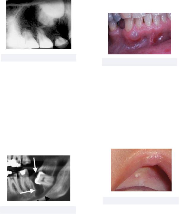
•Gingival Cyst (adults) - a gingival cyst formed near the mandibular canine and premolar regions
Figure 6.03 Adult Gingival Cyst
•Gingival Cyst (newborn) - raised nodules that appear as small, isolated, or multiple cysts
Must know: the rests of dental lamina epithelialize these lesions, making them true cysts, which have different names depending on where in the mouth they form
-Bohn’s nodules: lateral palate
-Epstein’s pearls: midline palate
Treatment: none needed as the cysts involute during aging
Figure 6.04 Newborn Gingival Cyst
•Keratocystic Odontogenic Tumor (KCOT) - a rare, aggressive, and recurrent cyst that commonly forms in the posterior ascending mandibular ramus
INBDE Booster | Booster PrepTM
INBDE ORAL PATHOLOGY NOTES
Must know: this tumor is characterized by thin corrugated parakeratinized epithelium, and is associated with the fatal Gorlin Syndrome (also known as nevoid basal cell carcinoma syndrome) that involves:
-Multiple KCOTs
-Multiple Basal Cell Carcinomas
-Calcified falx cerebri
Treatment: aggressive enucleation
Figure 6.05 KCOT
•Lateral Periodontal Cyst - a noninflammatory cyst forming on the lateral surface on the root of a vital tooth
Must know: this cyst is most common in the mandibular premolar lesion
Treatment: excision
Figure 6.06 Lateral Periodontal Cyst
• Primordial Cyst - a cyst that forms where a
33
•tooth should have formed (early on, in primordial matter) but is missing
Must know: this cyst is most common in the mandibular third molar region
Treatment: complete removal
Figure 6.07 Primordial Cyst
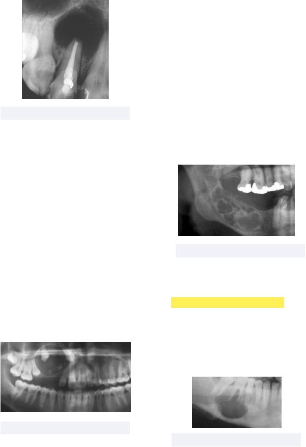
•Radicular Cyst - also called a periapical cyst, this most common odontogenic cyst originates from epithelial remnants of the periodontal ligament
Must know: radiolucency observed at the apex of the affected non-vital tooth
-Inflammation caused by pulp necrosis is responsible for the cyst. Acute inflammation leads to an abscess, while chronic inflammation leads to granuloma formation
-Epithelial Rests of Malassez (ERM), or residual cells from tooth development, from Hertwig’s Epithelial Root Sheath (HERS) encapsulate the lesion within the pocket of inflammation, allowing it to be a true cyst (HERS → ERM → cyst)
Treatment: root canal, apicoectomy, or tooth extraction with curettage
INBDE Booster | Booster PrepTM
INBDE ORAL PATHOLOGY NOTES
Figure 6.08 Radicular Cyst
Odontogenic Tumors
Odontogenic tumors are derived from the bone, and are unique to the jaw because they are derived from epithelial and mesenchymal cells involved in the formation of teeth. Note that these tumors do appear radiographically similar to odontogenic cysts, so use the images as more of a reference rather than exact representations of the tumors.
•Adenomatoid Odontogenic Tumor (AOT) - a rare, benign, painless, noninvasive, and slow growing lesion often misdiagnosed as an odontogenic cyst
Must know: this tumor contains epithelial duct-like spaces, hence the prefix adeno (meaning gland), as well as enameloid material
Treatment: surgical excision with good prognosis
34
•Ameloblastoma - one of the most commonly tested tumors that, while benign, behaves similar to an aggressive malignant tumor by eroding through tooth roots and the cortical plate
Must know: a typical differential diagnosis for a multilocular radiolucency (bubbly appearance, dark shadows) in the posterior mandible includes ameloblastoma, central giant cell carcinoma (CGCG), central odontogenic fibroma (COF), or keratocystic odontogenic tumor (KCOT)
Treatment: wide excision as too conservative can lead to high recurrence
Figure 6.10 Ameloblastoma
•Ameloblastic Fibroma - a rare benign mixed epithelial and mesenchymal tumor that often occurs in children and teens (adolescent amelobastic fibroma)
Must know: this tumor is most common in the posterior mandible, in myxomatous connective tissue, and can be called an ameloblastic fibro-odontoma if an odontoma is also present
Treatment: surgical excision
Figure 6.09 AOT
Figure 6.11 Ameloblastic Fibroma
INBDE Booster | Booster PrepTM
INBDE ORAL PATHOLOGY NOTES |
35 |
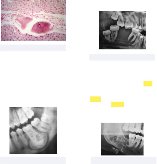
•Calcifying Epithelial Odontogenic Tumor (CEOT) - also called Pindborg tumor, this tumor is characterized by scattered-dense areas of calcification that appear as a “driven snow” radiolucency
Must know: histologically, Leisegang rings, or amorphous pink amyloid proteins with concentric calcifications, are observed
Treatment: surgical excision with good prognosis
•Central Odontogenic Fibroma (COF) - appearing as an asymptomatic expansion of the cortical plate of the mandible or maxilla, this tumor has dense collagen with strains of epithelium
Must know: has two forms, central or peripheral
-Central: occurs in bone and has a welldefined multiocular radiolucency (similar to ameloblastoma)
-Peripheral: occurs in gum tissue, so no radiolucency as no bone has been eroded away
Figure 6.12 CEOT Histology
•Cementoblastoma - neoplasm characterized by sheets of cementum that create a well circumscribed radiopaque mass
Must know: the root of the tooth becomes replaced by a ball of cementum and cementoblasts
Treatment: surgical excision and extraction
Figure 6.14 COF
•Odontogenic Myxoma - also known as myxofibroma, this tumor has myxomatous connective tissue that has a pulp-like material with very little collagen (slimy stroma)
Must know: radiographically, this has a messy radiolucency with a honeycomb pattern and unclear borders
Treatment: surgical excision with moderate recurrence
Figure 6.13 Cementoblastoma |
Figure 6.15 Odontogenic Myxoma |
INBDE Booster | Booster PrepTM
INBDE ORAL PATHOLOGY NOTES |
36 |
|||||||
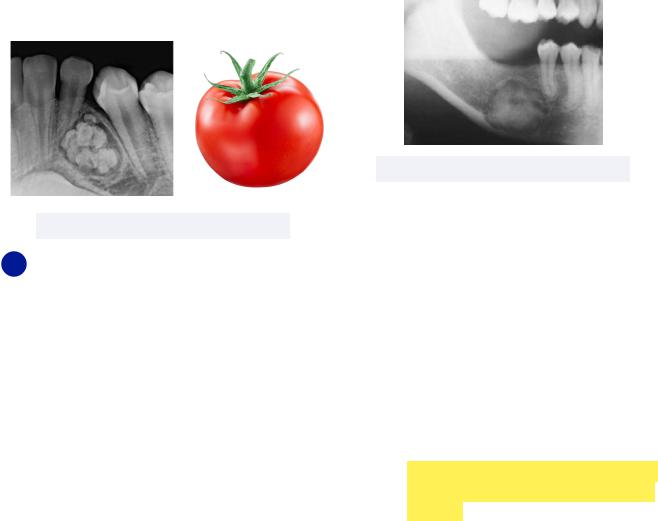
• Odontoma - a commonly tested tumor, this |
• between the premolar and molar regions of |
|||||||
is an opaque lesion composed of dental |
the mandible, this lesion is composed of |
|||||||
hard tissues that can block eruption and |
fibroblastic stroma (connective tissue) in |
|||||||
cause impaction |
which foci of mineralized products are |
|||||||
Must know: has two forms, compound |
formed |
|||||||
and complex |
Must know: there are two forms of this |
|||||||
- |
Compound: mostly in anterior region, |
condition, central and peripheral |
||||||
|
and is just a bunch of miniature teeth |
- Central: lesion occurs in bone, and has |
||||||
- |
Complex: mostly in posterior region, |
well-circumscribed radiolucency with |
||||||
|
and appears more like a messy |
ossification product in center → looks |
||||||
|
conglomerate mass of tissue |
similar to cementifying fibroma |
||||||
- Gardner Syndrome involves multiple |
- Peripheral: lesion occurs in gum with no |
|||||||
|
odontomas and intestinal polyps; |
radiolucency or radiopacity |
||||||
|
patients diagnosed with this syndrome |
- Also has a juvenile variant with |
||||||
|
have a higher risk of early colorectal |
aggressive and rapid growth |
||||||
|
cancer |
(think odonTOMATO |
→ |
the |
|
|
Treatment: surgical excision |
|
|
opaque lesion is buried in tissues like a |
|
|
|||||
|
tomato in a GARDEN) |
|
|
|||||
Figure 6.16 Odontoma
7 Bone Lesions
We’ve officially reached our final topic in INBDE oral pathology! We’ll be discussing fibro-osseous, giant cell, inflammatory, and malignant bone lesions as part of this final section. Starting with fibro-osseous lesions, these are benign tumors formed in fibrous tissue that contain newly-developing bony islands, and as a result, contain radiopaque elements.
• Central Ossifying Fibroma - occurring
Figure 7.01 Central Ossifying Fibroma
• Fibrous Dysplasia - condition in which scar-like fibrous tissue grows in place of normal bone, and usually does not stop growing until after puberty
Must know: radiographically, this has a ground-glass appearance with soft radiopaque areas throughout the lesion
-McCune-Albright Syndrome (MAS) is characterized by polyostic fibrous dysplasia (affecting multiple bones), as well as cutaneous café au lait spots, and endocrine abnormalities (such as precocious puberty)
Use mnemonic MASEBS to remember MAS affects Bone, Endocrine system, and Skin
INBDE Booster | Booster PrepTM
INBDE ORAL PATHOLOGY NOTES |
37 |
Treatment: surgical re-contouring, preferably after puberty when the lesion stops growing
Figure 7.02 Fibrous Dysplasia
•Osteoblastoma - a circumscribed radiopaque mass of osteoblasts and boneTreatment: surgical excision
Figure 7.03 Osteoblastoma
•Periapical Cemento-Osseous Dysplasia (PCOD) - this reactive process with unknown origin is characterized by fibrous tissue replacing normal bone tissue, commonly at the apices of mandibular anterior teeth
Must know: while the pulps may radiographically look necrotic, the affected teeth are vital
-This is more prevalent in middle-aged black females
-This condition starts out looking radiolucent, but transitions to having a more radiopaque border
Treatment: none
Figure 7.04 PCOD
Giant Cell Lesions
Giant cell bone lesions are termed as such for the characteristic large cell sizes, which are observed microscopically.
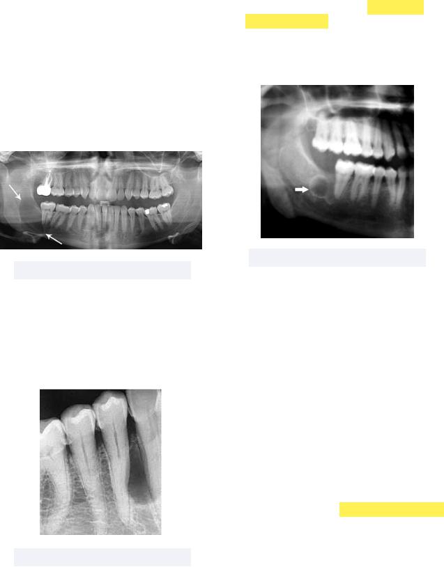
•Aneurysmal Bone Cyst - a benign bloodfilled pseudocyst that tends to expand or grow
Must know: this lesion has a multilocular radiolucency, and is commonly found in the posterior mandible
-An aspiration biopsy is the first step for diagnosing a condition like this to determine, if this is, in fact, a vascular bone cavity filled with blood
Treatment: surgical excision
Figure 7.05 Aneurysmal Bone Cyst
•Central Giant Cell Granuloma - an osteolytic neoplasm composed of fibroblasts and multinucleated giant cells
Must know: commonly found in the anterior mandible, this lesion has two forms
INBDE Booster | Booster PrepTM
INBDE ORAL PATHOLOGY NOTES |
|
38 |
||||||||||
- |
Central (CGCG): occurs in bone, and |
Must know: |
|
is the name of |
||||||||
Brown tumor |
||||||||||||
|
has radiolucency with |
thin wispy |
|
the lesion, |
which forms due to excess |
|||||||
|
separations |
|
|
osteoclast activity |
||||||||
- |
Peripheral: occurs in gum, and presents |
- |
As a result of excess osteoclast activity, |
|||||||||
|
as |
red/purple |
gingival mass |
|
elevated alkaline phosphatase levels are |
|||||||
Treatment: surgical excision |
|
observed |
||||||||||
|
|
|
|
|
|
- |
This condition can lead to |
Von |
|
NOT |
||
|
|
|
|
|
|
|
Recklinghausen’s disease of bone |
|||||
|
|
|
|
|
|
|
to be confused with Von |
|
||||
|
|
|
|
|
|
|
Recklinghausen’s Disease/ |
|||||
|
|
|
|
|
|
|
neurofibromatosis |
|||||
Figure 7.06 Central Giant Cell Granuloma
•Cherubism - a autosomal dominant disorder characterized by enlargement and prominence of the mandible and maxilla as bone is replaced by a fibrous granuloma contains multinucleated giant cells
Must know: this condition has symmetrical bilateral swelling (Bilateral cheruBism), and expansible bilateral multilocular radiolucencies that stop growing after puberty (unlike fibrous dysplasia that has unilateral swelling)
Figure 7.08 Hyperparathyroidism
•Langerhans Cell Disease - also known as idiopathic histiocytosis, this rare type of cancer involves the abnormal buildup of Langerhans cells (histiocytes) in certain parts of the body
Must know: this condition has discrete punched/scooped out radiolucencies that cause a floating teeth appearance (think: levitating Langerhans)
Treatment: surgical excision, radiation, and chemotherapy
Figure 7.07 Cherubism
• Hyperparathyroidism - excessive levels of |
Figure 7.09 Langerhans Cell Disease |
|
parathyroid hormone result in multiple |
||
|
||
bone lesions that look similar to CGCGs |
|
INBDE Booster | Booster PrepTM
INBDE ORAL PATHOLOGY NOTES
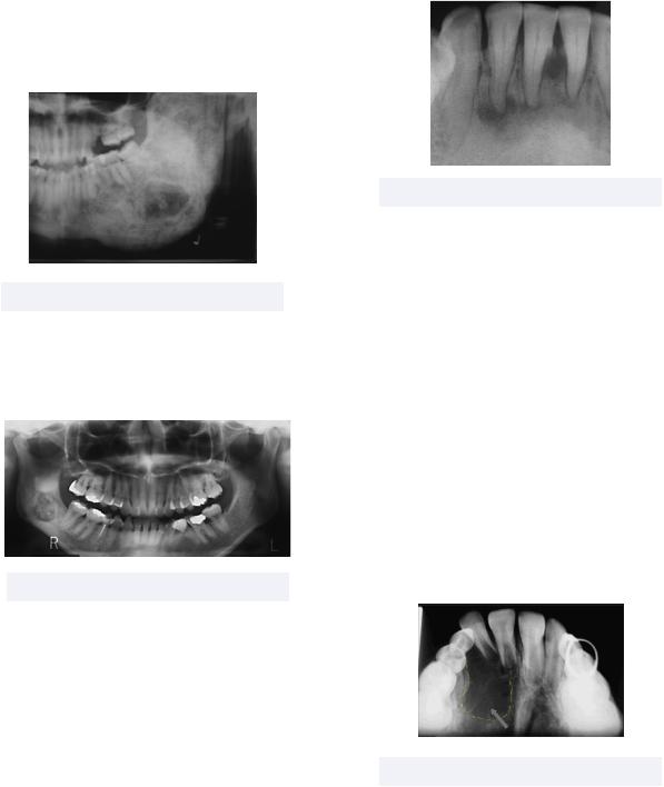
•Paget’s Disease - a progressive metabolic disturbance of different bones (skull, jaw, spine, femur), resulting in symmetrical enlargement with a cotton wool appearance
Must know: similar to hyperparathyroidism, elevated alkaline phosphatase levels are observed due to increased breakdown of bone
-Because of enlargement, dentures, or even hats, become too tight (think pinching Paget’s)
Treatment: bisphosphonates, calcitonin
Figure 7.10 “Cotton Wool” Skull
Bone Inflammatory Lesions
Inflammation of jaw bones is common, and most lesions are extensions of periodontal and periapical inflammation. Other lesions form in response to trauma.
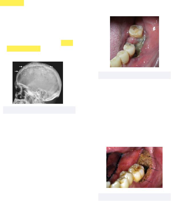
•Acute Osteomyelitis - a clinical term for a new infection in bone, the most common causes include odontogenic infection and trauma
Must know: infection and inflammation first start in the medullary space, involving cancellous, or spongey, bone. To observe any radiolucency, the infection needs to have spread to cortical bone, periosteum, and soft tissues, otherwise we won’t see much radiographically
- Noteworthy symptoms include deep
39
-pain, high or intermittent fever, and paresthesia or anesthesia of IAN. Note that teeth do NOT become loose — this is caused by periodontitis
Treatment: antibiotics and drainage
Figure 7.11 Acute Osteomyelitis
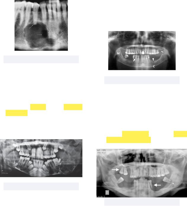
•Bisphosphonate-Related Osteonecrosis of the Jaws (BRONJ) - current or previous treatment of bisphosphonates that leads to exposed bone that does not heal quickly
Must know: this condition has a higher risk of development with IV administered bisphosphonates (drugs that end in -dronate)
Treatment: chlorhexidine (CHX) rinse, antibiotics, conservative surgery
Figure 7.12 BRONJ
•Chronic Osteomyelitis - a bone infection that doesn’t go away despite treatments, resulting in recurring drainage and intense pain
INBDE Booster | Booster PrepTM
INBDE ORAL PATHOLOGY NOTES |
40 |
||
Must know: has diffuse mottled |
• Diffuse Sclerosing Osteomyelitis - similar |
||
radiolucency, since it has now been able |
to the previous condition, but on a wider |
||
to impact cortical bone |
scale |
||
- Garre’s osteomyelitis is a condition that |
Must know: this condition may lead to |
||
involves chronic osteomyelitis and |
jaw fracture and osteomyelitis |
||
proliferative periostitis as the body tries |
Treatment: none, other than addressing |
||
to heal |
(think: GO COPP!) |
|
the cause of infection |
Treatment: |
antibiotics and |
debridement |
|
Figure 7.13 Proliferative Periostitis
•Focal Sclerosing Osteomyelitis (Condensing Osteitis) - a periapical lesion resulting from low-grade inflammation such as chronic pulpitis
Must know: a wall of diffuse dense bone forms to “wall off” the infection
Treatment: none, other than addressing the cause of infection, such as performing a root canal
Figure 7.15 Diffuse Sclerosing Osteomyelitis
Bone Malignant Lesions
We’ve reached the final section in our oral pathology notes! Malignancies occurring in bone encompass a lot of what we have already discussed, such as sarcomas, lymphomas, and leukemias. While we’ll get into specific symptoms as we discuss the conditions, know that paresthesia is the most common symptom and is also most frequently tested as related to bone malignancies on the INBDE.
•Chondrosarcoma - sarcoma of jaws when tumor cells produce new cartilage
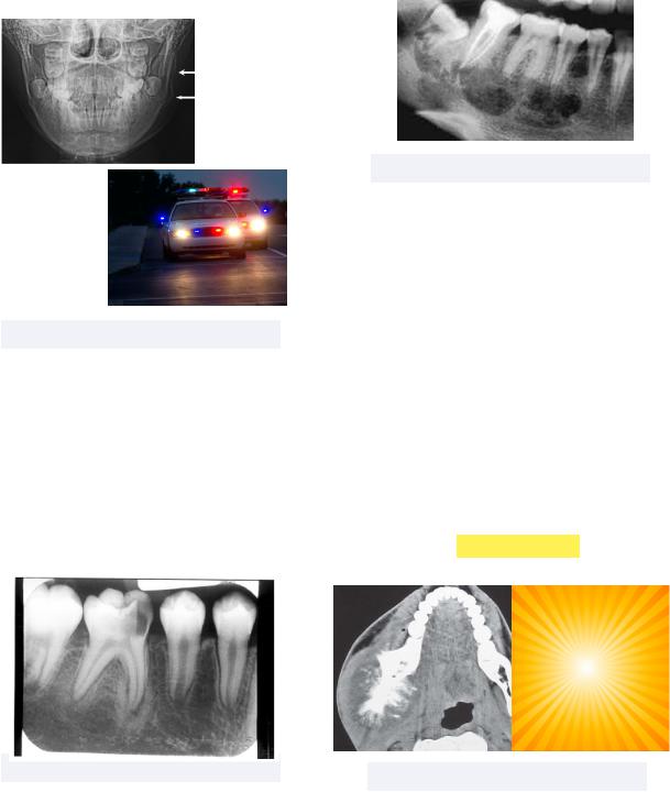
Must know: commonly involves the condyle due to its cartilaginous origin, and has a sunburst pattern
Figure 7.14 Condensing Osteitis |
Figure 7.16 Chondrosarcoma |
|
|
|
INBDE Booster | Booster PrepTM |