

Полезные материалы за все 6 курсов / Учебники, методички, pdf / INBDEBooster Orthodontics
.pdfORTHODONTICS |
1 |
DEVELOPMENT & GROWTH
Orthodontics is the branch of dentistry that focuses on the prevention, diagnosis and treatment of irregularities of the teeth and jaws. This section is combined with the paediatric section on the INBDE so overall, there are less questions on orthodontics compared to other sections.
In dentistry, growth is most relevant to the increased size of anatomy or increase in number of anatomic parts. Thus, it is more about quantity than it would be in development.
Meanwhile, development refers more to the greater complexity or specialization of functions. There is more of a qualitative focus on the physiological or behavioural effects on the body.
Both growth and development depend on pattern, timing, and variability of the individual.
1 Growth Curves
Cephalocaudal Growth
The cephalocaudal growth gradient refers to a pattern where early on in life, growth tends to occurs faster in parts that are closer to the cranium. Consequently, parts further way from the cranium experience more growth later in life.
•Observed in utero until adulthood (the head takes up less of the body as we grow into adulthood)
•Upper limbs grow earlier on in life, followed by the lower limbs
•Maxilla matures earlier than the mandible*
Human Growth Curve
The human growth curve uses terms of distance and velocity
•Distance - person’s height
•Velocity - person’s rate of change in height
 Generally the earlier the peak growth, the shorter the duration of the growth spurt
 Males and females exhibit different velocity
 Male - peaks at 14 years
  Female - peaks at 12 years
Figure 1.01 Human Growth Curve
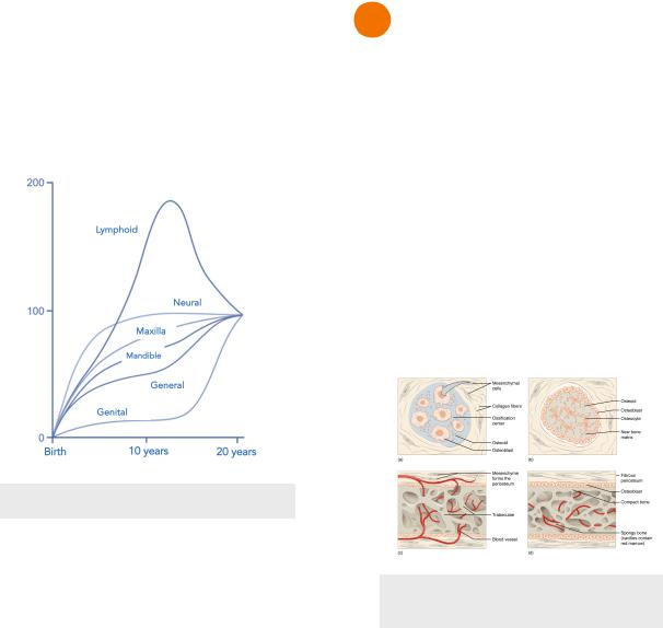
Scammon’s Growth Curve
Scammon’s growth curve describes the growth of variable body regions in terms of years and percentage of total adult body size.
•Notice that different regions have different growth patterns
INBDE Booster | Booster Prep™
ORTHODONTICS
•Brain and neural tissues grow rapidly after birth and reach near adult size by 6-7 years
•Lymphoid tissues grows rapidly to twice its adult size by 10 years, and then decreases until adulthood
•Maxilla more similar to neural growth curve
•Mandible more similar to general growth curve
Figure 1.02 Scammon’s Growth Curve
Timing
Timing of growth can be judged based on several different factors.
•Chronological age - not a good indicator of maturity due to variability
•Biologic age - based on markers of maturation, best indicators of growth and maturity
 Menstruation
 Secondary sex characteristics
•Skeletal age - based on maturity of cervical vertebrae or maturity of hand and wrist bones, better indicator of maturity
•Dental age - based on teeth that have exfoliated/erupted, generally not a good indicator of maturity
2
2 |
Growth Sites |
The location of growth can be described in sites or centres. Sites refer to the parts of the body where the growth is actually occurring. Centers are the sites of the body that are able to control its own growth.
•Synchondroses are the main growth centers for craniofacial structures
Intramembranous Growth
•Growth from the outside of bone from fibrous connective tissue = increased diameter of bone
•Controlled more by environmental factors
•Found in the following sites
 Sutures
 Cranial vault surface
Figure 2.01 Intramembranous
Ossification
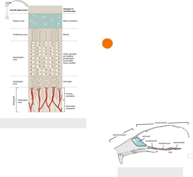
Endochondral Ossification
•Growth that occurs within cartilage that is eventually replaced by bone, 5 zones
 Zone of Resting Cartilage
 Zone of Proliferation
 Zone of Hypertrophy
 Zone of Calcification
 Zone of Ossification
•Results in increased bone length
•Controlled more by genetics
•Found in the following sites
 Epiphyseal plates (long bones)
 Synchondrosis of cranial base
 Condylar cartilage of the mandible
INBDE Booster | Booster Prep™
ORTHODONTICS
Figure 2.02 Endochondral Ossification
Growth Theories
No one theory can explain craniofacial growth and its control. Thus all three of the following theories should be considered.
1.Cartilage Theory
i.Created by James Scott
ii.Epigenetic control of growth where cartilage is the growth center
iii.Holds some validity
2.Suture Theory
i.Created by Harry Sicher
ii.Now known to be mostly false
iii.Genetics determine bone growth
iv.Sutures act as growth centres (but now we know they are growth sites)
3.Functional Matrix Theory
i.Created by Melvin Moss
ii.Environment (speaking, chweing, function) controls growth
a.Causes nasal and oral cavities to grow bigger
b.Primary determinant of growth in maxilla and mandible
3
Planes of Growth
1.Transverse - width, 10-12 years
2.Anteroposterior/sagittal - length, 14-16 years
3.Vertical - height, 18-20+ years
3 Craniofacial Growth
Cranial Base
•Cranial base - ethmoid, sphenoid and occipital bones
 Are cartilage at birth and go through endochondral ossification later on at snchondroses
 3 years, intersphenoid synchondrosis
 7 years, spheno-ethmoid synchondrosis inactive
 Later on, spheno-occipital synchondrosis inactive
Figure 3.01 Cranial Base
Cranial Vault
•Cranial vault - part of the skull that holds the brain
•Intramembranous ossification occurs
 At fontanelles and sutures, as the brain goes and pushes cranial bones apart
 Direct apposition of bone on external bone surface and resorption on internal bone surface
INBDE Booster | Booster Prep™
ORTHODONTICS
Maxilla
•Intramembranous ossification posterior and superior to the nasomaxillary complex at the sutures and for remodelling
 Apposition - palate, tuberosity, alveolar ridge
 Resorption - anterior maxilla
 Results in net forward and downward movement of maxilla from cranial base
Figure 3.02 Maxillary Growth
Mandible
• Embryonic growth
 Condylar cartilage from intramembranous ossification
 Embryonic corpus/ramus from endochondral ossification
 4 months (in utero) → Condylar cartilage and ramus fuse
• Adulthood
 Intramembranous ossification → surface remodelling
Apposition - ramus, coronoid, alveolar ridges, chin
Resorption - anterior ramus
 Endochondral ossification → proliferation and bone formation at condylar cartilage
  Results in net towards and downwards movement from cranial base
4
Figure 3.03 Maxillary Growth
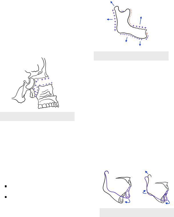
•Growth Rotation - mandible rotates during growth at the axis of the condyle
 Average growth rotation - condylar growth roughly equal to the amount of molar eruption
 Mandible does not rotate, just downward and forward translation
 Forward rotation - counter-clockwise growth
 Condylar growth exceeds the amount of molar eruption
 Tends to lead to shorter lower face height and deep bite
 Opening rotation - clockwise growth
 Condylar growth less than the amount of molar eruption
 Tends to lead to a longer lower face height and anterior open bite
Figure 3.03 Growth Rotation
INBDE Booster | Booster Prep™
ORTHODONTICS |
5 |
CRANIOFACIAL ANOMOLIES
Generally birth defects, including craniofacial anomalies, can be described as syndromes or sequences.
• Sequence - single etiology that results in predictable pattern of anomalies occurring together with recognizable presentation
• Syndrome - usually a single major anomaly effecting development of surrounding structures, resulting in more anomalies
1 Cranofacial Development
There are five stages of embryonic craniofacial development. Problems can arise during these stages to result in craniofacial abnormalities.
Problems with Neural Crest Cell Formation
1.Stage 1 - Germ Layer Formation
i.Time In Utero = Day 17
ii.Abnormal development
a.Fetal Alcohol Syndrome
2.Stage 2 - Neural Tube Formation
i.Time in Utero = Day 18-23
ii.Abnormal development
a.Anencephaly - absence of parts of the brain and skull
Figure 1.01 Anencephaly
3.Stage 3 - Neural Crest Cell Migration
i.Time in Utero - Day 19-28
ii.Abnormal development
a.Teacher Collins Syndrome (mandibulofacial dystosis)
b.Hemifacial microsomia
Lack of Fusion of Cells
4.Stage 4 - Formation of Organ Systems
i.Time in Utero = Week 4-5 (Day 28-38)
ii.Abnormal development → Cleft lip 4a. Primary Palate formation
i.Time in Utero = Week 6
ii.Abnormal development → Cleft palate 4b. Secondary Palate formation
i.Time in Utero = Week 8
ii.Abnormal development → Cleft palate
Problems with Suture Development
5.Stage 5 - Final Differentiation of Facial Tissues
i.Time in Utero = Day 50 - birth
ii.Abnormal development
a.Achondroplasia
b.Crouzon’s syndrome (Craniostynosis)
2 Cranofacial Anomalies
Down’s Syndrome (Trisomy 21)
•Extra chromosome 21 due to nondisjunction
•Upslanted palpebral fissures
•Increased risk of periodontal disease
•But no increased caries risk
•Midface deficiency*
Figure 2.01 Down’s Syndrome
INBDE Booster | Booster Prep™
ORTHODONTICS
Treacher Collins Syndrome
•Aka mandibulafacial dysostosis
•Caused by genetic mutation
 Affects neural crest cell development → abnormal development of facial bones and tissues
•Downslanted palpebral fissures
•Microtia (small ear)
•Cleft palate - 35% incidence
•Underdeveloped mandible*
 Hence the name of the syndrome
Figure 2.02 Treacher Collins Syndrome
Hemifacial Microsomia
•Due to neural crest cell loss during the migration stage
•Affected side exhibits deficient ear and mandibular ramus
Figure 2.03 Hemifacial Microsomia
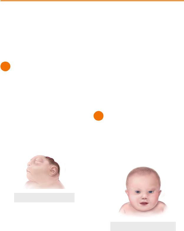
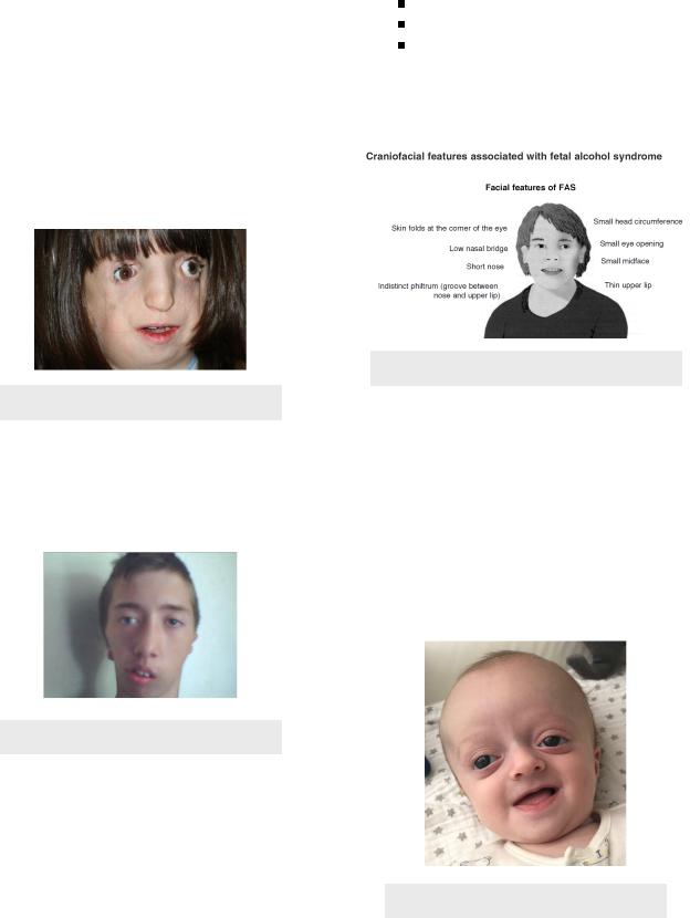
Fetal Alcohol Syndrome
•Early developing fetus exposed to high levels of ethanol (alcohol is a teratogen)
•Alcohol exposure to fetus can cause neural plate tissue deficiency
 Leads to abnormal brain development and microcephaly
 CNS problems
6
Communication difficulty
Learning problems
Hearing and vision problems
•Deficient midface* (important on exam)
•Smooth philtrum, small palpebral fissures, thin upper lip
•Cleft lip
Figure 2.04 Fetal Alcohol Syndrome
Crouzon Syndrome
•Autosomal dominant genetic disease
•Type of craniosynostosis
 Early closure of sutures of the skull
•Proptosis - bulging eyes
•Brachycephalic - short skull
•Frontal bossing - forehead prominent
•Hypertolerism* - wide set eyes
•Midface deficiency*
 Associated with Class III skeletal relationship
Figure 2.05 Crouzon Syndrome
INBDE Booster | Booster Prep™
ORTHODONTICS
Apert Syndrome
•Aka Acrocephalosyndactyly
•Exhibits craniosynostosis
•Similar to crouzon with exception of
•Byzantine arch - narrow palate and high palatal vault
•Acrocephalic - tall skull
•Syndactyly - fusion of fingers and toes → symmetric webbing of hands and feet
Figure 2.06 Syndactylyl in Apert
Syndrome
Hurler & Hunter Syndrome
•Aka mucopolysaccharidosis
•Two separate syndromes but usually grouped together
•Deficient in enzyme that breaks down glycosaminoglycans, resulting in their build up
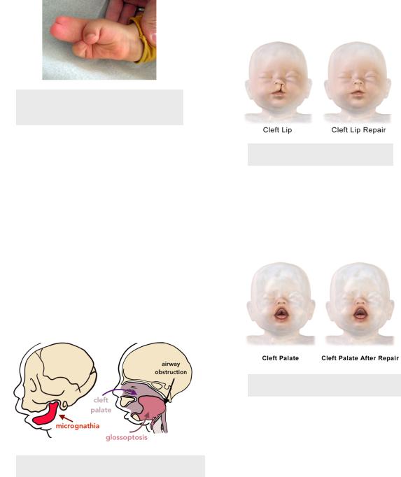
Pierre Robins Sequence
•Sequence: Micrognathia → Glossoptosis → Cleft palate
•Micrognathia - smaller mandible
•Glossoptosis - tongue displaced backwards
•Clossoptosis and cleft palate make breathing and feeding difficult
Figure 2.07 Pierre Robins Sequence
7
Cleft Lip & Palate
•Both due to failure of fusion of tissues during development
•Associated with Class III skeletal relationship + deficient maxilla
•Cleft Lip
 Occurs in utero at 4-6 weeks
 When medial nasal prominence and maxillary prominence fail to fused anteriorly
 Usually occurs off centre and is unilateral
  Bilateral less common
Figure 2.08 Cleft Lip
• Cleft Palate
 Occurs in utero at 6-8 weeks
 When medial nasal prominence and maxillary prominence fail to fused posteriorly
Figure 2.09 Cleft Palate
INBDE Booster | Booster Prep™
ORTHODONTICS |
8 |
DEVELOPMENT OF OCCLUSION
1 Birth and Primary Dentition
Gum Pads
•Infants only have gum tissue pads with no teeth present
 Birth - 6 months
•Ends with eruption of first primary tooth
•Positions of unerupted teeth can be predicted by elevation and grooves on alveolar ridges
 Areas with adjacent transverse grooves represent a developing tooth bud
 Lateral sulcus - more prominent groove on the gum pads dividing the primary canine and primary first molar
Figure 1.01 Gum Pads (Mandible)
Primary Dentition
•Begins with first primary tooth eruption and ends with first permanent tooth eruption
 Runs from 6 months to 6-years
•Minimal overbite and overjet found in young children
 Can be an edge to edge relationship
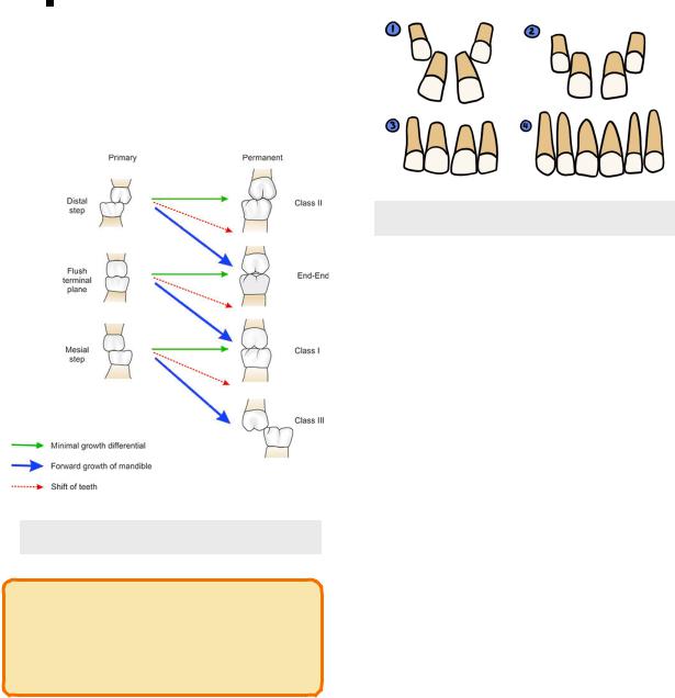
Terminal Plane Relationship
•Terminal Plane - relationship of the distal surface of primary second molars, will guide the eruption of the following:
 Flush Terminal Planes - mandibular and maxillary terminal planes are flush with each other
 37% of patients
 Distal Step - mandibular terminal plane is distal to maxillary terminal plane
14% of patients
 Mesial Step - mandibular terminal plane is mesial to maxillary terminal plane
49% of patients
Figure 1.02 Terminal Plane Relationships
Primary Dental Spacing
• Primate Space
 For maxilla, space between primary lateral incisor and primary canine
 For mandible, space between primary canine and primary first molar
Figure 1.03 Primate Space
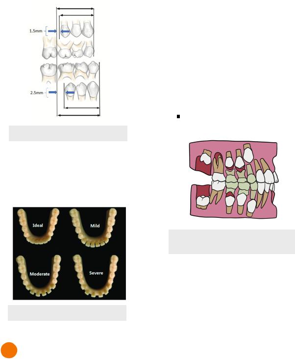
•Leeway Space - difference in combined mesiodistal width of primary canine, primary first molar and primary second molar and their subsequent underlying teeth (canine, first and second premolar)
 Premolars are smaller than primary molars = results in gain of space
 1.5mm gain of space per side in maxilla (3.0mm total)
 2.5 mm gain of space per side in mandible (5.0mm total)
INBDE Booster | Booster Prep™
ORTHODONTICS |
9 |
• Molar relationship will transition into Class I, II or III
• Anterior and Posterior transition occurs
• Individual will experience the Ugly Duckling Stage
Anterior Transition
• Incisors tend to erupt lingually
 Due to permanent tooth buds developing lingual and apical to primary teeth
 Maxillary central incisors are the exception
Are pushed labially by the tongue as they
erupt and can erupt laterally
Figure 1.04 Leeway Space
•Interdental Space - space between primary incisors
 Necessary to prevent crowding of permanent teeth due to increased size
 Incisor liability - difference in size between primary and permanent incisors
Figure 1.05 Interdental Spacing
2 Mixed Dentition
Mixed Dentition Stage
•Begins with eruption of first primary tooth and ends with exfoliation of last primary tooth
 6 years to 12 years
•Closure of dental spacing (interdental, primary and leeway)
Figure 2.01 Succedaneous Tooth
Eruption Positions
Posterior Transition
•Terminal plane relationship of primary second molars guide the position of the first permanent molars
•Mesial step → Class I (90%)
 Class III (10%)
•Distal step → Class II (nearly 100%)
•Flush Terminal Plane → End to end molar relationship (most often)
 End to End will eventually become Class I or II
 But Class I, II, III are all possible
•Class I molar relationship from flush terminal plane through differential jaw growth and teeth shift
 Late mandibular growth
INBDE Booster | Booster Prep™
ORTHODONTICS |
10 |
 Early mesial shift - permanent first molars erupt (~6 years) and close the primate space by shifting mesially
Usually more mesial shift in mandible than maxilla = net forward movement of mandibular molars = Class I relationship
 Late mesial shift - eruption of permanent second molars close leeway space ~12 years = pushes first permanent molars mesially
Figure 2.02 Posterior Transition
INBDE Pro-Tip:
For posterior transition, the INBDE will most likely ask questions about early and late mesial shift.
The Ugly Duckling Stage
•In maxillary teeth, diastema between central incisors begins to close with the eruption of lateral incisors and should close with the eruption of the canines
 Original diastema <2mm between central incisors is normal
 Diastema > 2mm may need additional treatment for closure
• Occurs around 11-12 years
Figure 2.03 Ugly Duckling Stage
Mixed Dentition Analysis
•Used to determine the amount of spacing or crowding in the permanent arch by estimating the mesiodistal width of the unerupted buccal segment of teeth
•Mathematical calculation subtracting space required from space available from the permanent dentition
 Spacing = positive value
 Crowding = negative value
 Permanent incisors should be erupted in order to calculate values
•Tanaka-Johnson Analysis
 Uses sum of width of mandibular incisors
 Maxillary buccal segment = Sum/2 + 11mm
 Mandibular buccal segment = Sum/2 + 10.5mm
• Moyer’s Analysis
 Uses sum of width of mandibular incisors to then refer to table of prediction values of buccal segments
INBDE Booster | Booster Prep™