

Полезные материалы за все 6 курсов / Учебники, методички, pdf / INBDEBooster Orthodontics
.pdfORTHODONTICS |
11 |
3 Permanent Dentition |
|
Permanent Dentition Stage
•Occurs from 12 years to edentulism
•Ideal occlusion
 10-20% overbite
 1-3mm overjet
 Class I occlusion
•Curve of Spee - curvature of arch in the sagittal plane
•Curve of Wilson - curvature of arch in the frontal plane
Figure 3.01 Arch Dimensions
Changes in Arch Dimension
•Arch dimensions change during the transition from mixed to permanent dentition
•Arch Length - ↓ during transition into permanent dentition
•Due to closure of the leeway space
•Arch Perimeter - slight ↑ in upper arch + significant ↓ in lower arch during transition into permanent dentition
•Net results of labial-lateral eruption of permanent teeth + loss of leeway space
•More loss of leeway space in mandible than maxilla, hence the difference in perimeter change
•Intercanine Width - ↑ during eruption of permanent teeth
 Stabilizes after canines erupt
•Intermolar Width - ↑ during eruption of permanent molars then stabilizes
•Greater in upper arch (molar erupt more divergently) than lower (molars erupt more conversantly) due to Curve of Wilson
Crowding of Incisors
•Late mandibular growth results in pressure between the lower lip and lower incisors = late lower incisor crowding increases as we age (between 20s to 40s)
INBDE Booster | Booster Prep™
ORTHODONTICS |
12 |
DIAGNOSIS & TREATMENT PLAN
1 Dentofacial Analysis
Orthodontics does not only involve the straightening of teeth, but also includes correcting malformations of the face in order to achieve overall harmony and function.
Ackerman & Profitt
Ackerman and Profitt were two orthodontists that first described the objective of orthodontic treatment through the soft tissue paradigm
1.Macroesthetic → face
2.Miniesthetics → smile
3.Microesthetics → teeth
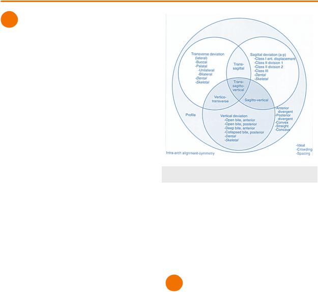
Ackerman and Profitt then continued to describe the features of orthodontic patients in 5 categories. These categories have lots of overlap and can be organized into a Venn diagram.
1.Facial Proportion & Esthetics
i.Facial Profile
ii.Smile arc
iii.Lip posture
2.Dental Alignment & Arch Symmetry
i.Rotations
ii.Crowding/spacing
3.Transverse Plane
i.Midline coincidence
ii.Posterior cross bite
4.Antero-Posterior Plane
i.Angles classification
ii.Overjet
5.Vertical Plane
i.Curve of Spee
ii.Overbite
Figure 1.01 Ackerman & Profit Classification
Orthodontic Examination
Orthodontic Exam evaluates 3 different aspects of the patient . We will cover them more in detail in the following sections
1.Extraoral examination
2.Intraoral examination
3.Cephalometric analysis
2 |
Intraoral Examination |
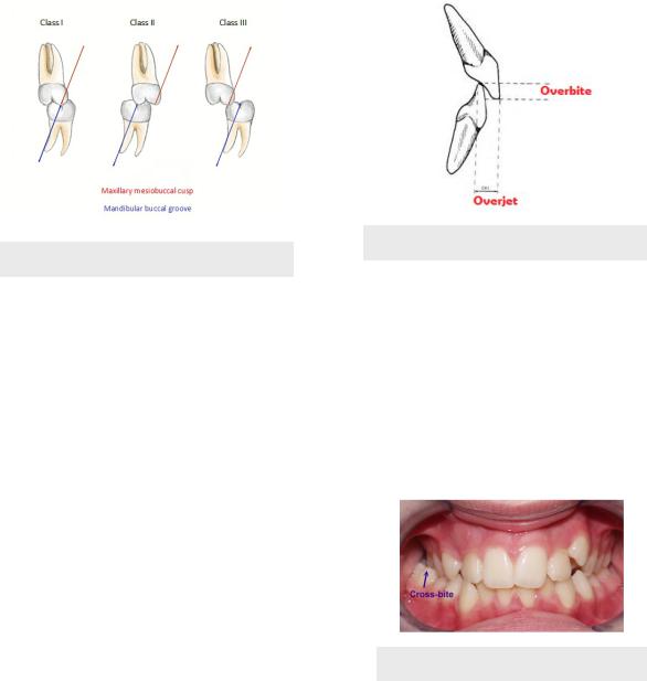
Molar Classification
1.Class I Normal Occlusion (30-35%)
i.MB cusp of maxillary first molars occludes with MB groove of mandibular first molar
ii.Teeth aligned along line of occlusion in the maxilla and mandible
2.Class I Malocclusion (50-55%)
1.MB cusp of maxillary first molars occludes with MB groove of mandibular first molar
2.Teeth do not align
INBDE Booster | Booster Prep™
ORTHODONTICS
3.Class II Malocclusion (15%)
i.MB cusp of maxillary first molars is mesial MB groove of mandibular first molar
ii.Maxillary arch displaced anteriorly
iii.Subdivisions - based on inclination of upper incisors
a.Subdivision 1 - proclined
b.Subdivision 2 - retroclined
4.Class III Malocclusion (1-5%)
i.MB cusp of maxillary first molars is distal MB groove of mandibular first molar
ii.Maxillary arch displaced posteriorly
Figure 2.01 Molar Classification
Malocclusion
65% of patients most often exhibit some form of malocclusion in their molar classification compared to normal occlusion (35%). Within the 65% of cases of malocclusion, 60% have an unknown cause, while 5% have a known cause.
•Malocclusion - any deviation of the teeth from ideal normal occlusion
•Spacing
•Rotations
•Molar relationship
•Overjet/overbite
•Cross bite
•Crowding
•15% adolescents and adults experience severe crowding
•Genetic predisposition
•Genetics of tooth size in relation to arch size
13
Overbite & Overjet
•Overbite - vertical overlap of incisor from incisal edge to incisal edge
•1-2mm overbite is normal
•Deep bite - too much vertical overlap
•Open bite - space between incisors
•Overjet - horizontal overlap of incisors from labial surface to labial surface of incisors
•2-3mm overjet is normal
•Excess overjet - overjet over 3mm
•Reverse overjet - lower incisors are in front of the upper incisors
Figure 2.02 Overbite & Overjet
Crossbite
•Crossbite - misalignment of teeth where upper teeth occlude behind lower teeth
•Anterior Crossbite - maxillary anterior teeth positioned lingual to mandibular anteriors
•Observed in reverse overjet
•Posterior Crossbite - maxillary posterior teeth positioned lingual to mandibular teeth or maxillary posterior teeth are more buccal to mandibular teeth (less common)
Figure 2.03 Posterior Crossbite
INBDE Booster | Booster Prep™
ORTHODONTICS |
14 |
Bolton Analysis
•Bolton Analysis - calculates tooth size discrepancy to measure relative mandibular or maxillary excess
•Compares mesiodistal widths of upper and lower teeth
•Smaller teeth may benefit from buildups
•Composites, veneers or crowns
•Larger teeth may benefit from interproximal reduction (IPR)
•More commonly preformed on lower incisors (for mandibular excess)
3 Extraoral Examination
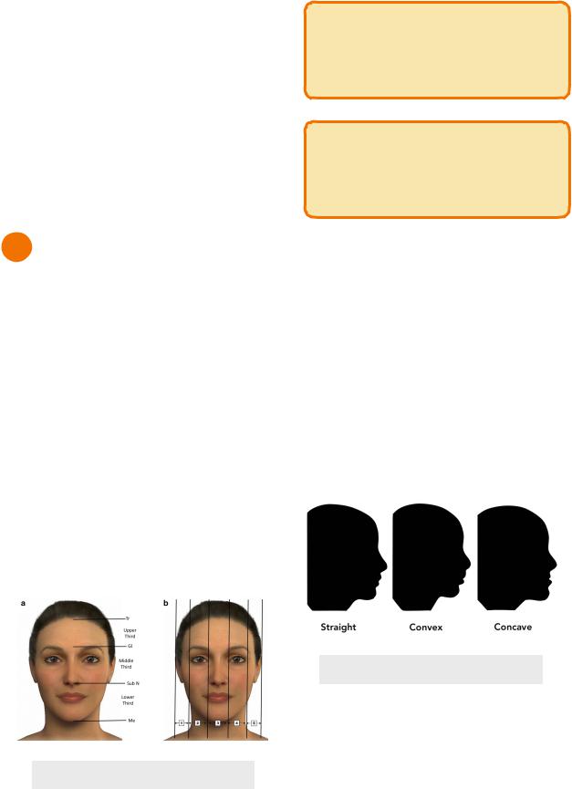
Face Proportions
•Facial thirds - vertical thirds
•Lower third - menton → subnasale
•Orthodontist can influence this third the most
•Can be divided into thirds
•Upper third - upper lip → subnasale
•Lower 2/3rds - mention → lower lip
•Middle third - subnasale → glabella
•Upper third - glabella → hairline
•Horizontal fifths
•Outer fifths - lateral helix → outer canthus
•Medial fifths - outer canthus → inner canthus
•Lines should be coincident with the angle of the mandible
•Middle fifth - inner canthus → inner canthus
•Lines should be coincident with alar base (sides of nose)
Figure 3.01 Face Proportions
INBDE Pro-Tip:
The interpupillary distance should ideally line up with the commissures (corners) of the mouth.
INBDE Pro-Tip:
The horizontal fifths can be memorized by parts of the face (ear → eye → nose → eye → ear).
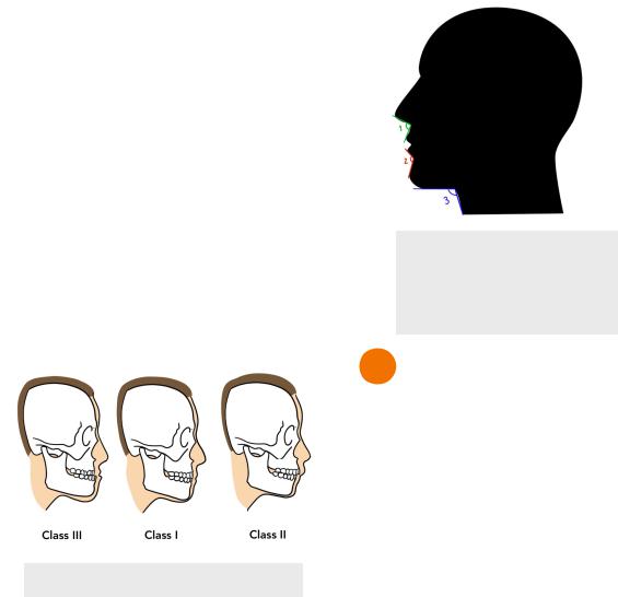
Facial Profile
Facial profile refers to the facial convexity of the facial plane, which is determined by the landmarks of the glabella, subnasale, and soft tissue pogonion.
1.Straight - 0-10° facial plane
i.Often associated with Class I occlusal relationship
2.Convex - >10° facial plane
i.Often associated with Class II malocclusion
3.Concave
i.Often associated with Class III malocclusion
Figure 3.02 Profile Types
INBDE Booster | Booster Prep™
ORTHODONTICS
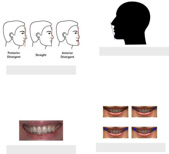
Facial Divergence
Facial divergence refers to the relationship of the lower face relative to the forehead. It can be determined by drawing a line straight down from the forehead/glabella and seeing where the subnasale and menton are relative to the line.
1.Straight - subnasale and soft tissue pogonion are close to the line
i.Associated with Class I occlusal relationship
2.Posterior divergent - subnasale is posterior to the line and the soft tissue pogonion is even more posterior
i.Associated with Class II malocclusion
3.Anterior divergent - subnasale is anterior to the line and the soft tissue pogonion is even more anterior
i.Associated with Class III malocclusion
Figure 3.03 Facial Divergence
Incisor Display
•Smiling
•Minimum = 75% of incisors visible
•Ideal = 100% incisor visible + 1-2mm gingiva visible
•Gummy smile >2mm gingiva showing
•At rest, ideal = 2-4mm of incised edge
Figure 3.04 Ideal Incisor Display
15
Lips
•Posture - the ability for lips to close without strain
•Competent - lips can close without strain at rest
•Incompetent - lips separated by 3-4mm at rest and muscles are strain upon closure
•Proportion - based on the amount of vermillion visible at rest
•Thick
•Thin
•Position - position relative to the tangent line that runs from the nose tip to chin (E-line)
•Prominent - lips in front of the line
•Retrusive - lips behind the line
Figure 3.05 E-line
Buccal Corridors
•Buccal corridor - the appearance of a gap of space during smiling, between the maxillary posterior teeth and the corner of the mouth
•Wide, medium or narrow
•Based on the amount of space visible
•Narrowing of buccal corridors can be achieved through palatal expansion
Figure 3.05 Buccal Corridors
INBDE Booster | Booster Prep™
ORTHODONTICS
Skeletal Classification
Skeletal classification refers to the relationship between the mandible and maxilla in an anteriorposterior position.
1.Skeletal Class I - normal relationship with the jaw
2.Skeletal Class II - maxilla more anterior to mandible
i.Prognathic/protrusive maxilla (10%)
ii.Retrognathic/retrusive mandible (85%)
iii.Both (5%)
3.Skeletal Class III - maxilla more posterior to mandible
i.Retrognathic/retrusive maxilla (20%)
ii.Prognathic/protrusive mandible (60%)
iii.Both (20%)
Figure 3.06 Skeletal Classification
Angles of the Facial Profile
1.Cervicomental angle - between menton → reflex point → base of the neck
i.Ideal = 90-120°
2.Mentolabial angle - soft tissue pognonion → mentolabial sulcus → lower lip
i.Aka mentolabial fold or mentolabial sulcus
ii.Ideal = 120°
3.Nasolabial angle - nose tip → subnasale → upper lip
i.Ideal = 90°
16
Figure 3.07 Angles
1)Nasolabial
2)Mentolabial
3)Cervicomental
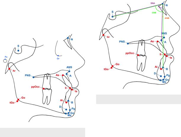
4 Cephalometrics
Cephalometric radiographs allows dentists and orthodontics to capture the hard and soft tissue structures from the side of the face. Analysis of these structures can then be judged based on the position of various soft tissue landmarks and their relationships to each other.
Additionally, cephalometric radiographs of the same patient over different time points can be superimposed to evaluate skeletal and dental changes that have occurred due to treatment or growth.
Soft Tissue Landmarks
•Bolton Point (Bo) - most superior point of the occipital bone
•Basion (Ba) - lowest point of the anterior margin of the foramen magnum
•Articulary (Ar) - inner section between zygomatic arch and posterior border of the ramus
•Porion (Po) - most superior point of the external auditory meatus
•Condylion (Co) - most poster-superior point of the condylar head
•Pterygomaxillary fissure (Ptm) - teardrop shaped fissure posterior to the maxilla
INBDE Booster | Booster Prep™
ORTHODONTICS |
17 |
•Sella (S) - midpoint of sella turcica
•Orbitale (Or) - lowest point on inferior margin of orbit
•Nasion (N) - anterior intersection of frontal and nasal bone
•Anterior Nasal Spine (ANS) - tip of the anterior projection of the maxilla
•Posterior Nasal Spine (PNS) - sharp posterior projection of palatine bone
 Usually underneath the Ptm
•A point - innermost point of the contour of the maxillary bone
•B point - innermost point of the contour of the mandibular bone
•Gonion (Go) - midpoint of contour connecting the ramus and body of the mandible
•Pogonion (Pog)- most anterior point of the chin
•Gnathion (Gn) - midpoint between the pogonion and menton
•Menton (Me) - most inferior point of the chin
are then compared to their norms according, to age, sex and ethic group.
• SNA - angle from Sella-Nasion-A point
 Represents relationships of maxilla to cranial base
 Larger angle = maxilla is more forward
• SNB - angle from Sella-Nasion-B point
 Represents relationships of mandible to cranial base
 Larger angle = mandible is more forward
•ANB - angle from A point-Nasion-B point
 Normal = 2°
 Skeletal Class III = 0° or less (negative)
 Skeletal Class II = 4° or more
Figure 4.02 Cephalometric Analysis
Figure 4.01 Cephalometric Landmarks
Analysis
As previously mention, landmarks taken from the cephalometric radiograph can be used to analyze the relationship of the jaws and teeth using linear and angular measurements. The measurements
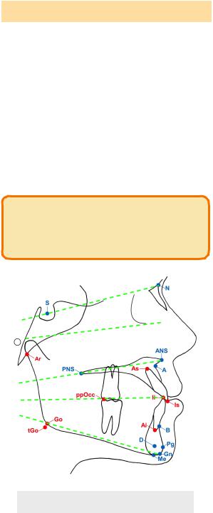
Reference Planes
Reference planes are created when connecting two landmark points together. All five of the following reference planes should intersect at the back of the head.
•If they intersect earlier, the patient is considered hyperdivergent
•If its is farther back, it is considered hypodivergent
INBDE Booster | Booster Prep™
ORTHODONTICS |
18 |
||
|
|
|
|
Landmarks |
Plane |
|
|
|
|
|
|
Go-Me |
Mandibular |
|
|
|
|
|
|
L6-L1 (occusal edge of |
Occlusal |
|
|
lower first molar - |
|
|
|
incisal edge of lower |
|
|
|
central incisor) |
|
|
|
|
|
|
|
ANS - PNS |
Palatal |
|
|
|
|
|
|
Porion-Orbitale |
Frankfort horizontal |
|
|
(Po-Or) |
|
|
|
|
|
|
|
Sella-Nasion (S-N) |
Cranial base |
|
|
|
|
|
|
INBDE Pro-Tip:
Questions asking about ANB measurements are common on the exam.
Figure 4.03 Reference Planes
INBDE Booster | Booster Prep™
ORTHODONTICS |
19 |
BIOLOGY OF TOOTH MOVEMENT
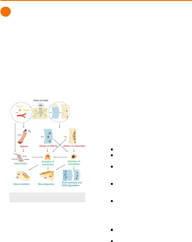
1 Factors of Tooth Movement
Orthodontic tooth movement is the involvement of the movement of a tooth within alveolar bone. The alveolar bone and the PDL play crucial roles in this process. The biology of tooth movement can be divided into 4 general steps.
1.Force applied to tooth
2.Stress of PDL
i.Compression = osteoclasts active
ii.Tension = osteoblasts active
3.Bone remodelling
4.Tooth movement
Figure 1.01 Tooth Movement
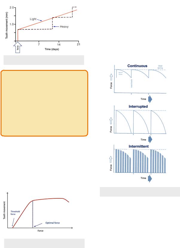
Light & Heavy Force
Force magnitude is important in determining the biological processes of tooth movement. Different sequences of events will occur depending on if there is light force or heavy force.
•Light Force (≤100g)
 Seconds
 No pain
 Blood vessels distorted - compression side = compressed, tension side = partially dilated
 Minutes
 Altered blood flow → O2 & CO2 levels change → trigger release of inflammatory mediators (↑PGE, ↑RANKL)
 Hours
Hours
 ↑ cAMP → cell differentiation within PDL
 2 days
 Osteoclasts recruited for frontal resorption - resorption lamina dura (outer surface of bone) to create Howship’s lacunae
 3-5 days
 Lamina dura resorbed as tooth moves
 1-2 weeks
 Tooth movement process continues and occurs at a steady pace
•Heavy Force (>100g)
 Seconds
Immediate pain from high pressure
Blood flow occluded
 Minutes
Blood flow ceases completely on compression side
 Hours
Hours
PDL hyalinized from sterile necrosis - cell death not caused by a pathogen
 3-5 days
Undermining resorption - osteoclasts recruited in nearby bone marrow → lamina dura collapse from the inside until area of bone is removed
 1-2 weeks
Tooth moves when undermining resorption ends
Tooth movement is not steady like frontal resorption - lag period of 1-2 weeks for
INBDE Booster | Booster Prep™
ORTHODONTICS |
20 |
resorption to take place, then tooth moves quickly
Figure 1.02 Light & Heavy Force
INBDE Pro-Tip:
It is important to note that light force causes frontal/direct resorption meanwhile heavy force causes undermining/indirect resorption. Light force is preferred in orthodontics because movement is steady and the patient experiences less discomfort, although it is difficult to achieve. Heavy force is more commonly encountered.
Duration of Force
•Threshold for tooth movement = 4-8h
 Associated with the amount of time required for cAMP to reach a level to trigger an inflammatory response in the PDL
•Orthodontic appliances must be worn for long enough each day, or else process needs to restart (patient compliance is key)
Figure 1.03 Force Duration
•Force Decay - natural process that occurs for the force to decrease/tire out an orthodontic appliance
1.Continuous - force is relatively constant
i.Example - light wires
a.Force declines slightly overtime, but replacement will restore the force
2.Interrupted - force slowly decreases to zero
i.Example - elastic chains
3.Intermittent - force abruptly ceases
i.Example - clear aligners
Figure 1.04 Force Decay Types
Tooth Movement Types
The force distribution to the tooth and the PDL is important for the process of orthodontic appliances causing tooth movement. Pressure (force/area) on the PDL, as a result of the force distribution, should be controlled to allow for
INBDE Booster | Booster Prep™