

Полезные материалы за все 6 курсов / Учебники, методички, pdf / INBDEBooster Endodontics Notes
.pdfENDODONTICS |
1 |
DENTAL PULP
Endodontics translates to the “knowledge of what is within teeth”. It is the field of dentistry concerning dental pulp and surrounding tissues of tooth roots. Studying endodontics grants clinicians an understanding of the etiology, diagnosis, and treatment of pulpal and periradicular conditions.
1 Pulp & Dentin
Composition
Dental pulp contains a variety of cells and connective tissues.
•Loose connective fibrous tissue
•Neuromuscular elements
 Blood vessels
 Lymph vessels
 Nerves
• Cells types
 Odontoblasts - produce primary and secondary dentin
Primary dentin - before complete root formation
Secondary dentin - after complete root formation
 Mesenchymal cells - can differentiate into secondary odontoblasts to produce tertiary dentin (response to injury)
 Fibroblasts
•Lack of collateral circulation = difficulty fighting infection
Dentin
Tooth pulp is surrounded by dentin. This hard dentin has limited expansion ability, thus creating an increase in pressure when infection occurs and the pulp and inflamed. There are different reactions that occur in dentin and pulp in response to damage.
•Secondary dentin/Reactionary dentin - produced in response to minor damage
•Tertiary dentin/Reparative dentin - produced in response to major damage
Figure 1.02 Tertiary dentin
INBDE Pro-Tip:
Pulp capping involves placing a calcium hydroxide liner that irritates odontoblasts, causing them to lay down secondary/tertiary dentin
•Sclerotic dentin - calcification of dentinal tubules
 Due to aging or a response to slowly advancing caries
•Pulp necrosis - occurs in rapid caries or severe damage
Figure 1.01 Tooth Anatomy
Figure 1.03 Sclerotic Dentin
INBDE Booster | Booster PrepTM
ENDODONTICS
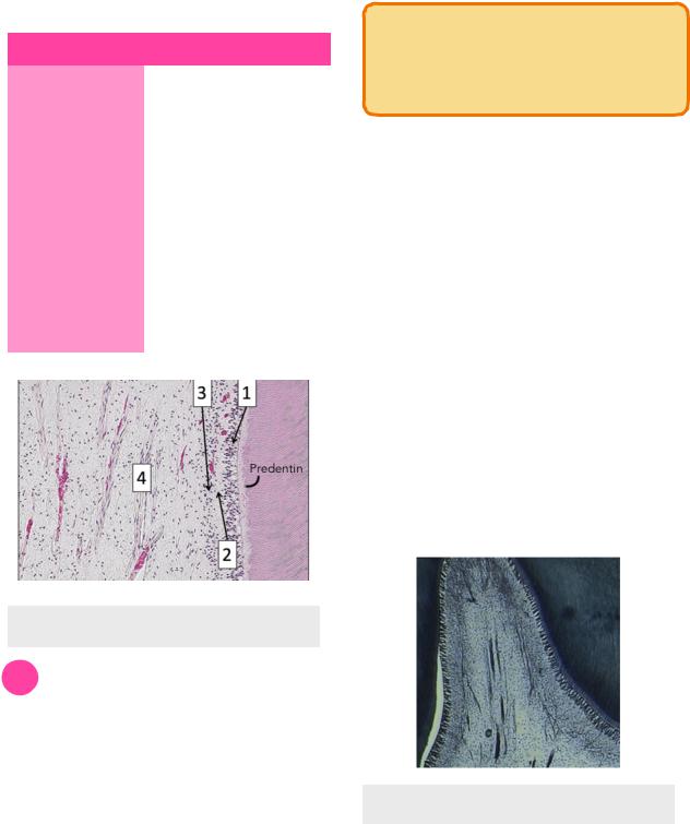
Histological Zones of Pulp
Layer |
Characteristics |
|
|
Predentin |
Unmineralized, inner |
|
dentin layer, directly |
|
adjacent to pulp |
|
|
Odontoblastic |
Location of odontoblasts, |
Layer (1) |
considered part of the |
|
pulp |
|
|
Cell-free zone of |
No nuclei/cells present, |
Weil (2) |
often seen with nerve |
|
bundles |
|
|
Cell-rich zone (3) |
Nuclei and cells present |
|
|
Pulp Core (4) |
Central part of pulp |
|
|
2
INBDE Pro-Tip:
In dentistry, referred pain from mandibular molars is often felt in the pre-auricular region (both have V3 innervation)
Pain from Pulpitis
•Pain conducted from C-fibres
 Afferent nerve
 Small diameter
 Unmyelinated
•Dull throbbing lingering pain
•Sensitive to heat
•Travel centrally through pulp
Pain from Dentin
•Pain conducted from A -fibres
 Afferent nerve
 Large diameter
 Myelinated
•Sensitive to cold
•Sharp transient pain
•Travel coronally in the pulp
 Hence, more easily provoked for pain sensation than central C-fibers
Figure 1.04 Pulp Histology
2 Pain
Altered Sensations
•Hyperalgesia - abnormal increase in sensitivity
•Anesthesianumbness
•Dysasthesiaunpleasant, abnormal sensation
• Allodynia - pain due to stimulus that does not |
Figure 2.01 Nerve Bundles |
normally cause pain |
|
•Referred pain - pain perceived to come from a location other than where it actually originates
INBDE Booster | Booster PrepTM
ENDODONTICS
DIAGNOSIS
Every tooth can be given a pulpal and periapical diagnosis. Pulpal diagnosis concerns the pulp of the tooth, whereas periapical diagnosis involves the tissues surrounding the tooth.
1 Pulpal Diagnosis
Possible Diagnosis
1.Normal
i.Normally asymptomatic
ii.Mild-moderate transient (short-lasting) response to thermal and electrical pulp test
2.Reversible pulpits
i.Symptomatic
ii.Cause = pulp irritant
a.Reversed by removal of irritant
b.No removal = may progress to irreversible pulpitis
iii.Not considered disease
iv.Positive cold test = hypersensitivity + transient, sharp pain
v.No spontaneous pain
3.Asymptomatic irreversible pulpitis
i.Asymptomatic
ii.Physiologically and microscopically comparable to symptomatic irreversible pulpitis, but without symptoms
4.Symptomatic irreversible pulpits
i.Symptomatic = spontaneous pain (intermittent or constant)
ii.Irreversible damage to pulp, will not fully heal with irritant removal
iii.Cold test = lingering pain
iv.EPT not useful
v.Radiographs usually insufficient
vi.Posture changes (bending, lying down) may exacerbate pain due to increased blood pressure
3
5.Necrotic pulp
i.Rarely symptomatic
ii.Often, occurs from long term lack of blood supply to the pulp
iii.Includes partial or total necrosis
iv.Anterior teeth may appear with crown discolouration
v.Untreated leads to PDL thickening, sensitivity to percussion and periapical disease
6.Previously treated pulp
i.Natural pulp tissue has been removed due to pulp therapy
Pulp Vitality Tests
1.Cold Test - application of cold substance on tooth to cause stimulation
i.Uses endo-ice (dichlorodifluromethane, -30ºC) sprayed onto a cotton pellet and applied onto the dried mid-facial surface of the tooth for 5s
ii.Pulpal diagnosis based on intensity and duration of response
Figure 1.01 Cold Test
2.Electric Pulp Test
i.Determines presence of vital sensory fibres in the pulp
ii.Can only indicate if the tooth is vital or non-vital (no severity)
iii.Least reliable pulp vitality test
i.False results (positive and negatives)
ii.No indication of vascular supply of pulp
iii.Cardiac pacemaker contraindicated
INBDE Booster | Booster PrepTM
ENDODONTICS
Figure 1.02 Electric Pulp Test
2 Periapical Diagnosis
Apical lesions that arise from pulpal origin is extension of pulpal disease into the apical tissues. Other causes include trauma, iatrogenic damage, and periodontal disease.
Possible Diagnosis
1.Normal
i.Asymptomatic = no pain on palpation and percussion
2.Asymptomatic Apical Periodontitis
i.Asymptomatic
ii.Radiographs useful = visualization of apical radiolucency
a. Confirms necrotic pulp
Figure 2.01 Apical Radiolucency
3.Symptomatic Apical Periodontitis
i.Symptomatic = pain on percussion (intense and throbbing)
ii.Inflammation around tooth apex
iii.PDL contains localized inflammatory infiltrate
iv.If tooth is vital → occlusal adjustment
v.If necrotic tooth → endodontic therapy
4
a.Due to belief that infection originated from pulp spreading to apical tissues
4.Acute Apical Abscess
i.Acute = rapid swelling + severe pain
ii.Apex contains purulent exudate/ liquefaction necrosis of tissue
5.Chronic Apical Abscess
i.No/less swelling or discomfort (than acute) due to the presence of draining sinus tract
a.Path and source of sinus tract can be located by inserting gutta percha cone into the tract until resistance is felt. Then, take a periapical radiograph
Tests for Periapical Diagnosis
1.Percussion Test
i.Done by tapping the tooth along its long axis using the end of the mirror handle
ii.Normal response should not have pain
Figure 2.02 Percussion Test
2.Apical Palpation
i.Palpation of gums/vestibular area around the area of the root apex of tooth
ii.Normal response should not have pain or feel swollen or bumpy
INBDE Booster | Booster PrepTM
ENDODONTICS
DENTAL TRAUMA
1 Dental Trauma Types
Fractures
• Uncomplicated fracture
Fracture that does not involve the pulp
Enamel only → smooth the edges of the tooth to prevent future damage Enamel and dentin involved → tooth restoration
Figure 1.01 Uncomplicated Fracture
• Complicated fracture
Fracture that involves pulp
Treatment according to timing
Less than 24h → direct pulp cap
±24h → partial pulpotomy (Cvek) ±72h → pulpotomy
Figure 1.02 Complicated Fracture
• Horizontal root fracture
Apical segment remains in place
Necrosis is rare
Displacement of coronal segment
25% of necrosis
Minimum 3 periapical + 1 occlusal radiograph
Fracture along one plane will need several radiographs at different angulations to visualize the fracture
5
Healing
Calcific metamorphosis can fuse close fragments through calcification
If tooth is necrotic perform root canal If tooth is vital, splint teeth immediately based on fracture site on root
Apical → flexible splint, 4 weeks
Middle → flexible splint, 4 weeks Coronal → flexible splint, 4 months
Figure 1.03 Apical Root Fracture
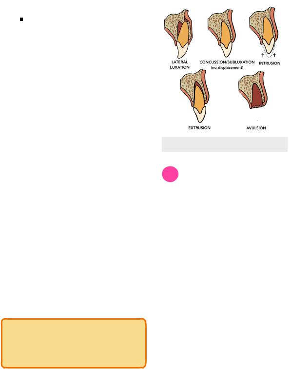
Luxation Injuries
•Concussion - minor injury, no tooth displacement or mobility
 PDL inflamed and sore
 No treatment needed, let tooth rest
•Subluxation - no tooth displacement, slight mobility
 PDL inflamed, may rip and bleed
 Closed apices = 6% chance of necrosis
 Open apices have better prognosis
 Treatment → flexible splint for up to 2 weeks
•Extrusion - displacement of tooth from socket in an extrusive direction
 Closed apices = 65% of necrosis
 Treatment
 Closed apices → reposition, flexible splint for up to 2 weeks, follow-up
INBDE Booster | Booster PrepTM
ENDODONTICS
Open apices → reposition, flexible splint, root canal treatment if necessary
•Lateral Luxation - tooth displaced from its long axis with apical end usually displaced labially and coronal end palatally
 Fracture of alveolar bone may occur in severe cases
 Close apices = 80% chance of necrosis
 Treatment → flexible splint, 4 weeks
•Intrusive Luxation - tooth pushed into socket/ apical displacement of tooth
 Closed apices = 95% chance of necrosis due to severing of blood vessels
 Treatment
 Closed apices → reposition, flexible splint or root canal
 Open apices → monitor and wait for tooth to re-erupt on its own
•Avulsion - tooth completely displaced from the alveolus
 Most serious of all dental injuries
 Extra-alveolar dry time (EADT) should be monitored
 Time the tooth has been dry and out of the mouth
 Prognosis is worse the longer the time
 Treatment (General)
 Re-implant tooth as soon as possible
 Flexible splint for up to 2 weeks
INBDE Pro-Tip:
The INBDE will most often ask questions regarding treatment when it comes to dental trauma injuries.
6
Figure 1.04 Luxation Injuries
2 Long-term Response to Trauma
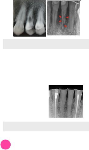
Internal Resorption
•Damage to odontoblastic layer initiates resorption in root canal system
•Inflammatory response to bacteria and their byproducts traveling from necrotic pulp to dentinal tubules, causing internal resorption
•Radiograph
 Does not move with radiograph angle change
 Margins well defined, sharp
•Better prognosis and easier treatment than external resorption
•Treat with root canal therapy to obturate and seal the canal
External Resorption
•Damage to cementoblastic layer initiates resorption in the periodontium
•Radiograph
 Radiographically moves with radiograph angle change
 Margins poorly defined & irregular
•Three subcategories
1.Replacement resorption - PDL replaced with bone (ankylosis)
i.Can result from splints being too rigid or being placed too long
INBDE Booster | Booster PrepTM
ENDODONTICS
2.Inflammatory root resorption - in response to bacteria and their byproducts traveling from necrotic pulp to dentinal tubules
3.Cervical resorption - due to trauma or non-vital bleaching causing subepithhelial sulcular infection
Figure 2.01 Internal & External Resorption
Calcific Metamorphosis
•Odontoblasts producing large amount of reparative dentin into the pulp space, induced by trauma
•Results in canal obliteration
•Yellow-orange
•Increased risk
 Open apices
 Intrusion
  Severe crown fractures
Figure 2.02 Calcified Canals
3 Dental Management
Classification
The Ellis Classification is commonly used to signify which traumatic injury has occurred.
•Class I = enamel only involved
•Class II = enamel + dentin involved
•Class III = enamel, dentin & pulp involved
•Class IV = tooth is non-vital from trauma
•Class V = luxation injury
•Class VI = avulsion
7
Protocol
The acronym “TRAVMA” can be used to remember to the appropriate steps in response for recent traumatic tooth injury
1.Tetanus booster
i.Within 48h
ii.For avulsions only, if tooth gets contaminated
2.Radiographs
i.At least 1 periapical radiograph
ii.Panoramic recommended for general trauma or suspected fracture
3.Antibiotics
i.For avulsion, only if the tooth is contaminated
4.Vitality tests
i.May give false negatives since sensory nerves can be disrupted for 2-8 weeks, but vascular supply is still in tact
5.More Appointments
i.Follow-up appointments recommended at 3 weeks, 3-, 6-, and 12-months after traumatic injury
Avulsion
An avulsed tooth should not be kept dry and should be kept in appropriate storage media before arriving to the dentist. The following are considered appropriate storage media:
•Milk
•Saline
•Hank’s balanced salt solution - best option
•Saliva
 Person can also hold the tooth in the area of the vestibule where the tooth is from
• Avoid storage in water!
 Hypotonic, incorrect balance of ions
INBDE Booster | Booster PrepTM

ENDODONTICS |
8 |
Avulsion (continued)
Dental protocol for avulsion of permanent tooth is more complicated, depending on apices and EADT.
Apices, EADT |
|
Treatment |
|
|
|
Closed, EADT < 1h |
Reimplant + splint |
|
|
|
|
Open, EADT < 1h |
Reimplant + splint |
|
|
Apexification if pulp |
|
|
infected |
|
|
No root canal |
|
|
|
|
Closed, EADT > 1h |
Reimplant + splint |
|
|
Root canal |
|
|
|
|
Open, EADT > 1h |
Options |
|
|
- |
Reimplant + splint |
|
- |
Root canal |
|
- |
Implants |
|
|
|
INBDE Booster | Booster PrepTM

ENDODONTICS |
9 |
VITAL & NON-VITAL PULP THERAPY
1 Introduction
Options for Vital pulp Therapy
Vital pulp therapy refers to treatment options that are suited for pulp that is still vital, but has some sort of damage. There are several different options.
•Indirect pulp cap
•Direct pulp cap
•Partial (Cvek) Pulpotomy
•Pulpotomy
•Apexogenesis
Options for Non-Vital pulp Therapy
•Pulpectomy
•Root Canal Therapy
•Apexification
•Extraction
Common Materials
• Calcium hydroxide - Ca(OH)2
 pH = 12.5 → bactericidal and cauterizes tissue
 Can stimulate mesenchymal cells to differentiate into secondary odontoblasts, which will create a dentin bridge (tertiary dentin) as a barrier to protect pulp
• MTA - mineral trioxide aggregate
 Stimulates cementoblasts to make hard tissue
 Contains
Calcium, silicon & aluminum
Bismuth oxide (opacifier) = radiopaque, but can leak and stain tissue
 Properties
Can sets in moisture
3h setting time
Nonresorbable & biocompatible - good sealant
Antimicrobial
2 Vital Pulp Therapy
Indirect Pulp Cap
•Indicated for deep caries, pulp is healthy and not exposed
•Dentin covering pulp lined with Ca(OH)2 or RGMI
 RGMI can also layer on top of Ca(OH)2
Direct Pulp Cap
• Indicated for healthy pulp that is exposed
 From caries of mechanical exposure <2mm
  From trauma that occurred within 24h
•Ca(OH)2 placed directly on top of exposed pulp
 Hard tissue barrier can form as early as 6 weeks
Figure 2.01 Pulp Capping
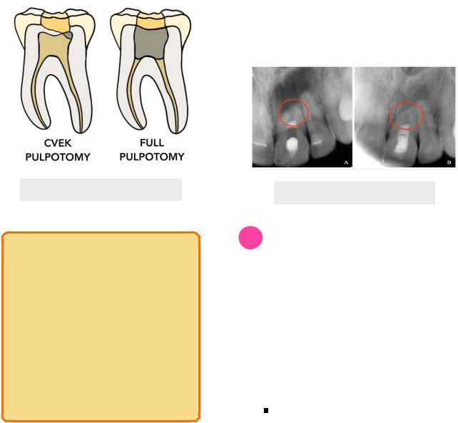
Partial (Cvek) Pulpotomy
•Indicated when there is a small amount of coronal diseased pulp
 From caries or mechanical exposure > 2mm
 From trauma that occurred more than 24h ago
•Removal of diseased pulp only as a means of reserving the remaining healthy coronal and radicular pulp
•Aka shallow pulpotomy
INBDE Booster | Booster PrepTM
ENDODONTICS
Full Pulpotomy
• Indicated for
 Trauma that occurred more than 72h ago
 For vital primary tooth with pulp exposure, but is restorable
 Traditionally place a ZOE crown and formocresel
•Removal of coronal diseased pulp as a means of preserving healthy radicular pulp tissue
Figure 2.02 Pulp Pulpotomy
INBDE Pro-Tip:
Buckley’s Formocresol is the form of formocresol that is asked about on the exam. It has bactericidal properties and is a fixative for pulp tissue to resist enzymatic breakdown. It can be toxic if used in large amounts, but should not be if used appropriately in the correct dilutions. The formulation is as follows:
•Formaldehyde - 19%
•Cresol - 35%
•Water - 31%
•Glycerine - 15%
10
Apexogenesis
•Performed to maintain pulp vitality and encourages root end closure by physiologic development of root end
•Occurs when Ca(OH)2 or MTA is placed on healthy or diseased pulp
•Contraindications
 Nonrestorable tooth
 Severe horizontal fracture
 Avulsed tooth
 Necrotic tooth
•Is a process that occurs when an indirect or direct pulp cap, Cvek of full pulpotomy is performed in an immature permanent tooth
Figure 2.03 Apexogenesis
3 Non-Vital Pulp Therapies
Pulpectomy
•Involves removing coronal and radicular pulp that is dead or dying
•Similar to root canal treatment, but filling with ZOE (instead of gutta percha)
•Indications
 Temporary treatment for irreversible pulpits (pain relief) until full RCT can be performed
 For asymptomatic non-vital primary tooth with exposed pulp, but the tooth is restorable
ZOE in crown and Ca(OH)2 in root (allows for resorption when the underlying permanent tooth erupts)
INBDE Booster | Booster PrepTM