


of Atlas
ultrasound musculoskeletal anatomy
244
Foot
(Figures 300 and 301)
•Plantar surface and sole of foot.
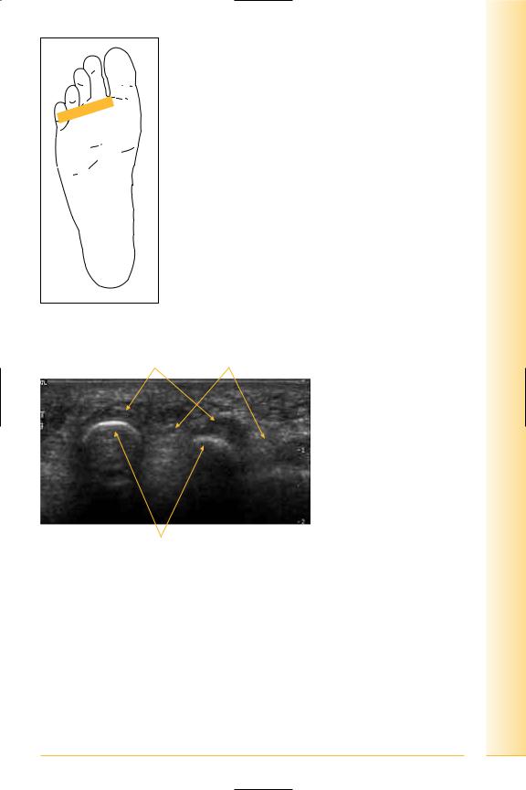
•Web space.
Contents: Flexor digitorum brevis and longus, quadratus plantae, lumbricals, flexor hallucis longus, abductor hallucis and interossei.
Notes
limb Lower
Foot
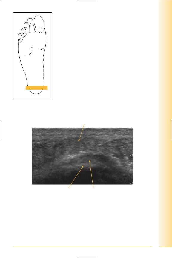
FIG. 300 TS web space, probe on plantar surface
Flexor tendons |
Intermetatarsal bursa/fat |
Lateral |
Metatarsal heads |
Medial FIG. 301 TS, web space |
245

of Atlas
ultrasound musculoskeletal anatomy
246
Flexor hallucis longus
(Figures 302–305)
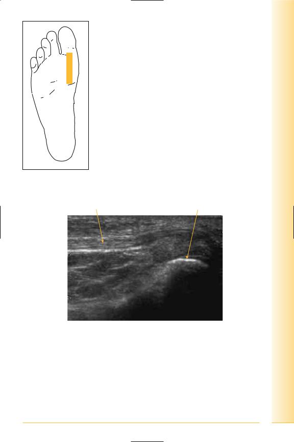
Tendon passes distally between the sesamoid bones and inserts in to the distal phalanx of the great toe.
Notes
limb Lower
Foot
FIG. 302 LS, probe over first metatarsal head. Dynamic examination using flexion/extension of the great toe
Tendon |
Metatarsal head |
Proximal |
Distal |
FIG. 303 Flexor hallucis longus in forefoot
247
of Atlas
ultrasound musculoskeletal anatomy
248
Proximal |
Tendon |
Distal |
Metatarsal |
Proximal phalanx |
Terminal phalanx |
FIG. 304 LS, flexor hallucis longus
Flexor tendons |
Medial and lateral sesamoids |
Fifth metatarsal |
Plantar interossei |
First metatarsal |
Lateral |
|
Medial |
FIG. 305 TS panorama, plantar foot – metatarsals
Flexor hallucis brevis
(Figures 306–311)
•Origin: medial plantar surface of the cuboid and lateral cuneiform.
•Insertion: splits in two around flexor hallucis longus and inserts either side into the proximal phalanx. Each tendon contains a sesamoid bone.

limb Lower
Foot
FIG. 306 LS, probe over medial sesamoid
Medial sesamoid
Distal |
Tendon, muscle of flexor |
Proximal |
|
hallucis brevis |
|
FIG. 307 LS, flexor hallucis brevis
249
of Atlas
ultrasound musculoskeletal anatomy
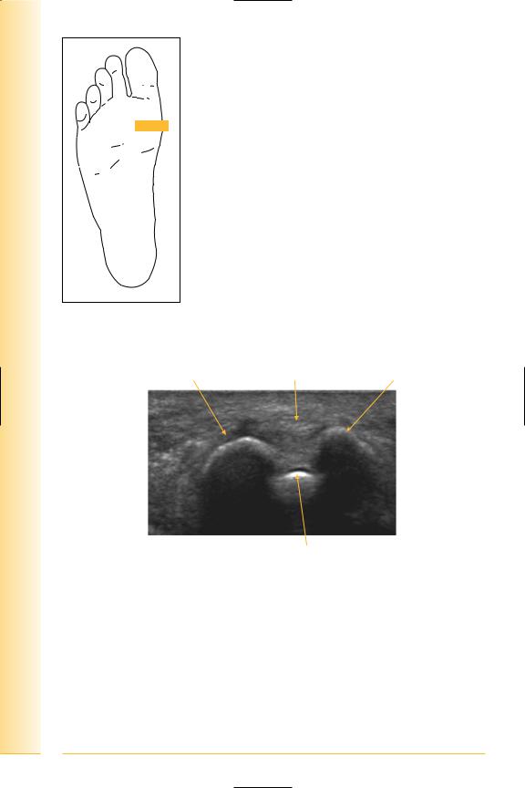
FIG. 308 TS, probe over sesamoids
Lateral sesamoid |
Flexor hallucis longus |
Medial sesamoid |
Lateral |
Metatarsal |
Medial |
FIG. 309 TS, sesamoids
250
limb Lower
Foot
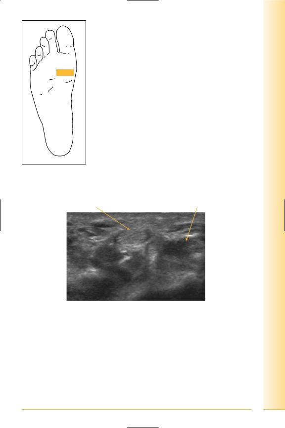
FIG. 310 TS, probe proximal to sesamoids
Flexor hallucis longus tendon |
Flexor hallucis brevis muscle |
Lateral |
Medial |
FIG. 311 TS, flexor hallucis – proximal to sesamoids
251

of Atlas
ultrasound musculoskeletal anatomy
252
Plantar fascia
(Figures 312–315)
Three bundles – medial, lateral and middle.
Attaches proximally to the medial process of the calcaneum and fans into five slips to merge with the flexor digitorum sheaths to attach to the transverse intermetatarsal ligaments and the base the proximal phalanges. Strong septa pass from this fascia laterally to divide flexor digitorum from abductor digiti minimi, and medially from abductor hallucis.
Notes
limb Lower
Foot
FIG. 312 TS, probe over heel pad
Heel pad
Lateral |
Calcaneum |
Plantar fascia |
Medial |
FIG. 313 TS, plantar fascia
253
of Atlas
ultrasound musculoskeletal anatomy
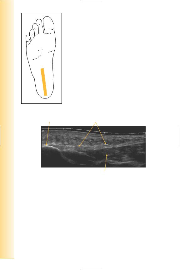
FIG. 314 LS, probe midline over plantar surface
Calcaneum |
Plantar fascia |
Proximal |
Flexor digitorum brevis |
Distal |
FIG. 315 LS panorama, plantar fascia
|
Plantar muscles mid-foot |
|
(Figures 316 and 317) |
|
There are four layers. |
|
• Superficial: abductor hallucis, abductor digiti minimi, flexor digitorum |
|
brevis. |
|
• Second layer: flexor digitorum longus, quadratus plantae, lumbricals, |
|
flexor hallucis longus. |
|
• Third layer: flexor hallucis brevis, flexor digiti minimi, adductor hallucis |
254 |
transversus, adductor hallucis obliquus. |
• Fourth layer: interossei, tendons of tibialis posterior and peroneus longus. |
limb Lower
Foot
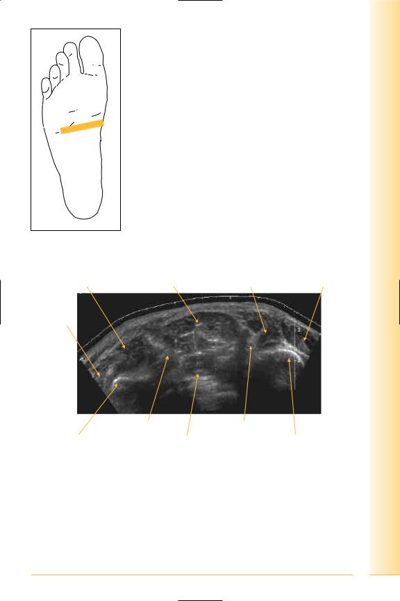
FIG. 316 TS, probe mid-foot
Flexor digiti minimi Flexor digitorum brevis Flexor hallucis brevis Abductor hallucis
Opponens digiti minimi
Lateral |
|
Medial |
|
Lateral plantar nerve |
Tendon of flexor |
Fifth metatarsal |
Plantar interossei |
hallucis longus |
First metatarsal |
FIG. 317 TS panorama, plantar mid-foot
255

of Atlas
ultrasound musculoskeletal anatomy
256
Dorsum of foot
(Figures 318 and 319)
Notes
limb Lower
Foot
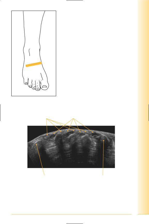
FIG. 318 TS, probe over mid-dorsum of foot
Extensor tendons |
Dorsal interosseus muscles |
Medial |
Lateral |
First metatarsal |
Fifth metatarsal |
FIG. 319 TS panorama, dorsum foot
257
of Atlas
ultrasound musculoskeletal anatomy
258
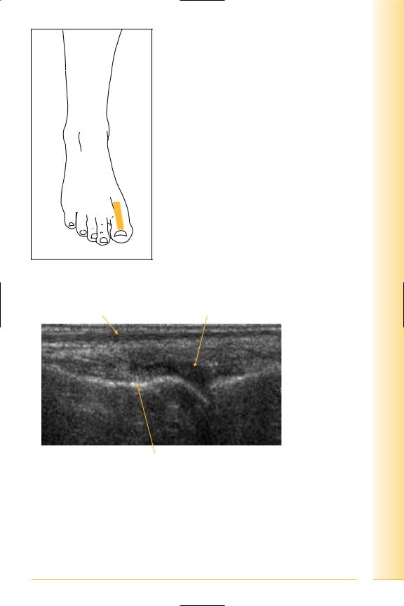
Extensor hallucis longus
(Figures 320–322)
•Origin: anterior surface of fibula, interosseous membrane.
•Insertion: passes distally to insert into the terminal phalanx of the great toe.
Notes
limb Lower
Foot
FIG. 320 LS, probe over dorsum great toe. Dynamic examination using flexion/extension of the great toe
Extensor tendon |
Synovium/capsule |
 Proximal phalanx
Proximal |
First metatarsal |
Distal |
FIG. 321 LS, extensor great toe
259

of Atlas
ultrasound musculoskeletal anatomy
260
Tendon |
Terminal phalanx |
Proximal |
First metatarsal |
Proximal phalanx |
Distal |
FIG. 322 LS panorama, extensor hallicus longus