


of Atlas
ultrasound musculoskeletal anatomy
82
Wrist
Anterior
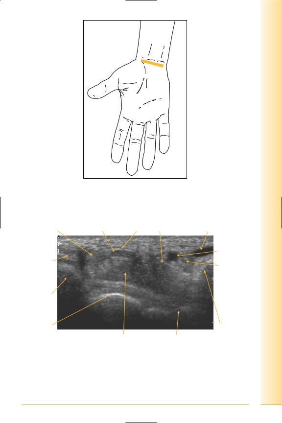
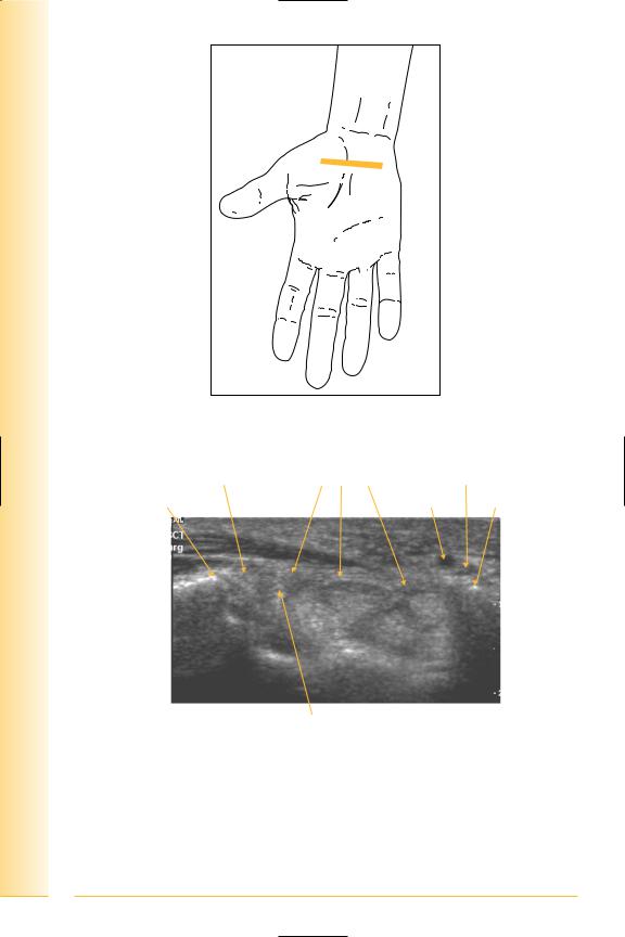
Carpal tunnel
(Figures 96–101)
The roof of the tunnel is formed by the flexor retinaculum, which is attached on the radial side to the tuberosity of the scaphoid and ridge of the trapezium, and on the ulnar side to the pisiform and hook of the hamate. The carpal bones form the floor.
From lateral to medial, the major contents are: flexor carpi radialis, flexor pollicis longus (deep to median nerve), flexor digitorum superficialis and profundus. Palmaris longus, if present, passes superficial to the retinaculum.
The ulnar nerve lies on the retinaculum alongside the pisiform, medial to the ulnar artery. Both are covered by a superficial part of the retinaculum, forming Guyon’s canal.
Notes
limb Upper
Wrist
FIG. 96 TS, probe transverse to volar aspect of wrist, level of proximal carpal tunnel
Flexor pollicis longus |
Median nerve Flexor retinaculum |
Flexor carpi ulnaris |
|
|
Ulnar artery |
Flexor carpi |
|
and nerve |
|
in Guyon’s |
|
radialis |
|
canal |
Scaphoid |
|
|
Lunate |
|
|
Pisiform |
|
|
|
|
Lateral |
Flexor digitorum tendons |
Triquetral |
Medial |
FIG. 97 TS, proximal carpal tunnel
83
of Atlas
ultrasound musculoskeletal anatomy
FIG. 98 TS, probe transverse to volar aspect of wrist, level of distal carpal tunnel
Flexor pollicis longus Flexor retinaculum |
Ulnar nerve |
Trapezium |
Ulnar artery Hook of hamate |
Flexor carpi  radialis
Lateral |
Median nerve |
Medial |
FIG. 99 TS, distal carpal tunnel
84
limb Upper
Wrist
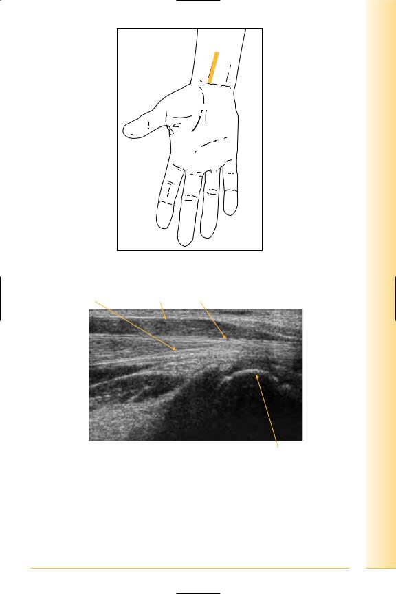
FIG. 100 LS, flexor tendons
Flexor digitorum |
Flexor digitorum superficialis |
profundus tendon |
muscle and tendon |
Proximal |
Distal |
Lunate
FIG. 101 LS, flexor tendons at wrist
85

of Atlas
ultrasound musculoskeletal anatomy
86
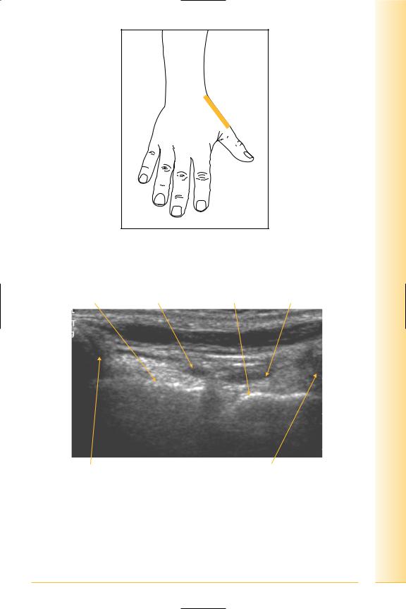
Posterior
Anatomical snuffbox
(Figures 102–107)
Proximally, the snuffbox is demarcated by the radial styloid, and distally by the base of the thumb metacarpal. Its radial boundary is formed by two tendons (extensor pollicis brevis and abductor pollicis longus) and on the ulnar aspect by extensor pollicis longus. The floor of the snuffbox is formed by the scaphoid proximally and the trapezium distally. It contains the radial artery and cephalic vein.
Notes
limb Upper
Wrist
FIG. 102 LS, probe longitudinal to snuffbox, radial aspect of wrist. Ulnar deviation of the wrist with extension of the thumb
Scaphoid |
Radial artery |
Trapezium |
Cephalic vein |
Radial styloid |
Base of thumb metacarpal |
Proximal |
Distal |
FIG. 103 LS, snuffbox
87
of Atlas
ultrasound musculoskeletal anatomy
FIG. 104 TS, probe transverse to snuffbox, radial aspect of wrist. Ulnar deviation of the wrist with extension of the thumb
Radial artery and cephalic vein
Extensor pollicis longus |
Bony floor |
Abductor pollicis longus/ |
|
|
extensor pollicis brevis |
Medial |
|
Lateral |
FIG. 105 TS, anatomical snuffbox
88
limb Upper
Wrist
FIG. 106 LS, probe longitudinal to extensor pollicis longus tendon, radial side of wrist. Ulnar deviation of the wrist with extension of the thumb. Dynamic examination with flexion and extension of interphalangeal joint of thumb
Radius |
Scaphoid |
Trapezium Thumb metacarpal |
Proximal |
Extensor pollicis longus tendon |
Distal |
FIG. 107 LS, extensor pollicis longus tendon
89

of Atlas
ultrasound musculoskeletal anatomy
90
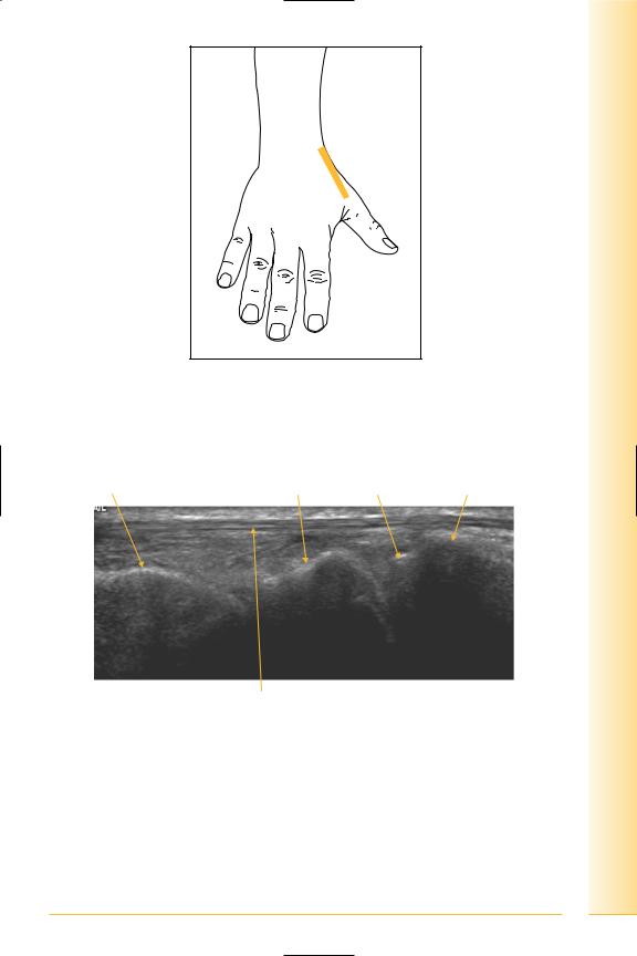
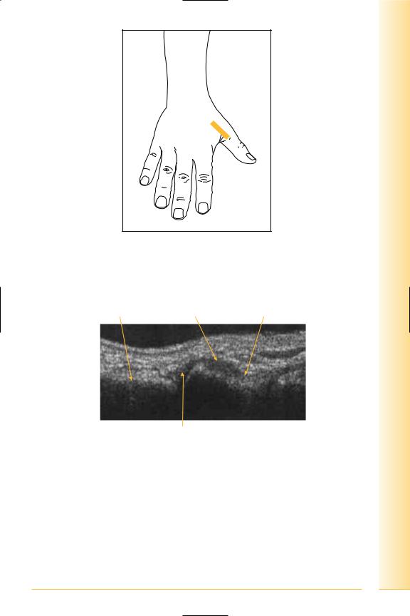
Thumb carpometacarpal joint
(Figures 108 and 109)
Notes
limb Upper
Wrist
FIG. 108 LS, probe longitudinal to thumb carpometacarpal joint
Abductor pollicis longus and extensor pollicis brevis tendons
Base of thumb metacarpal
Trapezium 
Proximal |
Distal |
Capsule
FIG. 109 LS, thumb carpometacarpal joint
91

of Atlas
ultrasound musculoskeletal anatomy
92
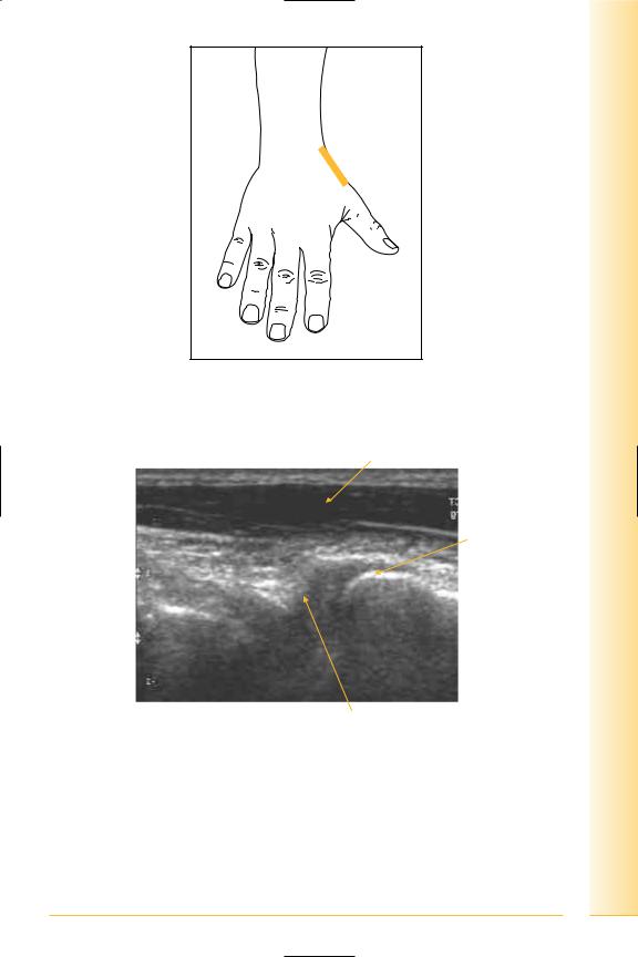
Ulnar collateral ligament
(Figures 110 and 111)
Stabilizing ligament along the ulnar side of the metacarpophalangeal joint of the thumb.
Notes
limb Upper
Wrist
FIG. 110 LS, probe over metacarpophalangeal joint. Dynamic examination using abduction at this joint
Metacarpal |
Joint capsule |
Proximal phalanx |
Proximal |
Distal |
Collateral ligament
FIG. 111 LS, ulnar collateral ligament
93

of Atlas
ultrasound musculoskeletal anatomy
94
Interosseous scapholunate ligament
(Figures 112 and 113)
The dorsal aspect of this ligament is seen as a high reflectivity linear structure in the scapholunate space.
Notes
limb Upper
Wrist
FIG. 112 TS, probe transverse to dorsal aspect of wrist, level of proximal carpal row
Extensor digitorum tendons |
Scapholunate ligament |
Extensor pollicis longus |
 Extensor carpi radialis
Medial |
Lunate |
Scaphoid |
Lateral |
FIG. 113 TS, dorsal scapholunate ligament
95