

Книги по МРТ КТ на английском языке / Normal Findings in CT and MRI
.pdfWrist 195
Wrist
The bones comprising the wrist present a normal configuration.
The radial joint angle is normal. The carpal bones show normal shape and relationship to one another and to the radiocarpal and carpometacarpal joints.
The articular surfaces are smooth and congruent with normal cortical thickness and normal width of the joint spaces. There are no osteophytes and no subchondral signal changes. The bone marrow signal is normal.
The ulnar (triangular) disk exhibits normal configuration and normal signal characteristics. The interosseous ligaments also appear normal. The carpal tunnel is of normal width and transmits tendons that are normal in width and position. The median and ulnar nerves appear normal.
The metacarpals and phalanges have normal margins and normal bonemarrow signal intensity. The soft tissues are normal.
Interpretation
The wrist and hand appear normal.
Checklist
Bony structures ! Radius
!Ulna (configuration, no shortening)
!Carpal bones (proximal and distal rows)
!Metacarpals
!Radiocarpal angle (see below)
!Carpal bones:
—Shape and position (see below)
!Metacarpals and phalanges:
—Five digital rays
—Shape
—Normal bone marrow signal
!Articular surfaces, especially of radiocarpal and carpometacarpal joints:
—Smooth
—Congruent
!Normal cortical thickness
!No marginal osteophytes
!No subchondral signal changes
!Normal width of joint space (see below)
Moeller, Normal Findings in CT and MRI © 2000 Thieme
All rights reserved. Usage subject to terms and conditions of license.

196 MRI: Joints
Ligamentous |
! Ulnar (triangular) disk: |
||
structures |
|
— |
Configuration (see below) |
|
|
— Margins |
|
|
|
— Internal structure (hypointense expansion to |
|
|
|
|
styloid attachment and to radial end of ulna |
|
|
|
with central rarefaction) |
|
|
— |
No signal abnormalities |
|
|
— |
No discontinuities |
|
! Interosseous ligaments: scapholunate and luna- |
||
|
|
totriquetral ligaments and ligaments of the distal |
|
|
|
row of carpal bones |
|
|
|
— Shape |
|
|
|
— |
Signal intensity |
|
! |
— |
Contours (smooth, intact) |
|
Extrinsic ligaments: |
||
|
|
— Shape |
|
|
|
— |
Signal intensity |
Carpal tunnel |
|
— |
Contours (smooth, intact) |
! Width (see below) |
|||
|
! Tendons (tendon sheaths in six compartments, |
||
|
|
thickness, position, symmetry) |
|
|
! Flexor retinaculum (no palmar convexity) |
||
|
! No circumscribed widening of tendons |
||
|
! No thickening of tendon sheath walls |
||
|
! No increase of fluid in tendon compartment |
||
|
! No fluid-filled cyst |
||
Median nerve |
! |
No ganglion |
|
! |
Course |
||
|
! |
Width |
|
|
! No impingement, especially in the carpal tunnel |
||
|
|
(axial image) |
|
|
! No diffuse or circumscribed swelling (e.g., com- |
||
|
|
mon at level of pisiform bone) |
|
|
! No narrowing (e.g., common at level of hamate |
||
|
! |
bone) |
|
Ulnar nerve |
No signal changes |
||
! |
Width |
||
|
! |
Course |
|
|
! |
No expansion |
|
Soft tissues |
! |
No bony impingement |
|
! No subcutaneous nodules |
|||
|
|
|
|
Moeller, Normal Findings in CT and MRI © 2000 Thieme
All rights reserved. Usage subject to terms and conditions of license.
Wrist 197
1a
2
.
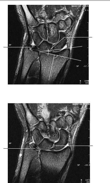
Coronal image
5b
6
5a 
Coronal image
Moeller, Normal Findings in CT and MRI © 2000 Thieme
All rights reserved. Usage subject to terms and conditions of license.
198 MRI: Joints
Important Data
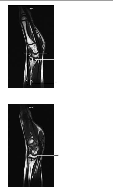
1Radiocarpal angle: a Coronal: 10−30° b Lateral: 10−15°
2Ulnar (triangular) disk or triangular fibrocartilage complex (TFC):
!Maximum thickness: 1.6 cm ± 0.5 cm
3Inclination of lunate bone relative to long axis (lateral view):
!0−30°
4Inclination of scaphoid bone relative to long axis (sagittal view):
!30−60°
5Joint spaces:
a Distal radioulnar joint: ca. 3 mm b Other joints: ca. 2 mm
6Distal radioulnar length relation:
!1−5 mm
!> 5 mm = ulnar shortening
!< 1 mm = ulnar lengthening
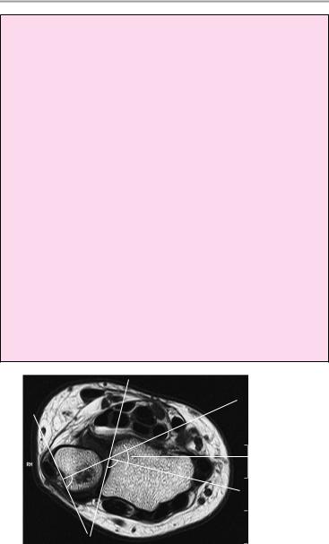
7Lines are drawn tangent to the corners of the radial ulnar notch and to the base points of the roughly triangular cross section of the distal ulna. Lines are drawn perpendicular to these tangents, and the angle between them is measured:
!In neutral position +15° − +45°, in supination approx. +100°. Always compare with the opposite side.
. |
|
7 |
|
||
|
|
.
Axial image at level of distal radioulnar joint
Moeller, Normal Findings in CT and MRI © 2000 Thieme
All rights reserved. Usage subject to terms and conditions of license.
Wrist 199
.

 1b
1b
3
Sagittal image
4
Sagittal image
Moeller, Normal Findings in CT and MRI © 2000 Thieme
All rights reserved. Usage subject to terms and conditions of license.

200 MRI: Joints
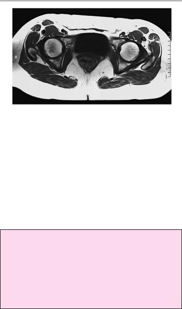
Hip Joint
The femoral heads and acetabula are of normal shape, and the femoral heads are well covered by the acetabular margins. The joint spaces are of normal width.
The articular surfaces are smooth and congruent and show normal cortical thickness. There are no marginal osteophytes or subchondral signal changes.
The bone marrow shows normal signal intensity, especially in the femoral head and neck. Each femoral shaft has normal margins and contains a normal bone marrow signal.
The imaged muscles and the lesser pelvis show no abnormalities.
Interpretation
The hip joints appear normal.
Checklist
Hip joint |
! |
Femoral heads: |
|
|
— Shape |
|
! |
— Bilateral symmetry |
|
Acetabula: |
|
|
|
— Shape |
|
|
— Roundness |
|
! |
— Symmetry |
|
Position: |
|
|
|
— Femoral heads articulate with the acetabula |
|
|
— Femoral heads are well covered by the |
|
|
acetabular margins (see below) |
|
! Normal width of joint space |
|
|
! |
Articular surfaces: |
|
|
— Contours (smooth and congruent) |
|
! Normal cortical thickness on the articular sur- |
|
|
! |
faces |
|
No marginal osteophytes |
|
|
! No subchondral signal changes |
|
|
! Femoral head and neck: |
|
|
|
— Shape |
|
|
— Position |
— Normal femoral neck angle (CCD angle) (see below)
Moeller, Normal Findings in CT and MRI © 2000 Thieme
All rights reserved. Usage subject to terms and conditions of license.

Hip Joint 201
!Bone marrow signal:
—Homogeneous
—Fat-equivalent intensity
—No circumscribed “double line sign” (femoral
head necrosis) or patchy bone marrow edema
Other structures ! Femoral shaft:
—Smooth margins
—Normal cortical thickness
—Bone marrow signal appropriate for age (“ad olescent” signal before age 25) and homogeneous
!Musculature:
—Anatomy
—Course
—Bilateral symmetry
—Homogeneous signal intensity
—No circumscribed hypointense or hyperintense areas
!Major nerves and blood vessels:
—Course
—No circumscribed expansion
!No lymphadenopathy
!Structures of the lesser pelvis (bladder, prostate, and seminal vesicles or uterus and adnexa, intestinal structures, lymph node stations)
Moeller, Normal Findings in CT and MRI © 2000 Thieme
All rights reserved. Usage subject to terms and conditions of license.
202 MRI: Joints
4
1
2 |
|
|
3 |
|
|
||||
|
|
|
|
|
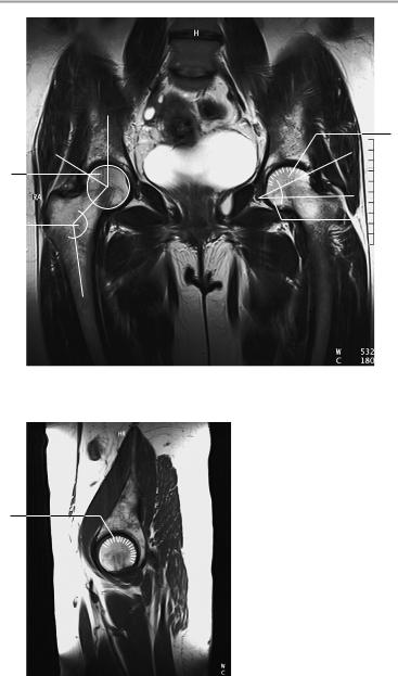
T2-weighted coronal image
Sagittal image
4
Moeller, Normal Findings in CT and MRI © 2000 Thieme
All rights reserved. Usage subject to terms and conditions of license.
Hip Joint 203
Axial image
Important Data
1Center-edge angle of Wiberg:
!26−30°
2CCD angle:
!Approximately 125−135°
3Slope of acetabular roof:
!<10°
4Femoral head coverage by the acetabulum:
!Approximately 70% of articular surface
Moeller, Normal Findings in CT and MRI © 2000 Thieme
All rights reserved. Usage subject to terms and conditions of license.

204 MRI: Joints
Knee Joint
The bones comprising the knee joint show normal configuration and position. The bone marrow signal is normal, with a normal trabecular pattern and normal epiphyseal lines.
The cortex shows smooth contours and normal thickness with no subchondral signal changes.
The cartilage covering the patella, femoral condyles, and tibial plateau is of normal thickness and has normal signal characteristics. The cartilaginous surface is smooth.
The medial and lateral menisci of the knee joint present a normal triangular configuration on axial images and have a homogeneous internal structure of low signal intensity. The anterior horn, midportion, and posterior horn each display a smooth, intact surface.
The anterior and posterior cruciate ligaments are intact and are normal in their width and signal characteristics. The collateral ligaments are intact and of normal width.
The soft tissues surrounding the knee joint and the imaged vascular structures are unremarkable.
Interpretation
The knee joint appears normal.
Checklist
Configuration |
! |
Femur |
and position |
! |
Tibia |
|
! |
Fibula |
Bone marrow |
! Patella (shape, centering—see below) |
|
! |
Fat-equivalent |
|
signal |
! |
May be slightly patchy |
|
! Adolescent bone marrow signal before age 25 |
|
|
|
years |
|
! No bone marrow edema |
|
|
! |
No contusions |
|
! |
Normal trabecular pattern |
Cortex |
! Epiphyseal plate closure after age 18 |
|
! |
Thickness |
|
|
! |
Contours (smooth) |
|
! No subchondral signal changes in the bone mar- |
|
|
|
row |
Moeller, Normal Findings in CT and MRI © 2000 Thieme
All rights reserved. Usage subject to terms and conditions of license.