

Книги по МРТ КТ на английском языке / Normal Findings in CT and MRI
.pdfBreast 125
Breast
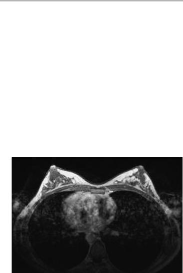
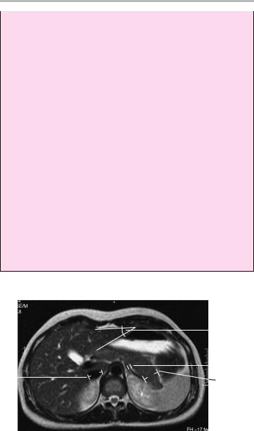
The anatomy of the glandular breast tissue is symmetrical and normal for age. The breast parenchyma is uniformly subdivided by fatty tissue. Unenhanced MR images show no lesions that are hypointense or hyperintense to the breast parenchyma or fat.
Following contrast administration, a significant, abnormal rise in signal intensity is not observed in any segment of the breast.
The skin and subcutaneous tissues show no abnormalities.
Interpretation
The breasts appear normal.
Checklist
Breast paren- |
! |
Size |
chyma |
! |
Symmetry |
|
! Extent of breast parenchyma in relation to fat |
|
|
|
(note physiological involution of the paren- |
|
|
chyma with aging) |
|
! Symmetrical development of glandular breast |
|
|
|
tissue |
Noncontrast |
! Uniform subdivision by fat |
|
! No lesions that are hypointense or hyperintense |
||
images |
|
to the breast parenchyma or fat (cysts, solid |
Postcontrast |
|
tumors, stellate densities) |
! No significant abnormal enhancement (more |
||
images |
|
than about 70% of initial signal intensity in the |
|
|
early phase after contrast administration) |
|
! No abnormal enhancing structures on delayed |
|
|
|
images |
|
! Early, intense enhancement of the nipple area |
|
Skin and subcu- |
! |
(confirms proper injection technique) |
Thickness |
||
taneous tissues |
! |
No retraction |
Axilla (unless |
! |
No circumscribed expansion |
! |
No lymphadenopathy |
|
obscured by |
|
|
motion artifacts) |
|
|
Moeller, Normal Findings in CT and MRI © 2000 Thieme
All rights reserved. Usage subject to terms and conditions of license.
126 MRI: Chest
Lungs (unless |
! |
Complete aeration |
obscured by |
! |
No pulmonary nodules |
motion artifacts) |
! |
Bony structures (ribs and sternum, unless ob- |
|
|
scured by motion artifacts): |
|
|
— Contours |
|
|
— Shape |
|
|
— No voids or expansion |
|
|
— Retrosternal structures (lymph nodes along |
|
|
internal thoracic artery) appear grossly nor- |
Heart (unless |
! |
mal |
Shape |
||
obscured by |
! |
Size |
motion artifacts) |
! |
Position |
|
! |
Enhancement characteristics |
|
|
|
Axial image through the center of the breasts following contrast administration (GdDTPA: gadolinium diethylenetriaminepentaacetate)
Moeller, Normal Findings in CT and MRI © 2000 Thieme
All rights reserved. Usage subject to terms and conditions of license.
Breast 127
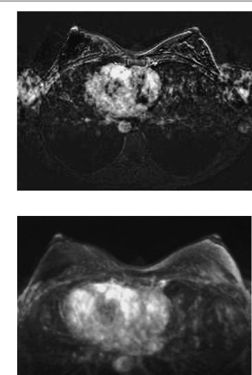
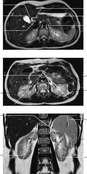
Subtraction image of the breasts
3-D MIP (Maximum Intensity Projection) rendering of subtraction images
Moeller, Normal Findings in CT and MRI © 2000 Thieme
All rights reserved. Usage subject to terms and conditions of license.

128
MRI: Abdomen
Upper Abdominal Organs
The liver is normally positioned and has normal size and smooth borders. Its internal structure is normal, with no focal abnormalities of signal intensity. The intrahepatic and extrahepatic bile ducts are not distended. The porta hepatis appears normal.
The gallbladder displays a normal size, smooth borders, and homogeneous contents.
The spleen is orthotopic and of normal size. It has smooth outer contours and a homogeneous internal structure.
The pancreas is normal in size and position. The head, body, and tail of the organ have smooth, lobulated outer contours and normal internal structure. The pancreatic duct is unobstructed.
Both kidneys are normal in size and position. The renal parenchyma shows normal width and structure.
The renal pelvis and calices are normal. The urinary drainage tract is unobstructed.
Both adrenal glands are normal in position and size, and the adrenal crura are normally developed. The adrenal compartment is unremarkable.
Major vessels and the para-aortic region appear normal, with no evidence of lymphadenopathy.
Imaged portions of the lung and soft tissues show no abnormalities.
Interpretation
The upper abdominal organs appear normal.
Moeller, Normal Findings in CT and MRI © 2000 Thieme
All rights reserved. Usage subject to terms and conditions of license.

Upper Abdomial Organs 129
Checklist
Liver |
! |
Position |
|
! |
— Directly below the right hemidiaphragm |
|
Size (see below) |
|
|
! |
Borders: |
|
|
— Smooth |
|
! |
— Sharp |
|
No focal abnormalities |
|
|
! |
Intrahepatic bile ducts: |
|
|
— Course (toward porta hepatis) |
|
|
— Width |
|
|
— No calculi |
|
! |
— No air |
|
Extrahepatic bile ducts: |
|
|
|
— Course (from porta hepatis to head of pan- |
|
|
creas) |
|
|
— Width (see below) |
|
|
— Homogeneous contents of fluid-equivalent |
|
|
signal intensity |
|
|
— No calculi |
|
! |
— No air |
|
Gallbladder: |
|
|
|
— Size (see below) |
|
|
— Contours (smooth) |
|
|
— Wall thickness (see below) |
|
! |
— No pericholecystic fluid |
|
Gallbladder contents: |
|
|
|
— Homogeneous |
|
|
— Fluid-equivalent signal intensity |
|
|
— No calculi (hypointense or hyperintense) |
|
|
— No air |
|
! Porta hepatis occupied by the hepatic artery, |
|
|
! |
common bile duct, and portal vein |
|
No masses |
|
Spleen |
! |
No lymphadenopathy |
! |
Size (see below) |
|
|
! |
Smooth outer contours |
Pancreas |
! |
Homogeneous internal structure |
! |
Size normal for age (see below) |
|
|
! |
Normal lobulation |
|
! |
Smooth outer contours |
Moeller, Normal Findings in CT and MRI © 2000 Thieme
All rights reserved. Usage subject to terms and conditions of license.

130 MRI: Abdomen
|
! Pancreatic duct unobstructed (see below) |
|
Kidneys |
! |
No peripancreatic fluid |
! |
Paired |
|
|
! |
Position (see below) |
|
! |
Size (see below) |
|
! |
Smooth contours |
|
! Width of cortex and medulla |
|
|
! Renal pelvis (presence, symmetry, size, no |
|
|
|
widening, homogeneous fluid contents) |
|
! Calices (shape, width, homogeneous contents) |
|
Ureters |
! Enhancement characteristics (see below) |
|
! |
Not duplicated |
|
|
! |
Course |
Adrenal glands |
! No obstruction of urinary drainage |
|
! |
Shape |
|
|
! |
Size (see below) |
|
! Slender crura (no asymmetric widening) |
|
|
! No circumscribed hypointense (T1: cyst, ade- |
|
Intestinal |
! |
noma), isointense, or hyperintense expansion |
Colon haustrations |
||
structures |
! |
Small bowel |
|
! |
Wall thickness |
|
! Homogeneous opacification with oral contrast |
|
|
|
medium (if administered) |
|
! No free extraintestinal or intra-abdominal air or |
|
|
|
fluid |
|
! Para-aortic region: |
|
|
|
— Major vessels (position, size, fluid signal) |
|
|
— Soft tissues (no masses) |
Lungs |
! |
— No lymphadenopathy |
Clear and expanded |
||
Costophrenic |
! Clear and aerated on both sides |
|
sinus |
|
|
Soft tissues |
|
|
|
|
|
Moeller, Normal Findings in CT and MRI © 2000 Thieme
All rights reserved. Usage subject to terms and conditions of license.
Upper Abdomial Organs 131
Important Data
Dimensions
1Liver:
a Left lobe (anteroposterior diameter on the left paravertebral line): up to 5 cm
b Caudate lobe/right lobe (CL/RL) = 0.37 ± 0.16 (e.g., 0.88 ± 0.2 in cirrhosis). Reference lines [from medial side]: line I is tangent to the medial border of the caudate lobe; line II is parallel to I and tangent to the lateral aspect of the portal vein; line III is tangent to the lateral hepatic border and perpendicular to a line midway between the portal vein and inferior vena cava and perpendicular to I and II.
c Angle of hepatic border: ca. 45° on the left side (formed by left lateral and inferior hepatic borders)
2Gallbladder:
a Horizontal diameter up to 5 cm (> 5 cm is suspicious for hydrops)
b Width of gallbladder wall: 1−3 mm
3Width of common bile duct:
!! 8 mm (! 10 mm after cholecystectomy)
4Spleen:
a Depth: 4−6 cm b Width: 7−10 cm
c Length: 11−15 cm
Splenic index: D × W × L = 160−440
IIIII I
 1a
1a
4a
1b |
|
rL |
Lc |
|
4b
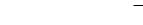
Axial image
Moeller, Normal Findings in CT and MRI © 2000 Thieme
All rights reserved. Usage subject to terms and conditions of license.
132 MRI: Abdomen
5Pancreas:
a Head: up to 3.5 cm b Body: up to 2.5 cm c Tail: up to 2.5 cm
Pancreatic duct: width 1−3 mm
6Adrenal glands (variable):
!Crural thickness < 10 mm
7Kidneys:
a Craniocaudal diameter: 8−13 cm
b Anteroposterior diameter: ca. 4 cm c Transverse diameter 5−6 cm Position of superior poles of kidneys: d Right: superior border of L1
d Left: inferior border of T12
f Transverse renal axis: posteriorly divergent angle of 120° g Width of renal cortex: 4−5 mm
Time to corticomedullary equilibrium: 1 minute
Contrast excretion into the pyelocaliceal system: 3 minutes Gerota fascia (thickness): 1−2 mm
Width of ureter: 4−7 mm
8Diameter of abdominal aorta:
!Approximately 18−30 mm
9Inferior vena cava:
!Transverse diameter up to 2.5 cm
Lymph nodes larger than 1 cm are suspicious for pathology.
|
|
|
|
|
1c |
|
|
|
|
|
|
||
9 |
|
|
|
|
6 |
|
|
|
|||||
|
|
|
|
5c |
||
|
|
|
|
|||
|
|
|
|
|
||
Axial image
Moeller, Normal Findings in CT and MRI © 2000 Thieme
All rights reserved. Usage subject to terms and conditions of license.
|
|
|
|
Upper Abdomial Organs 133 |
||||
2b |
|
|
|
|
|
5a |
||
|
|
|
|
|
||||
2a |
|
|
|
|
||||
|
|
|
|
|||||
|
|
|
|
5b |
||||
|
|
|
|
|||||
3 |
|
|
|
|
|
8 |
||
|
|
|
|
|||||
|
|
|
|
|
|
|
|
|
|
|
|
|
|
|
|
|
|
Axial image
7f |
|
7b |
|
7c
7g
Axial image
7e
4c
7d 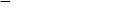
7a
7a
Coronal image
Moeller, Normal Findings in CT and MRI © 2000 Thieme
All rights reserved. Usage subject to terms and conditions of license.

134 MRI: Abdomen
Liver
The liver is orthotopic and presents normal size and smooth borders. It has a normal internal structure with no focal abnormalities. The intrahepatic and extrahepatic bile ducts are not dilated.
The gallbladder appears normal, displaying smooth borders and homogeneous contents.
The porta hepatis shows no abnormalities.
Other visualized upper abdominal organs are unremarkable.
Interpretation
The liver appears normal.
Checklist
Liver |
! |
Position |
|
! |
— Directly below the right hemidiaphragm |
|
Size (see below) |
|
|
! |
Borders: |
|
|
— Smooth |
|
! |
— Sharp |
|
No focal abnormalities |
|
|
! |
Intrahepatic bile ducts: |
|
|
— Course (toward porta hepatis) |
|
|
— Width |
|
|
— No calculi |
|
! |
— No air |
|
Extrahepatic bile ducts: |
|
|
|
— Course (from porta hepatis to head of pan- |
|
|
creas) |
|
|
— Width (see below) |
|
|
— Homogeneous contents of fluid-equivalent |
|
|
signal intensity |
|
|
— No calculi |
|
|
— No air |
Moeller, Normal Findings in CT and MRI © 2000 Thieme
All rights reserved. Usage subject to terms and conditions of license.