

Книги по МРТ КТ на английском языке / Neurosurgery Fundamentals Agarval 1 ed 2019
.pdf4.6 Spinal Cord
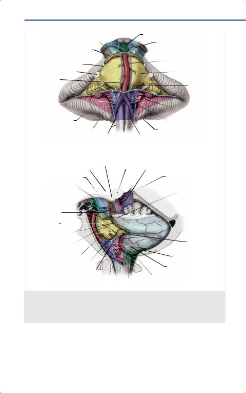
Fig. 4.20 The infrafacial collicular safe entry zone (shaded area inside green dashed lines) is limited medially by the medial longitudinal fasciculus, laterally by the nucleus ambiguus, superiorly by the facial colliculus, and inferiorly by the hypoglossal trigone.
Thus, the lateral border of the infrafacial collicular safe entry zone corresponds to the most medial point of attachment of the tela choroidea along the lower margin of the lateral recess, whereas the rostral and caudal borders are the same as the upper and lower edges of the lateral recess. (Reproduced from Spetzler R, Kalani M, Nakaji P et al, Color Atlas of Brainstem Surgery, 1st edition, ©2017, Thieme Publishers, New York.)
4.5 Cisterns
The cisterns are areas where the pia matter and arachnoid are separated and filled with CSF. Additionally, these spaces are also occupied by blood vessels and cranial nerves. Fig. 4.21 summarizes the different cisterns and their relationship with the anatomical structures in the posterior fossa.
4.6 Spinal Cord
4.6.1 Surface Anatomy
The spinal cord is the most caudal segment of the CNS. It is located in the superior two-third of the spinal canal, ending at the level of L2 (second lumbar vertebrae). Along its length, the spinal cord has two regions where its maximal diameter increases. One of these regions lies in its
cervical portion and one in the lumbar portion, representing areas where the brachial and lumbar plexi are respectively located.
Generally, there are 31 spinal cord segments, each of which give off a pair of spinal nerves:
•Eight pairs of cervical spinal nerves, the first spinal nerve (C1) exits between the occipital bone and the atlas or first cervical vertebra. For this reason, there are eight cervical pairs, the last being located between C7 and T1.
•Twelve thoracic spinal nerves. The first thoracic nerve pairs (T1) start below the first thoracic vertebrae, this order continues for the remaining segments.
•Five lumbar pairs.
•Five sacral pairs.
•One coccygeal pair.
59
Agarwal, Neurosurgery Fundamentals (ISBN 978-1-62623-822-0), copyright © 2019 Thieme Medical Publishers. All rights reserved. Usage subject to terms and conditions of license.
Neuroanatomy
Posterior cerebral artery Oculomotor nerve (CN III) 
Superior cerebellar artery
Basilar artery
Trochlear nerve (CN IV)
Interpeduncular cisternCrural cistern
Ambient cistern
Prepontine cistern
Trigeminal nerve (CN V) |
|
|
Cerebellopontine |
||
Anterior inferior |
|
|
cistern |
||
|
|
|
|||
cerebellar artery |
|
|
Abducens nerve |
||
|
|
|
|
(CN VI) |
|
Facial and |
|
|
|
|
|
vestibulocochlear |
|
|
Cerebellomedullary |
||
nerves (CN VII & VIII) |
|
|
cistern |
||
Glossopharyngeal |
|
|
Posterior inferior |
||
and vagus nerves |
|
|
|||
|
|
Vertebral cerebellar artery |
|||
(CN IX & X) |
Hypoglossal nerve |
|
|||
a |
(CN XII) |
Accessory |
Premedullary artery |
||
|
nerve (CN XI) |
cistern |
|||
|
Posterior cerebral |
|
|||
|
Crural cistern |
artery |
Ambient cistern Trochlear nerve (CN IV) |
||
Interpeduncular cistern |
|
|
Quadrigeminal cistern |
||
Oculomotor nerve |
|
|
|
||
(CN III) |
|
|
|
Cerebellopontine |
|
Superior cerebellar |
|
|
cistern |
||
artery |
|
|
|
|
|
Prepontine cistern |
|
|
|
||
Trigeminal nerve |
|
|
|
||
(CN V) |
|
|
|
|
|
Basilar artery |
|
|
|
|
|
Facial and |
|
|
|
|
|
vestibulocochlear |
|
|
|
||
nerves (CN VII & VIII) |
|
|
Glossopharyngeal |
||
Anterior inferior |
|
|
|||
cerebellar artery |
|
|
and vagus nerves |
||
Abducens nerve (CN VI) |
|
|
(CN IX & X) |
||
|
|
Cisterna magna |
|||
Premedullary cistern |
|
|
|||
|
|
Posterior inferior |
|||
Hypoglossal nerve (CN XII) |
|
||||
|
cerebellar artery |
||||
b |
Accessory nerve (CN XI) |
|
Cerebellomedullary cistern |
||
Vertebral artery |
|||||
|
|
|
|||
Fig. 4.21 The brainstem and the cisterns that are associated with the cranial nerves.
(a) Ventral view and (b) lateral view. (Reproduced from Spetzler R, Kalani M,
Nakaji P et al, Color Atlas of Brainstem Surgery, 1st edition, ©2017, Thieme Publishers,
New York.)
60
Agarwal, Neurosurgery Fundamentals (ISBN 978-1-62623-822-0), copyright © 2019 Thieme Medical Publishers. All rights reserved. Usage subject to terms and conditions of license.
4.7 Vertebral Column
Above C7, the spinal nerves exit above the vertebra with the same name, except for C8, which exits below C7 and above T1. From T1 and below, the spinal nerves exit below the vertebra with the same name.
Topographically, the spinal cord its divided into three columns on each side:
•Anterior column, between the anterior median fissure and anterolateral sulcus.
•Lateral column, between the anterolateral and posterolateral sulci. These sulci represent the exit of the ventral (motor) and the entry of the dorsal (sensory) nerve roots that forms the spinal nerve.
•Posterior column, between the posterolateral sulcus and posterior median fissure. From T6 and above, the posterior column is further divided into two tracts by the posterior intermediate sulcus. A medial tract (fasciculus gracile), limited medially by the posterior median fissure and laterally by the posterior intermediate sulcus, and a lateral
tract (fasciculus cuneate) limited medially by the posterior intermediate sulcus and laterally by the posterolateral sulcus.
Fixation of the Spinal Cord
The spinal cord is maintained in its position via the following structures:
•Superiorly,brainstem. its continuation with the
•Laterally, spinal nerves exiting through the intervertebral foramens.
•Dura mater, two attachments: Filum terminalis with the anterior coccyx and sacrum, and the periosteum of the skull.
•Dentate ligaments, located between the ventral and dorsal nerve roots.
These are extensions of the pia matter and arachnoid towards the dura mater.
4.6.2 Internal Configuration
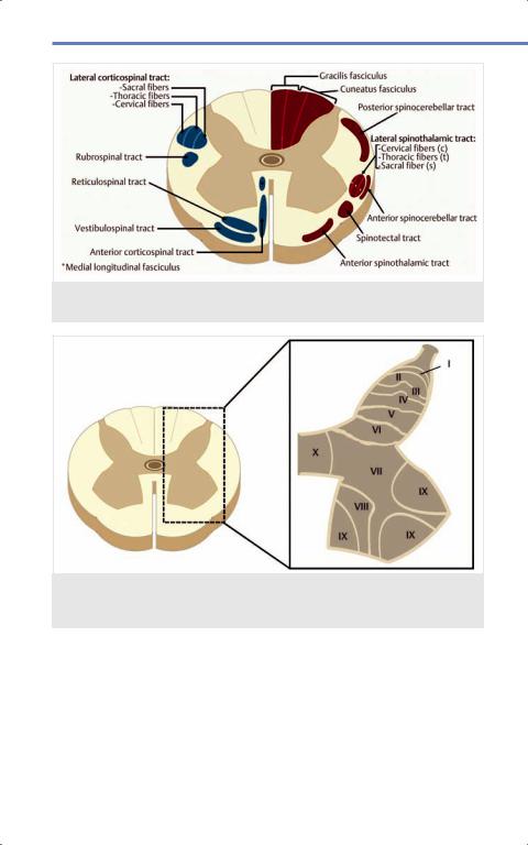
Understanding the anatomical arrangement and physiology of the spinal cord is essential for determination of different pathological syndromes that may affect it. Opposite to the brain, white matter surrounds the gray matter in the spinal cord. Spinal gray matter lies centrally around the central canal, assuming the shape of an “H.” Fig. 4.22 and Fig. 4.23 show anaxial cut of a spinal cord segment, the Rexed laminae configuration and the ascending and descending white matter tracts ( Table 4.2). Fig. 4.24 shows the myotomes and dermatomes.
4.7 Vertebral Column
The spine is composed of 33–35 vertebrae in the following distributions:
•7 cervical.
•12 thoracic.
•5 lumbar.
•3–5 coccygeal.
4.7.1 Vertebrae Constitution
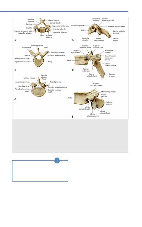
Except for the first (C1 or atlas) and second (C2 or axis) vertebrae, six segments can be identified in a standard vertebra ( Fig. 4.25):
1.Vertebral body: Forms the anterior portion of the vertebra and has two horizontal portions (superior and inferior) in which the intervertebral disc sits. The posterior wall forms the anterior arch of the vertebral canal.
2.Pedicles: There are two pedicles (left and right) extending from the posterolateral aspect of the vertebral body.
61
Agarwal, Neurosurgery Fundamentals (ISBN 978-1-62623-822-0), copyright © 2019 Thieme Medical Publishers. All rights reserved. Usage subject to terms and conditions of license.
Neuroanatomy
Fig. 4.22 Cross-sectional cut of the cervical spinal cord. Blue descending fibers, red ascending fibers. (Illustration by Joao T. Alves Belo, MD.)
Fig. 4.23 Cross-sectional cut of the spinal cord with a close-up view of the anatomical classification of the gray matter. For a description, please see Table 4.2. (Illustration by Joao T. Alves Belo, MD.)
body. They serve as attachments for |
foramina (spaces where the nerve |
the transverse processes, the laminae, |
roots exit the spinal canal). |
and the facets (articular processes). |
3. Laminae: Extend from the pedicles in a |
The superior and inferior borders of |
posteromedial direction relative to the |
the pedicles form the inferior and |
spinous processes. They form the |
superior borders of the intervertebral |
posterior arch of the spinal canal. The |
62
Agarwal, Neurosurgery Fundamentals (ISBN 978-1-62623-822-0), copyright © 2019 Thieme Medical Publishers. All rights reserved. Usage subject to terms and conditions of license.

|
|
|
4.7 Vertebral Column |
|
|
|
|
|
|
|
|
|
|
|
|
Table 4.2 Configuration of the gray matter in the spinal cord |
|
||
|
Rexed laminae |
Classical terminology |
Function |
|
|
I |
Posteromarginal nucleus |
Exteroceptive sensations |
|
|
II |
Substantia gelatinosa |
|
|
|
III |
Nucleus proprius |
|
|
|
IV |
|
|
|
|
V |
Neck of the posterior |
Proprioceptive sensations |
|
|
|
horn |
|
|
|
VI |
Base of the posterior |
|
|
|
|
horn |
|
|
|
VII |
Two groups: |
1. Medial, thoracic nucleus: from |
|
|
|
1. Medial, thoracic nucleus |
C8–L3 receive information from |
|
|
|
2. Lateral, divided in two |
the muscle spindle and Golgi |
|
|
|
other nuclei: |
tendon organ |
|
|
|
a) Intermediomedial |
2. Lateral: |
|
|
|
zone |
a) Intermediomedial zone: γ mo- |
|
|
|
b) Intermediolateral |
tor neurons involved in motor |
|
|
|
zone |
reflexes |
|
|
|
|
b) Intermediolateral zone: motor |
|
|
|
|
visceral function. From C8– |
|
|
|
|
L2–3 sympathetic thoracolum- |
|
|
|
|
bar column. S2–S4 parasympa- |
|
|
|
|
thetic sacral nuclei |
|
|
VIII |
Commissural nucleus |
Regulates skeletal muscle |
|
|
|
|
contraction |
|
|
IX |
Ventral horn |
Main motor area composed by α |
|
|
|
|
motor neurons |
|
|
X |
Grisea centralis/substantia |
Contains motor nuclei from the auto- |
|
|
|
gelatinosa centralis |
nomic nervous system |
|
internal surface of the laminae serves as an attachment for the yellow ligament.
4.Spinous process: Starts from the point where laminae join in the midline and follows a posterior trajectory. The inferior and superior edges of the spinous processes serve as an attachment for the interspinous ligament. The supraspinous ligament runs over the tip or free edge of the spinous process in the midline.
5.Facet or articular process: There are four facets, two on each side (superior
and inferior) which articulate with their superior and inferior vertebral counterparts.
6.Transverse process: Attached to the pedicles on either side. Their shape varies according to the segment of the spine they originate from.
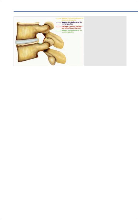
Intervertebral Foramen
As previously mentioned, this is the space where the spinal nerves exit the spinal
63
Agarwal, Neurosurgery Fundamentals (ISBN 978-1-62623-822-0), copyright © 2019 Thieme Medical Publishers. All rights reserved. Usage subject to terms and conditions of license.
Neuroanatomy
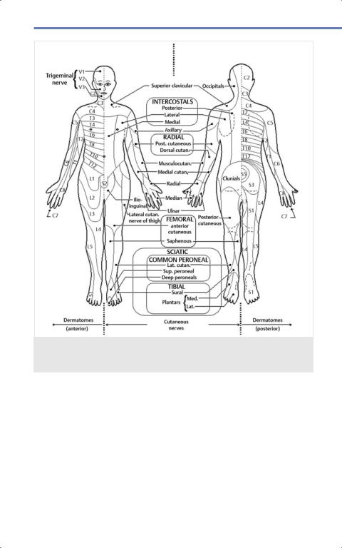
Fig. 4.24 Dermatomes and sensory distributions of peripheral nerves. (Reproduced from Baaj A, Mummaneni P, Uribe J et al, Handbook of Spine Surgery, 2nd edition,
©2016, Thieme Publishers, New York.)
canal. Its limits are important from a surgical standpoint ( Fig. 4.26):
•Superior: Inferior border of the overlying vertebral pedicle.
•Inferior: Superior border of the underlying vertebral pedicle.
•Anterior: Posterolateral aspect of the intervertebral disc.
•Posterior: The capsule of the facets and the yellow ligament.
Cervical Vertebrae
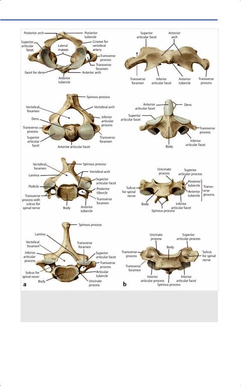
The vertebrae of the cervical spine have unique characteristics differentiating them from the thoracic and lumbar vertebrae ( Fig. 4.27). The first and second vertebras possess unique morphology including presence of the transverse foramina on the transverse processes.
64
Agarwal, Neurosurgery Fundamentals (ISBN 978-1-62623-822-0), copyright © 2019 Thieme Medical Publishers. All rights reserved. Usage subject to terms and conditions of license.
4.7 Vertebral Column
Fig. 4.25 (a) Anatomic drawing of a typical cervical vertebra from a superior projection.
(b)Anatomic drawing of a typical cervical vertebra from a lateral projection.
(c)Anatomic drawing of a typical thoracic vertebra from a superior projection.
(d)Anatomic drawing of a typical thoracic vertebra from a lateral projection.
(e)Anatomic drawing of a typical lumbar vertebra from a superior projection.
(f)Anatomic drawing of a typical lumbar vertebra from a lateral projection.
(Reproduced from Schuenke, Schulte, and Schumacher, Atlas of Anatomy, 2nd edition,
©2014, Thieme Publishers, New York. Illustration by Karl Wesker.)
Thevertebralarteriesandveinsrunwithin the transverse foramina of C6 to C1 and C7 to C1 respectively, entering the cranial vault via the foramen magnum.
Some of these special features are summarized here:
•C1 (Atlas): The first cervical vertebra lacks a body. Instead, it possesses two
lateral masses that articulate with the occipital condyles superiorly, and
C2 (axis) inferiorly. From these lateral masses, two arches (one anterior and one posterior) enclose anteriorly and posteriorly the spinal canal at this level. In the midline, the interior surface of the anterior arch of C1 articulates with the anterior segment of the odontoid process of C2 (axis). The anterior surface
65
Agarwal, Neurosurgery Fundamentals (ISBN 978-1-62623-822-0), copyright © 2019 Thieme Medical Publishers. All rights reserved. Usage subject to terms and conditions of license.
Neuroanatomy
of the anterior arch also possesses an anterior tubercle, which serves as the insertion point for the longus colli muscle. The posterior arch possesses a posterior tubercle on its exterior surface, in the midline, which serves as an insertion for the rectus capitis posterior minor muscle. The medial aspect of the lateral masses has tuberculae where the transverse ligament originates and runs posteriorly to the odontoid process before attaching to C1. The transverse process and foramen of C1 are located on the lateral surface of the C1 masses.
•C2 (Axis): This vertebra is characterized by its odontoid process (dens). The dens articulates with the anterior arch of the atlas. The left and right occipitoodontoid ligaments (alar ligaments) attach firmly to either side of the dens. The spinous process of the axis serves as an attachment for the rectus capitis posterior major muscle and the obliquus capitis inferior muscle.
•C6: Unique to this vertebra is the presence of a tubercle at the anterior aspect of its transverse process. This tubercle is called carotid tubercle or Chassaignac tubercle. It denotes the entry of the vertebral artery into the transverse foramen.
•C7: This vertebra possesses features similar to the thoracic vertebrae. Its
66
Fig. 4.26 Lateral view of two adjacent vertebral segments showing the boundaries of the interver tebral foramen. (Illustration by Joao T. Alves Belo, MD.)
spinous process is prominent and its transverse foramen is smaller or absent, and is occupied by the vertebral vein.
Thoracic Vertebrae
The most distinguishing feature of the thoracic vertebrae is the presence of transverse costal facets, which articulate with the ribs.
Lumbar Vertebrae
The lumbar vertebral bodies are larger than those of other segments; this is functionally related with their weight-bearing role.
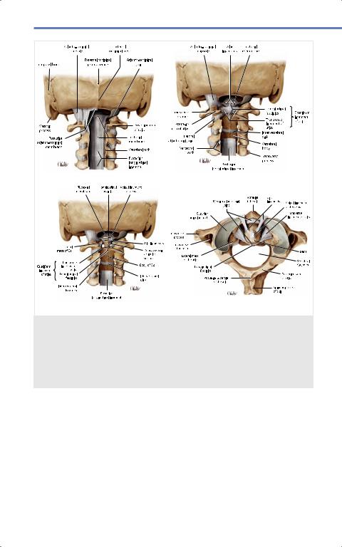
4.7.2 Ligaments of the Occipitoatlantoaxial Junction
Transverse and Cruciate Ligaments
The transverse ligament is a short ligament attached laterally to the transverse tubercle, at the medial aspect of the lateral mass of C1 ( Fig. 4.28). It runs posteriorly to the dens of the axis and for this reason it has an anterior concave trajectory. At the
Agarwal, Neurosurgery Fundamentals (ISBN 978-1-62623-822-0), copyright © 2019 Thieme Medical Publishers. All rights reserved. Usage subject to terms and conditions of license.
4.7 Vertebral Column
Fig. 4.27 (a) Superior and (b) anterior view of the first, second, fourth, and seventh cervical vertebrae. (Reproduced from Schuenke, Schulte, and Schumacher, Atlas of Anatomy, 2nd edition, ©2014, Thieme Publishers, New York. Illustration by Karl Wesker.)
67
Agarwal, Neurosurgery Fundamentals (ISBN 978-1-62623-822-0), copyright © 2019 Thieme Medical Publishers. All rights reserved. Usage subject to terms and conditions of license.
Neuroanatomy |
|
a |
b |
c |
d |
Fig. 4.28 Craniocervical ligaments. Views of the upper part of the vertebral canal with the spinous processes and parts of the vertebral arches removed to expose the ligaments on the posterior vertebral bodies viewed posteriorly (a) before and (b) and (c) after removal of the posterior longitudinal and transverse atlas ligaments. (d) The ligaments of the median atlantoaxial joint are shown from a superior view. (Reproduced from Schuenke, Schulte, and Schumacher, Atlas of Anatomy, 2nd edition, ©2014,
Thieme Publishers, New York. Illustration by Karl Wesker.)
midline are two more bands of vertically oriented ligaments: The superior and inferior longitudinal bands constitute the transverse and cruciate ligaments. The superior band ends at the anterior edge of the foramen magnum, whereas the inferior extension ends attach to the body of C2.
Alar Ligaments
These are two short and strong ligaments (left and right) connecting the superolateral segments of the dens of the axis to the medial segment of the occipital condyles.
68
Agarwal, Neurosurgery Fundamentals (ISBN 978-1-62623-822-0), copyright © 2019 Thieme Medical Publishers. All rights reserved. Usage subject to terms and conditions of license.