

Книги по МРТ КТ на английском языке / MRI for Orthopaedic Surgeons Khanna ed 2010
.pdf110 II Upper Extremity
A B
Fig. 4.23 (A,B) Coronal oblique T2-weighted images of the left shoulder showing the normal low signal intensity of the biceps tendon (arrow on each).
is particularly evident on sagittal oblique images obtained at the level of the intracapsular portion of the long head of the biceps tendon.21 Biceps tendinosis frequently is associated with impingement syndrome and rotator cu tears. There-
Fig. 4.24 A sagittal oblique T2-weighted image showing the normal low signal intensity of the biceps tendon (arrow) as it courses through the glenohumeral joint.
fore, MRI often visualizes the associated pathologic findings, including joint e usion and increased fluid in the bicipital synovial sheath, which is nonspecific for inflammation of the long head of the biceps tendon.72,73 However, fluid in the tendon sheath of the long head of the biceps is abnormal if it completely surrounds the tendon in the absence of a glenohumeral joint e usion.61,62
Biceps tendinosis is the earliest stage of biceps tendon disease, but it can progress to partial or complete tears of the tendon. Transverse partial tears are di cult to diagnose and usually are shown by a sudden change in the cross-sectional diameter of the tendon.72 However, a longitudinal tear is easier to identify because the tendon surrounds the high signal intensity tear on T2-weighted images (Fig. 4.26).72 More often, complete rupture occurs proximally, at the level of the proximal portion of the extracapsular segment, within the bicipital groove.74 Any axial image that definitively shows no tendon in the bicipital groove and no medial dislocation is diagnostic of rupture of the long head of the biceps tendon (Fig. 4.27).72 Additional MRI findings include atrophy and distal retraction of the tendon and fluid within the tendon sheath.21,72,73 These findings are best shown on axial sections. Intraarticular tears of the biceps tendon frequently are associated with rotator cu tears, and are visualized on MRI as the absence of the intraarticular portion of the tendon and the associated rotator cu lesion.21
Biceps subluxations, although rare, can be seen in disease processes in which loss of the integrity of the rotator cu has occurred or in which the biceps tendon loses the supporting structures that contain it in within the bicipital

4 The Shoulder 111
Fig. 4.25 A sagittal oblique T2-weighted image showing a thickened biceps tendon (biceps tendinosis) with increased T2 signal (arrow). Note also the glenohumeral joint e usion.
groove (i.e., the transverse humeral ligament).72 Because of the forces acting on the biceps tendon, displacement always occurs in the medial direction.72 The subluxation is best visualized on axial sections (Fig. 4.28). In addition to subluxation, frank dislocation of the biceps tendon can occur. The
Fig. 4.26 An axial fat-suppressed proton-density image of the right shoulder showing a longitudinal tear of the biceps tendon (arrow).
MRI findings of a dislocated biceps tendon include visualization of the dislocated tendon medial to an empty bicipital groove (Fig. 4.29).72 This condition also is best visualized on axial images.72,73,75,76 On sagittal oblique images, dislocation of the biceps tendon is identified as a more medial position
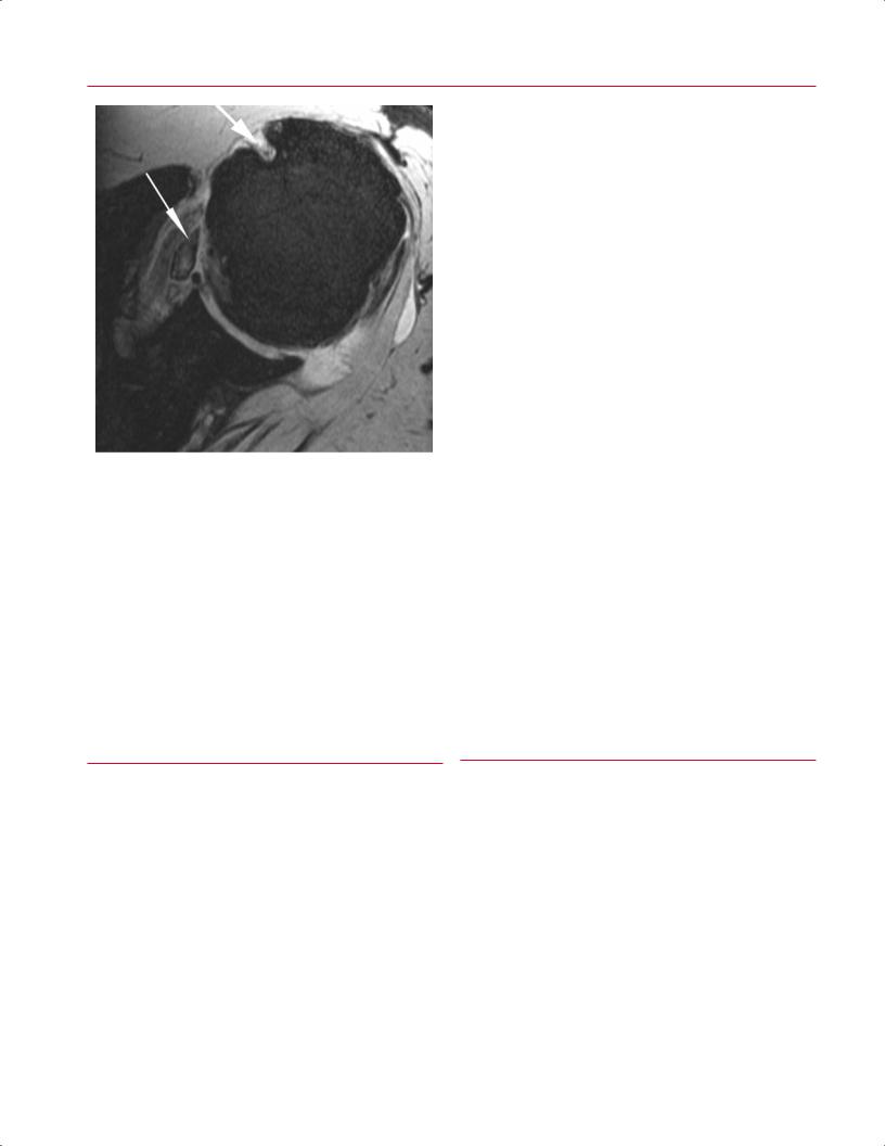
Fig. 4.27 An axial fat-suppressed T2-weighted image of the right shoulder showing absence of the biceps tendon within the bicipital groove (thin arrow) and complex fluid in the biceps tendon sheath (thick arrow), which represents hemorrhage related to complete rupture.
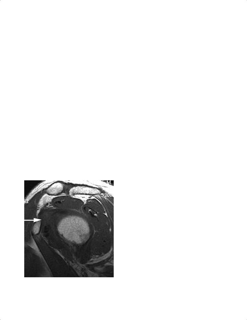
Fig. 4.28 An axial fat-suppressed proton-density image of the right shoulder showing subluxation of the biceps tendon (thin arrow) and the bicipital groove (thick arrow). Note also the subscapularis tear.
112 II Upper Extremity
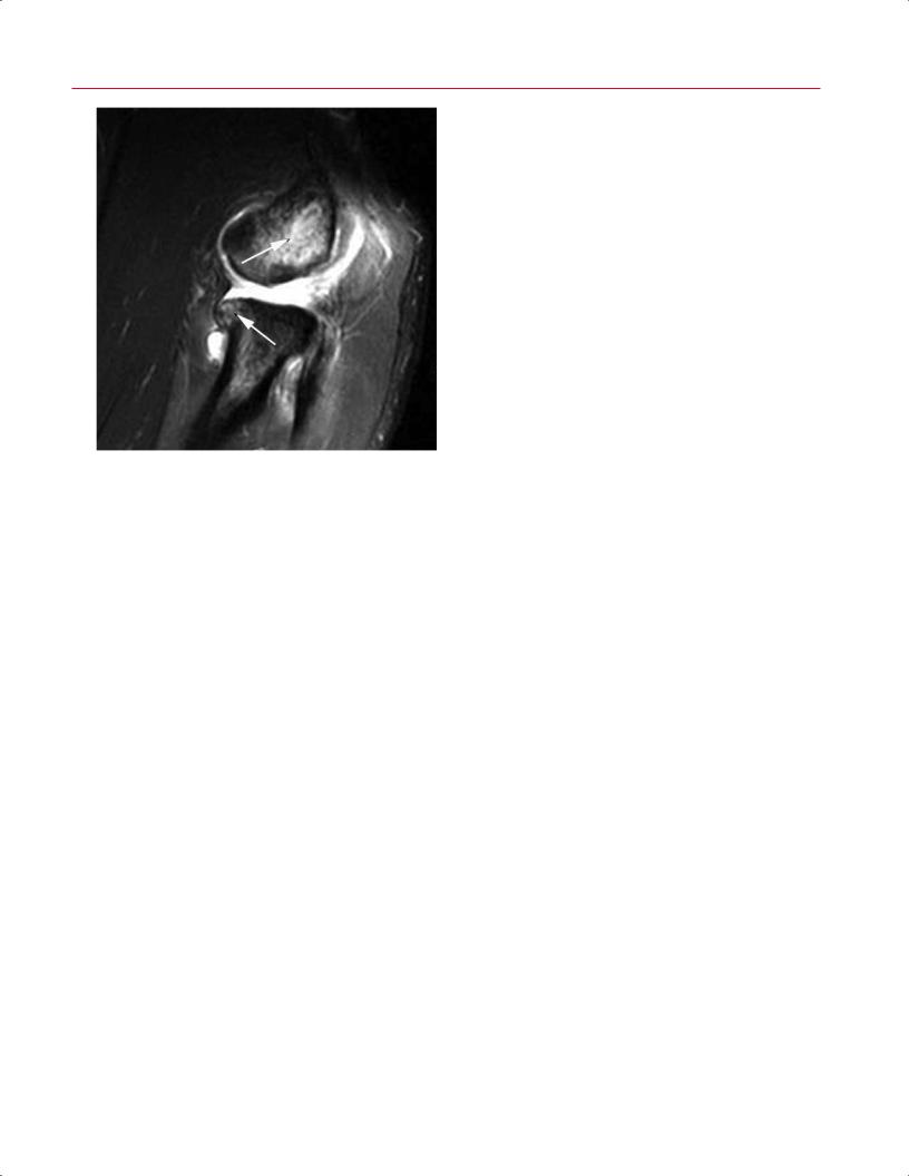
Fig. 4.29 An axial fat-suppressed T2-weighted image of the left shoulder showing the dislocated biceps tendon (thin arrow) medial to an empty bicipital groove (thick arrow). Note the ruptured subscapularis tendon and the resulting intraarticular entrapment of the biceps tendon.
of the descending tendon and as an abnormal course of the intraarticular portion of the tendon.72 Associated findings may include a shallow bicipital groove and tears of the coracohumeral ligament, subscapularis tendon, and supraspinatus tendon. The biceps tendon is located medial and anterior to the subscapularis tendon, with disruption of the transverse ligament and an intact subscapularis. If the subscapularis tendon is detached from its insertion on the lesser tuberosity, however, the biceps tendon becomes entrapped in an intraarticular position.21,73
This condition usually occurs in patients older than those with septic knees and hips. Patients with glenohumeral septic arthritis often have substantial comorbidities, including the following77:
•Coronary artery disease
•Congestive heart failure
•Cancer
•RA
•Alcoholism
•Cirrhosis
•Diabetes mellitus
•End-stage renal disease
•Intravenous drug abuse
•HIV
Frequently, there is a history of recent shoulder surgery, intraarticular cortisone injections, or another infectious source, such as endocarditis, urinary tract infections, or pneumonia.77,78
The diagnosis usually is made on the basis of physical examination, laboratory evaluation, and joint fluid aspiration with subsequent microbiologic analysis. Conventional radiographs usually are not useful in establishing the diagnosis, especially early in the course of the infection77,78; MRI may be useful in ambiguous cases of septic arthritis or to determine the extent of the bone and soft-tissue infection.78,79 The joint e usion and synovitis associated with septic arthritis are shown on T2-weighted and fat-suppressed T2-weighted MRI sequences.78 In the acute stage of the infection, bone marrow edema also may be visualized with MRI.78 If gadolinium contrast is administered intravenously, a prominent rim enhancement of the thickened synovium can be seen on the postgadolinium T1-weighted images.78,80 If untreated, cartilage destruction, marginal erosions, joint space narrowing, and subchondral cyst formation are eventually seen on both MR images and conventional radiographs.78
■Infectious Conditions: Septic Arthritis
The glenohumeral joint is the third most common site for septic arthritis, after the hip and knee.77 As do patients with septic arthritis in other joints, patients with septic arthritis in the glenohumeral joint present with a combination of findings, including the following:
•Pain
•Decreased range of motion
•Fevers
•Focal swelling
•Warmth
•Tenderness
•Erythema
■ Other Pathologic Conditions
Certain pathologic conditions that a ect the shoulder, such as adhesive capsulitis and calcific tendinitis, cannot be classified as traumatic, degenerative, or infectious.
Adhesive Capsulitis
Adhesive capsulitis occurs when inflammation, thickening, and contracture of the joint capsule and synovium result in capsular fibrosis that causes pain and a global limitation of glenohumeral motion.81,82 Patients with diabetes mellitus are at increased risk for this condition.83 Patients present with a history of shoulder pain and sti ness. Physical examination reveals a global loss of active and passive range of motion. Although this condition is primarily a clinical diagnosis, MRI
|
4 The Shoulder 113 |
||
can be useful in confirming clinical suspicions and ruling out |
is not indicated routinely. If MRI is obtained, the calcifica- |
|
|
concomitant abnormalities. Studies have found that a com- |
tions appear as areas of decreased signal intensity on T1- |
||
bined thickness of more than 4 mm for the joint capsule and |
weighted and T2-weighted images; T2-weighted images |
||
synovium, as assessed on the coronal oblique images at the |
frequently show a perifocal band of increased signal inten- |
||
level of the axillary recess, is a useful MRI criterion for the di- |
sity surrounding the lesion, compatible with edema.86,87 |
||
agnosis of adhesive capsulitis.81,84 Furthermore, a thickened |
|
|
|
region of soft-tissue signal intensity in the rotator interval |
|
|
|
that encases the middle and superior glenohumeral liga- |
|
|
|
■ Postoperative MRI Findings |
|||
ments and extends to the biceps anchor is another common |
|||
finding in patients with adhesive capsulitis; this soft tissue |
The accuracy of MRI of the postoperative shoulder is limited |
||
enhances with gadolinium and is best assessed on sagittal |
|||
by several factors, including the following80: |
|||
oblique images (Fig. 4.30).84,85 |
|||
• Surgical distortions of native anatomy |
|||
|
|||
Calcific Tendinitis |
• Changes in the signal intensity of tissues secondary to |
||
surgical trauma |
|||
Calcific tendinitis of the shoulder is an acute or chronically |
|||
• Image degradation caused by metallic artifacts associ- |
|||
painful and self-limiting condition of unknown cause that |
ated with surgical implants |
||
is associated with inflammation around calcium deposits |
The key questions to consider with MRI evaluation of the |
||
located in or around the rotator cu tendons.86,87 This con- |
|||
postoperative shoulder are the following: |
|||
dition predominantly a ects middle-aged women.86,87 The |
|||
|
|
||
most common site of occurrence is within the supraspinatus |
• What imaging sequences limit the e ects of artifact? |
||
tendon and at a location 1.5 to 2.0 cm proximal to its inser- |
• What are the expected MRI findings in the postopera- |
||
tion on the greater tuberosity.87 Patients usually exhibit spe- |
tive shoulder? |
||
cific tenderness over the greater tuberosity and symptoms |
• What MRI findings suggest new/recurrent pathology? |
||
similar to those of subacromial impingement syndrome. The |
Furthermore, it is optimal that the person interpreting the |
||
diagnosis is made by history, physical examination, and ra- |
|||
MR images, whether a radiologist or an orthopaedic surgeon, |
|||
diographic workup. Calcium deposits usually are visualized |
|||
be familiar with the details of the shoulder’s operative pro- |
|||
on conventional radiographs, and therefore MRI evaluation |
|||
cedure. Finally, during image interpretation, preoperative |
|||
|
|||
|
and postoperative images should be correlated closely.80 |
||
|
MR images of the postoperative shoulder are often af- |
||
|
fected by metallic artifacts because of the presence of fer- |
||
|
romagnetic surgical implants such as anchors, screws, tacks, |
||
|
or shoulder arthroplasty prostheses.88,89 These artifacts are |
||
|
most pronounced on images obtained with a long TE, on |
||
|
gradient-echo images, and on fat-suppression sequences.80,90 |
||
|
Certain pulse sequences can minimize the e ects of these |
||
|
artifacts and thus maximize the accuracy of MRI. To avoid |
||
|
magnetic susceptibility artifacts, inversion recovery may be |
||
|
used instead of fat suppression, and FSE sequences may be |
||
|
used instead of conventional SE sequences; gradient-echo |
||
|
sequences may be avoided.80,91,92 These specialized pulse |
||
|
sequences usually are prescribed by a radiologist with ex- |
||
|
pertise in musculoskeletal MRI, which highlights the need |
||
|
for good communication between the orthopaedic surgeon |
||
|
and the radiologist during MRI evaluation of patients with |
||
|
complex processes a ecting the shoulder joint. |
||
|
The most frequently performed surgical procedures that |
||
|
are evaluated postoperatively with MRI of the shoulder in- |
||
|
clude the following93: |
||
|
• Subacromial decompression (impingement syndrome) |
||
|
• Rotator cu repair |
||
Fig. 4.30 A sagittal T1-weighted image showing decreased signal in |
• Glenohumeral instability treatment |
||
the subcoracoid triangle (arrow). |
• Arthroplasty |
||

114 II Upper Extremity
Expected MRI findings after arthroscopic subacromial decompression include morphologic changes in the acromion and coracoacromial ligament and widening of the AC distance. The acromion, as viewed on the sagittal images, usually changes from a hook or curve to a more flat and slightly tapered configuration.80 Decreased signal intensity in the distal acromion on T1-weighted, T2-weighted, and protondensity sequences usually is present and represents fibrosis in the acromial bone marrow.93,94 After resection, the coracoacromial ligament often is replaced by fatty tissue; this site also may have abnormal signal intensity and an irregular morphology caused by scar tissue and metallic artifacts.80 In addition, bursitis-like signal abnormalities at the bursal surface of the rotator cu are common.95 Finally, if a Mumford procedure (distal clavicle excision) is performed, widening of the AC joint by 1 to 2 cm may be visualized.93
Expected MRI findings after rotator cu repair include signal intensity changes in the repaired tendon and regular or irregular tendon morphology, depending on the procedure performed and the quality of the remaining tendon.92,93 The signal intensity changes include intermediate signal intensity (representing granulation tissue) or low signal intensity (representing fibrosis).92,93 Irregularities of the tendon surfaces do not have the same relevance as those in preoperative assessments because even perfect tendon repairs are associated with tendon distortion and variable diameters.91 In one study, only 10% of repaired rotator cu tendons in asymptomatic patients had a normal appearance on MRI.96 Fluid in the subacromial space is another nonspecific, postoperative finding that may be associated with a functional but not watertight repair, a recurrent tear in the rotator cu , or an otherwise normal postoperative rotator cu .96 Nonvisualization of the subacromial fat and the presence of a joint e usion are other common findings.96 Granu-
lation tissue may also be seen around the sutures, resulting in a high-signal abnormality on T2-weighted images, which may mimic a recurrent tear.91 After rotator cu repair, susceptibility artifacts are common on MRI, especially in the presence of bone anchors.91 The criteria for MRI diagnosis of a recurrent full-thickness rotator cu tear are nonvisualization of a portion of the rotator cu tendon and the presence of a fluid-like signal in the tendinous gap on T2-weighted sequences.91,93 Recurrent partial-thickness rotator cu tears are di cult to distinguish from granulation tissue, fluid trapped in sutures, and recurrent full-thickness tears of the rotator cu .80 MR arthrography would help distinguish these possibilities.
Expected MRI findings after a procedure for glenohumeral instability, such as a Bankart repair, include the presence of paramagnetic artifacts from anchors and the restoration of normal anatomy, including the reattachment of the capsulolabral complex to the glenoid rim.80 The labrum-like structure found after Bankart repair usually is rounded and may have an inhomogeneous signal.91 In addition, thickening of the anterior capsule often is visualized on MRI images after surgical repair of instability and labral tears.91,97 MR arthrography is useful for optimal evaluation of the rotator cu , capsulolabral structures, and tendon defects in the postoperative shoulder.80
MRI evaluation of the painful shoulder arthroplasty (total arthroplasty or hemiarthroplasty) is limited by the signal loss associated with imaging adjacent to the metallic prosthesis. Despite this limitation, MRI may be useful in evaluating the integrity of the rotator cu tendons and residual glenoid articular cartilage (in the presence of hemiarthroplasty).98 To optimize the value of MRI, pulse sequences should be manipulated to reduce the susceptibility artifact from the shoulder prosthesis.98
References
1.Kassarjian A, Bencardino JT, Palmer WE. MR imaging of the rotator cu . Radiol Clin North Am 2006;44:503–523
2.Chung CB, Lektrakul N, Gigena L, Resnick D. Magnetic resonance imaging of the upper extremity: advances in technique and application. Clin Orthop Relat Res 2001;383:162–174
3.Kazár B, Relovszky E. Prognosis of primary dislocation of the shoulder. Acta Orthop Scand 1969;40:216–224
4.McFarland EG, Torpey BM, Curl LA. Evaluation of shoulder laxity. Sports Med 1996;22:264–272
5.Baker CL, Uribe JW, Whitman C. Arthroscopic evaluation of acute initial anterior shoulder dislocations. Am J Sports Med 1990;18: 25–28
6.Bankart ASB. The pathology and treatment of recurrent dislocation of the shoulder-joint. Br J Surg 1938;26:23–29
7.Chandnani VP, Yeager TD, DeBerardino T, et al. Glenoid labral tears: prospective evaluation with MRI imaging, MR arthrography, and CT arthrography. AJR Am J Roentgenol 1993;161:1229–1235
8.Stoller DW, Wolf EM. The shoulder. In: Stoller DW, ed. Magnetic Resonance Imaging in Orthopaedics and Sports Medicine. 2nd ed. Philadelphia: Lippincott-Raven; 1997:597–742
9.Sanders TG, Miller MD. A systematic approach to magnetic resonance
imaging interpretation of sports medicine injuries of the shoulder. Am J Sports Med 2005;33:1088–1105
10.Neviaser TJ. The anterior labroligamentous periosteal sleeve avulsion lesion: a cause of anterior instability of the shoulder. Arthroscopy 1993;9:17–21
11.Hill HA, Sachs MD. The grooved defect of the humeral head. A frequently unrecognized complication of dislocations of the shoulder. Radiology 1940;35:690–700
12.Denti M, Monteleone M, Trevisan C, De Romedis B, Barmettler F. Magnetic resonance imaging versus arthroscopy for the investigation of the osteochondral humeral defect in anterior shoulder instability. A double-blind prospective study. Knee Surg Sports Traumatol Arthrosc 1995;3:184–186

4 The Shoulder 115
13.Richards RD, Sartoris DJ, Pathria MN, Resnick D. Hill-Sachs lesion and normal humeral groove: MR imaging features allowing their di erentiation. Radiology 1994;190:665–668
14.Tirman PFJ, Steinbach LS, Feller JF, Stau er AE. Humeral avulsion of the anterior shoulder stabilizing structures after anterior shoulder dislocation: demonstration by MRI and MR arthrography. Skeletal Radiol 1996;25:743–748
15.Wolf EM, Cheng JC, Dickson K. Humeral avulsion of glenohumeral ligaments as a cause of anterior shoulder instability. Arthroscopy 1995;11:600–607
16.Hottya GA, Tirman PFJ, Bost FW, Montgomery WH, Wolf EM, Genant HK. Tear of the posterior shoulder stabilizers after posterior dislocation: MR imaging and MR arthrographic findings with arthroscopic correlation. AJR Am J Roentgenol 1998;171:763–768
17.Gusmer PB, Potter HG, Schatz JA, et al. Labral injuries: accuracy of detection with unenhanced MR imaging of the shoulder. Radiology 1996;200:519–524
18.Loredo R, Longo C, Salonen D, et al. Glenoid labrum: MR imaging with histologic correlation. Radiology 1995;196:33–41
19.Monu JUV, Pope TL Jr, Chabon SJ, Vanarthos WJ. MR diagnosis of superior labral anterior posterior (SLAP) injuries of the glenoid labrum: value of routine imaging without intraarticular injection of contrast material. AJR Am J Roentgenol 1994;163:1425–1429
20.Jee WH, McCauley TR, Katz LD, Matheny JM, Ruwe PA, Daigneault JP. Superior labral anterior posterior (SLAP) lesions of the glenoid labrum: reliability and accuracy of MR arthrography for diagnosis. Radiology 2001;218:127–132
21.Beltran J, Jbara M, Maimon R. Shoulder: labrum and bicipital tendon. Top Magn Reson Imaging 2003;14:35–49
22.Mohana-Borges AVR, Chung CB, Resnick D. Superior labral anteroposterior tear: classification and diagnosis on MRI and MR arthrography. AJR Am J Roentgenol 2003;181:1449–1462
23.Rao AG, Kim TK, Chronopoulos E, McFarland EG. Anatomical variants in the anterosuperior aspect of the glenoid labrum: a statistical analysis of seventy-three cases. J Bone Joint Surg Am 2003;85: 653–659
24.Wall MS, O’Brien SJ. Arthroscopic evaluation of the unstable shoulder. Clin Sports Med 1995;14:817–839
25.Williams MM, Snyder SJ, Buford D Jr. The Buford complex—the “cordlike” middle glenohumeral ligament and absent anterosuperior labrum complex: a normal anatomic capsulolabral variant. Arthroscopy 1994;10:241–247
26.Tuite MJ, Cirillo RL, De Smet AA, Orwin JF. Superior labrum anteriorposterior (SLAP) tears: evaluation of three MR signs on T2-weighted images. Radiology 2000;215:841–845
27.Snyder SJ, Karzel RP, Del Pizzo W, Ferkel RD, Friedman MJ. SLAP lesions of the shoulder. Arthroscopy 1990;6:274–279
28.Mileski RA, Snyder SJ. Superior labral lesions in the shoulder: pathoanatomy and surgical management. J Am Acad Orthop Surg 1998;6:121–131
29.Ma et MW, Gartsman GM, Moseley B. Superior labrum-biceps tendon complex lesions of the shoulder. Am J Sports Med 1995;23:93– 98
30.Cartland JP, Crues JV III, Stau er A, Nottage W, Ryu RKN. MR imaging in the evaluation of SLAP injuries of the shoulder: findings in 10 patients. AJR Am J Roentgenol 1992;159:787–792
31.Connell DA, Potter HG, Wickiewicz TL, Altchek DW, Warren RF. Noncontrast magnetic resonance imaging of superior labral lesions.
102 cases confirmed at arthroscopic surgery. Am J Sports Med 1999;27:208–213
32.Hunter JC, Blatz DJ, Escobedo EM. SLAP lesions of the glenoid labrum: CT arthrographic and arthroscopic correlation. Radiology 1992;184:513–518
33.Nam EK, Snyder SJ. The diagnosis and treatment of superior labrum, anterior and posterior (SLAP) lesions. Am J Sports Med 2003;31:798– 810
34.Tirman PFJ, Feller JF, Janzen DL, Peterfy CG, Bergman AG. Association of glenoid labral cysts with labral tears and glenohumeral instability: radiologic findings and clinical significance. Radiology 1994;190:653–658
35.Snyder SJ, Banas MP, Karzel RP. An analysis of 140 injuries to the superior glenoid labrum. J Shoulder Elbow Surg 1995;4:243–248
36.Kim TK, Queale WS, Cosgarea AJ, McFarland EG. Clinical features of the di erent types of SLAP lesions: an analysis of one hundred and thirty-nine cases. J Bone Joint Surg Am 2003;85:66–71
37.Almekinders LC. Impingement syndrome. Clin Sports Med 2001;20:491–504
38.Bigliani LU, Morrison DS, April EW. The morphology of the acromion and its relationship to rotator cu tears. [Abstr] Orthop Trans. 1986;10:216
39.Ozaki J, Fujimoto S, Nakagawa Y, Masuhara K, Tamai S. Tears of the rotator cu of the shoulder associated with pathological changes in the acromion. A study in cadavera. J Bone Joint Surg Am 1988;70:1224– 1230
40.Morrison DS, Ofstein R. The use of magnetic resonance imaging in the diagnosis of rotator cu tears. Orthopedics 1990;13:633–637
41.Panni AS, Milano G, Lucania L, Fabbriciani C, Logroscino CA. Histological analysis of the coracoacromial arch: correlation between age-re- lated changes and rotator cu tears. Arthroscopy 1996;12:531–540
42.Uri DS. MR imaging of shoulder impingement and rotator cu disease. Radiol Clin North Am 1997;35:77–96
43.Kieft GJ, Bloem JL, Rozing PM, Obermann WR. Rotator cu impingement syndrome: MR imaging. Radiology 1988;166(1 Pt 1):211–214
44.Seeger LL, Gold RH, Bassett LW, Ellman H. Shoulder impingement syndrome: MR findings in 53 shoulders. AJR Am J Roentgenol 1988;150:343–347
45.Hawkins RJ, Kennedy JC. Impingement syndrome in athletes. Am J Sports Med 1980;8:151–158
46.Neer CS II, Welsh RP. The shoulder in sports. Orthop Clin North Am 1977;8:583–591
47.Miniaci A, Salonen D. Rotator cu evaluation: imaging and diagnosis. Orthop Clin North Am 1997;28:43–58
48.Rafii M. Shoulder. In: Firooznia H, Golimbu CN, Rafii M, Rauschning W, Weinreb JC, eds. MRI and CT of the Musculoskeletal System. St. Louis: Mosby-Year Book; 1992:465–549
49.Ogawa K, Yoshida A, Inokuchi W, Naniwa T. Acromial spur: relationship to aging and morphologic changes in the rotator cu . J Shoulder Elbow Surg 2005;14:591–598
50.Mirowitz SA. Normal rotator cu : MR imaging with conventional and fat-suppression techniques. Radiology 1991;180:735–740
51.Erickson SJ, Cox IH, Hyde JS, Carrera GF, Strandt JA, Estkowski LD. E ect of tendon orientation on MR imaging signal intensity: a manifestation of the “magic angle” phenomenon. Radiology 1991;181: 389–392
52.Jerosch J, Müller T, Castro WHM. The incidence of rotator cu rupture. An anatomic study. Acta Orthop Belg 1991;57:124–129

116 II Upper Extremity
53.Quinn SF, Sheley RC, Demlow TA, Szumowski J. Rotator cu tendon tears: evaluation with fat-suppressed MR imaging with arthroscopic correlation in 100 patients. Radiology 1995;195:497–500
54.Ellman H. Shoulder arthroscopy: current indications and techniques. Orthopedics 1988;11:45–51
55.McConville OR, Iannotti JP. Partial-thickness tears of the rotator cu : evaluation and management. J Am Acad Orthop Surg 1999;7:32–43
56.Farley TE, Neumann CH, Steinbach LS, Jahnke AJ, Petersen SS. Fullthickness tears of the rotator cu of the shoulder: diagnosis with MR imaging. AJR Am J Roentgenol 1992;158:347–351
57.Hodler J, Kursunoglu-Brahme S, Snyder SJ, et al. Rotator cu disease: assessment with MR arthrography versus standard MR imaging in 36 patients with arthroscopic confirmation. Radiology 1992;182:431– 436
58.Hodler J, Kursunoglu-Brahme S, Flannigan B, Snyder SJ, Karzel RP, Resnick D. Injuries of the superior portion of the glenoid labrum involving the insertion of the biceps tendon: MR imaging findings in nine cases. AJR Am J Roentgenol 1992;159:565–568
59.Patten RM. Tears of the anterior portion of the rotator cu (the subscapularis tendon): MR imaging findings. AJR Am J Roentgenol 1994;162:351–354
60.Reinus WR, Shady KL, Mirowitz SA, Totty WG. MR diagnosis of rotator cu tears of the shoulder: value of using T2-weighted fat-saturated images. AJR Am J Roentgenol 1995;164:1451–1455
61.Kaplan PA, Bryans KC, Davick JP, Otte M, Stinson WW, Dussault RG. MR imaging of the normal shoulder: variants and pitfalls. Radiology 1992;184:519–524
62.Kaplan PA, Helms CA, Dussault R, Anderson MW, Major NM. Shoulder. In: Kaplan PA, et al., eds. Musculoskeletal MRI. Philadelphia: WB Saunders; 2001:175–223
63.Matsen FA III, Rockwood CA Jr, Wirth MA, Lippitt SB. Glenohumeral arthritis and its management. In: Rockwood CA Jr, Matsen FA III, eds. The Shoulder. 2nd ed. Philadelphia: WB Saunders; 1998:840–964
64.Disler DG, Recht MP, McCauley TR. MR imaging of articular cartilage. Skeletal Radiol 2000;29:367–377
65.Gold GE, Reeder SB, Beaulieu CF. Advanced MR imaging of the shoulder: dedicated cartilage techniques. Magn Reson Imaging Clin N Am 2004;12:143–159
66.Bredella MA, Tirman PFJ, Peterfy CG, et al. Accuracy of T2-weighted fast spin-echo MR imaging with fat saturation in detecting cartilage defects in the knee: comparison with arthroscopy in 130 patients. AJR Am J Roentgenol 1999;172:1073–1080
67.Potter HG, Linklater JM, Allen AA, Hannafin JA, Haas SB. Magnetic resonance imaging of articular cartilage in the knee. An evaluation with use of fast-spin-echo imaging. J Bone Joint Surg Am 1998;80:1276– 1284
68.Needell SD, Zlatkin MB, Sher JS, Murphy BJ, Uribe JW. MR imaging of the rotator cu : peritendinous and bone abnormalities in an asymptomatic population. AJR Am J Roentgenol 1996;166:863–867
69.de Abreu MR, Chung CB, Wesselly M, Jin-Kim H, Resnick D. Acromioclavicular joint osteoarthritis: comparison of findings derived from MR imaging and conventional radiography. Clin Imaging 2005;29:273–277
70.Stein BES, Wiater JM, Pfa HC, Bigliani LU, Levine WN. Detection of acromioclavicular joint pathology in asymptomatic shoulders with magnetic resonance imaging. J Shoulder Elbow Surg 2001;10:204–208
71.Sethi N, Wright R, Yamaguchi K. Disorders of the long head of the biceps tendon. J Shoulder Elbow Surg 1999;8:644–654
72.Tuckman GA. Abnormalities of the long head of the biceps tendon of the shoulder: MR imaging findings. AJR Am J Roentgenol 1994;163:1183–1188
73.Erickson SJ, Fitzgerald SW, Quinn SF, Carrera GF, Black KP, Lawson TL. Long bicipital tendon of the shoulder: normal anatomy and pathologic findings on MR imaging. AJR Am J Roentgenol 1992;158:1091– 1096
74.Van Leersum M, Schweitzer ME. Magnetic resonance imaging of the biceps complex. Magn Reson Imaging Clin N Am 1993;1:77– 86
75.Cervilla V, Schweitzer ME, Ho C, Motta A, Kerr R, Resnick D. Medial dislocation of the biceps brachii tendon: appearance at MR imaging. Radiology 1991;180:523–526
76.Chan TW, Dalinka MK, Kneeland JB, Chervrot A. Biceps tendon dislocation: evaluation with MR imaging. Radiology 1991;179:649– 652
77.Cleeman E, Auerbach JD, Klingenstein GG, Flatow EL. Septic arthritis of the glenohumeral joint: a review of 23 cases. J Surg Orthop Adv 2005;14:102–107
78.Weishaupt D, Schweitzer ME. MR imaging of septic arthritis and rheumatoid arthritis of the shoulder. Magn Reson Imaging Clin N Am 2004;12:111–124
79.Learch TJ, Farooki S. Magnetic resonance imaging of septic arthritis. Clin Imaging 2000;24:236–242
80.Mohana-Borges AVR, Chung CB, Resnick D. MR imaging and MR arthrography of the postoperative shoulder: spectrum of normal and abnormal findings. Radiographics 2004;24:69–85
81.Emig EW, Schweitzer ME, Karasick D, Lubowitz J. Adhesive capsulitis of the shoulder: MR diagnosis. AJR Am J Roentgenol 1995;164:1457– 1459
82.Warner JJP. Frozen shoulder: diagnosis and management. J Am Acad Orthop Surg 1997;5:130–140
83.Kordella T. Frozen shoulder & diabetes. Frozen shoulder a ects 20 percent of people with diabetes. Proper treatment can help you work through it. Diabetes Forecast 2002;55:60–64
84.Connell D, Padmanabhan R, Buchbinder R. Adhesive capsulitis: role of MR imaging in di erential diagnosis. Eur Radiol 2002;12:2100– 2106
85.Lefevre-Colau MM, Drapé JL, Fayad F, et al. Magnetic resonance imaging of shoulders with idiopathic adhesive capsulitis: reliability of measures. Eur Radiol 2005;15:2415–2422
86.Hurt G, Baker CL Jr. Calcific tendinitis of the shoulder. Orthop Clin North Am 2003;34:567–575
87.Uhtho HK, Loehr JW. Calcific tendinopathy of the rotator cu : pathogenesis, diagnosis, and management. J Am Acad Orthop Surg 1997;5:183–191
88.McMenamin D, Koulouris G, Morrison WB. Imaging of the shoulder after surgery. Eur J Radiol 2008;68:106–119
89.Potter HG, Jawetz ST, Foo LF. Imaging of the rotator cu following repair: human and animal models. J Shoulder Elbow Surg 2007;16(5, Suppl):S134–S139
90.Guermazi A, Miaux Y, Zaim S, Peterfy CG, White D, Genant HK. Metallic artefacts in MR imaging: e ects of main field orientation and strength. Clin Radiol 2003;58:322–328
91.Zanetti M, Hodler J. MR imaging of the shoulder after surgery. Magn Reson Imaging Clin N Am 2004;12:169–183
92.Zlatkin MB. MRI of the postoperative shoulder. Skeletal Radiol 2002;31:63–80

4 The Shoulder 117
93.Resnick D, Kang HS. Shoulder. In: Resnick D, Kang HS, eds. Internal Derangement of Joints. Emphasis on MR Imaging. Philadelphia: WB Saunders; 1997:163–333
94.Owen RS, Iannotti JP, Kneeland JB, Dalinka MK, Deren JA, Oleaga L. Shoulder after surgery: MR imaging with surgical validation. Radiology 1993;186:443–447
95.Zanetti M, Jost B, Hodler J, Gerber C. MR imaging after rotator cu repair: full-thickness defects and bursitis-like subacromial abnormalities in asymptomatic subjects. Skeletal Radiol 2000;29:314–319
96.Spielmann AL, Forster BB, Kokan P, Hawkins RH, Janzen DL. Shoulder after rotator cu repair: MR imaging findings in asymptomatic indi- viduals—initial experience. Radiology 1999;213:705–708
97.Vahlensieck M, Lang P, Wagner U, et al. Shoulder MRI after surgical treatment of instability. Eur J Radiol 1999;30:2–4
98.Sperling JW, Potter HG, Craig EV, Flatow E, Warren RF. Magnetic resonance imaging of painful shoulder arthroplasty. J Shoulder Elbow Surg 2002;11:315–321

5 The Elbow
Lance M. Brunton, Mark W. Anderson, and A. Bobby Chhabra
■Specialized Pulse Sequences and Imaging Protocols
MRI of the elbow has improved with the development of high-field systems and enhanced surface coil technology.1 Most centers image the elbow with the patient in the supine position and the arm comfortably positioned at the patient’s side, with the elbow in full extension. This position limits rotation of the proximal radioulnar joint with respect to the distal humerus and minimizes motion artifact compared with imaging the patient prone with the arm overhead. Axial, coronal, and sagittal images are obtained routinely.2
Most centers use a standard screening examination, consisting of an array of specified imaging planes and pulse sequences.3 In addition to standard T1-weighted and T2weighted images, fat-suppressed T2-weighted and STIR sequences are extremely sensitive for detecting marrow and soft-tissue pathology by accentuating the increased signal of fluid and edema relative to the surrounding structures. These sequences are acquired with an FSE technique that produces the desired contrast in a fraction of the time needed for conventional T2-weighted sequences. Gradient-echo sequences are often used to evaluate ligaments and articular cartilage (see Chapter 14) and to delineate loose bodies in the elbow joint. However, these sequences should be avoided after elbow surgery (especially in the presence of metal hardware) because of the increased artifact inherent to the gradient-echo technique (see Chapter 1). A proton-density pulse sequence is occasionally used, often with fat suppression, and combines characteristics of T1-weighted and T2-weighted images. FSE pulse sequences have substantially reduced the length of examination, a great benefit for claustrophobic patients. Imaging parameters can be refined additionally for optimal cartilage visualization when indicated (see Chapter 14).4
MR arthrography is performed by injecting approximately 5 to 10 mL of diluted gadolinium contrast agent into the radiocapitellar joint from a lateral approach and acquiring fat-suppressed T1-weighted and T2-weighted images.5 Although somewhat controversial, MR arthrography of the elbow may help evaluate undersurface collateral ligament tears, capsular disruption, OCD, and intraarticular loose bodies. When investigating infectious, neoplastic, or synovial disorders involving the elbow, intravenous gadolinium is
often used to evaluate for synovial hypertrophy or enhancement of pathologic tissues. Intravenous contrast is also useful for detecting soft-tissue abscesses, which show a thick enhancing wall surrounding the nonenhancing central fluid. Additional refinement of conventional MRI pulse sequences may eventually render these invasive techniques obsolete.6
■ Traumatic Conditions
Most patients with elbow trauma may be evaluated and treated adequately without the use of advanced imaging. However, for select patients, MRI may facilitate the diagnosis of suspected occult fractures, loose bodies, ligamentous injuries, and tendinous pathology.
Occult Fractures
Conventional radiographs often are unrevealing in a patient presenting with elbow pain after acute trauma. In this setting, a joint e usion is often detected clinically, and an elevated posterior fat pad sign may be evident on conventional radiographs. Prospective studies in adults and children have shown that the presence of a posterior fat pad sign correlates with an occult fracture in more than 75% of patients.7,8 However, localizing the injury is a challenging endeavor, and patients often may be overtreated with immobilization in the absence of advanced imaging.
MRI may be used in the setting of elbow trauma to localize and characterize osseous injury when conventional radiographs are unrevealing or equivocal. Fat-suppressed T2-weighted (or STIR) pulse sequences are the most sensitive images for detecting radiographically occult traumatic or stress fractures. Fractures show a linear pattern of signal change on T1-weighted (decreased signal) or T2-weighted (increased signal) images, whereas an osseous contusion often has a more nonspecific appearance (Fig. 5.1A). The most frequent site of occult fracture is the radial head; other sites, such as the lateral epicondyle or olecranon, are much less common.7 Radial head fractures may be accompanied by associated injuries that are most e ectively detected by MRI, such as ligament or capsular disruptions, OCD, loose bodies, or capitellar bone contusions.9
118
5 The Elbow 119
A 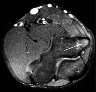 B
B
Fig. 5.1 Osseous injury. (A) Bone contusions. A sagittal STIR image reveals high signal intensity within the capitellum and radial head, indicative of marrow edema (arrows). No discrete low signal fracture lines are evident. (B) Epiphysiolysis (Little Leaguer’s elbow). An axial STIR image of the right elbow showing di use high signal intensity within
In children with a traumatic injury to the elbow and radiographic evidence of only a joint e usion, MRI is a sensitive and accurate modality for identifying or excluding an occult fracture.10 MRI reveals a broad spectrum of elbow pathology in this setting, including the following11:
•Bone bruises
•Ligamentous disruptions
•Muscle injury
MRI also may be e ective in evaluating pediatric physeal injuries, especially in young children with poorly visualized ossification centers.12 Finally, MRI has a role in the detection of Little Leaguer’s elbow, a valgus stress injury in skeletally immature throwing athletes in which epiphysiolysis occurs at the medial epicondylar apophysis (Fig. 5.1B).13
Loose Bodies
The clinical manifestation of loose bodies within the elbow joint is typically pain with motion, often accompanied by mechanical symptoms, such as catching or locking. Loose bodies may originate from a traumatic shear injury to the osteochondral surface, synovial disorders, or OCD. The definitive treatment of symptomatic loose bodies is arthroscopic or open removal, but accurate diagnosis is imperative to prevent unnecessary surgery.
Although conventional radiographs or CT may detect the presence of osseous loose bodies in the elbow joint, MRI is
the medial epicondyle consistent with epiphysiolysis from repetitive valgus stress to the elbow (arrow). Part A is adapted with permission from Brunton LM, Anderson MW, Pannunzio ME, Khanna AJ, Chhabra AB: Magnetic resonance imaging of the elbow: update on current techniques and indications. J Hand Surg Am 2006;31:1001–1011.
especially useful for detecting intraarticular cartilaginous or osteocartilaginous loose bodies. Loose bodies are found most commonly in the olecranon or coronoid fossae on T2weighted images (Fig. 5.2). Osteophytes and synovial hypertrophy may mimic loose bodies on MRI images.3 The use of MRI for detecting loose bodies in the elbow has been disputed, and an overall low specificity and sensitivity, similar to that of conventional radiography, when compared with arthroscopic findings have been reported.14 MR arthrography may be e ective in this setting, but it has not been studied adequately.
MCL Injury
The MCL is composed of the anterior, posterior, and transverse bundles. The anterior bundle is the most important stabilizer against valgus stress, and it can be divided into anterior and posterior bands. The anterior band is the primary restraint with the elbow in extension, and the posterior band is the primary restraint with the elbow in increasing amounts of flexion. MCL injuries have received tremendous attention because of their prevalence in throwing athletes of all ages and at all levels of competition.15 Accurate diagnosis of a full-thickness or partial-thickness tear of the MCL has important implications for treatment, as does the di erentiation between MCL pathology and other causes of medialside elbow pain, such as ulnar neuropathy, stress fracture, and flexor-pronator mass injury.