

Атлас анатомии человека в трех томах. Том II. (Билич, Крыжановский)
.pdf Рис. 419.
Опускание яичка в мошонку и формирование его оболочек (А - положение яичка в период закладки, Б - яичко
у внутреннего кольца пахового канала, В - яичко в мошонке) (схема):
1 - Testis; 2 - Peritoneum; 3 - Testicular artery; 4 - Epididymis; 5 - Ductus deferens; Vas deferens; 6 - Peritoneum, vaginal process; 7 - Guide's ligament of egg; 8 - Dartos fascia; Superficial fascia of scrotum; 9 - Skin; 10 - Internal spermatic fascia; 11 - Serosa cavity of
testis; 12 - Tunica vaginalis, parietal layer; 13 - Tunica vaginalis, visceral layer
 Рис. 420.
Развитие внутренних мужских половых органов (левое яичко представлено на более ранней стадии, правое прошло через паховый канал и находится в мошонке) (схема):
1 - Appendix of testis; 2 - Appendix of epididymis; 3 - Prostate; 4 - Prostatic utricle; 5 - Ductus deferens; Vas deferens; 6 - Urinary bladder; 7 - Ureter; 8 - Aorta; 9 - Kidney; 10 - Paramesonephricus (Mullerian) duct; 11 - Mesonephricus canaliculus; 12 - Mesonephricus inferior canaliculus; 13 - Inguinal canal; 14 - Mesonephricus duct; 15 - Seminal gland; Seminal vesicle; 16 - Gubernaculum testis;
17 - Bulbo-urethral gland; 18 - Rectum
 Рис. 421.
Аномалии мужской уретры
А. Аномалии расположены на нижней стороне полового члена и мошонки.
Б. Возможные участки открытия уретры при гипоспадиях (вид сбоку).
Если половые валики не соединяются полностью во время половой дифференцировки, возникает аномалия уретры в виде щели, которая может открываться на нижней поверхности полового члена (гипоспадия) или на его дорсальной поверхности (эписпадия). Гипоспадия встречается гораздо чаще (3:3000 случаев), чем эписпадия (3:300 000). Чаще всего встречается гипоспадия головки. Тело полового члена обычно укорочено и отклонено вниз из-за наличия вентральных фиброзных тяжей. Хирургическая коррекция обычно проводится между шестым месяцем и вторым годом жизни.
 Рис. 422.
Развитие мужских (I) и женских (II) наружных половых органов (А - индифферентная стадия - эмбрион 7 нед., Б - эмбрион 12 нед., В - плод 9 мес) (схема):
1 - Tail; 2 - Pudendal tubercle; 3 - Pudendal fold; 4 - Pudendal torus; 5 - Urogenital sinus; 6 - Anus; 7 - Glans penis; 8 - Male urethra, sulcus; 9 - Scrotum; 10 - Raphe of scrotum; 11 - Male urethra, suture; 12 - Vaginal orifice; 13 - Glans of clitoris; 14 - Labium majus;
15 - External urethral orifice; 16 - Labium minus; 17 - Hymen
ЖЕНСКАЯ ПОЛОВАЯ СИСТЕМА
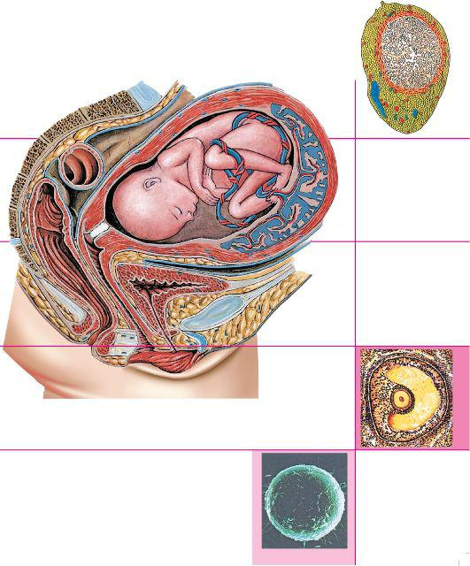 Таблица 22. Женские половые органы


Рис. 423.
Строение и происхождение женской половой системы (схема):
1 - Bulb of vestibule; 2 - Crus of clitoris; 3 - Vagina; 4 - Round ligament of uterus; 5 - Ligament of ovary; 6 - Ovary; 7 - Vesicular appendices; 8 - Infundibulum; 9 - Epoophoron; 10 - Uterine tube; 11 - Paroophoron; 12 - Epoophoron, longitudinal duct, transverse ductules; 13 - Kidney; 14 - Renal pelvis; 15 - Ureter; 16 - Uterus; 17 - Urinary bladder; 18 - Median umbilical ligament; 19 - Female urethra; 20 - Glans of clitoris; 21 - External urethral orifice; 22 - Vaginal orifice; 23 - Greater vestibular gland
 Рис. 424.
Женская половая система, сагиттальный распил, вид слева:
1 - Bulb of vestibule; 2 - Labium minus; 3 - Labium majus; 4 - Corpus cavernosum of clitoris; 5 - Fascia of clitoris; 6 - Clitoris; 7 - Crus of clitoris; 8 - Dorsal artery and vein of clitoris; 9 - Suspensory ligament of clitoris; 10 - Pubis; 11 - Urinary bladder; 12 - Transverse vesical fold; 13 - Round ligament ofuterus; 14 - Uterus; 15 - Right uterine tube; 16 - Ovary; 17 - Right external iliac vein; 18 - Right external iliac artery; 19 - Ovarian plexus; 20 - Promontory; 21 - Right ureter; 22 - Left uterine tube; 23 - Ligament of ovary; 24 - Rectum; 25 - Broad ligament of uterus; 26 - Parametrium; 27 - Recto-uterine fold; 28 - Recto-uterine
pouch; 29 - Vesico-uterine pouch; 30 - Left ureter; 31 - Uterine artery; 32 - Pelvic diaphragm; Pelvic floor; 33 - Peritoneum; 34 - External anal sphincter; 35 - Anorectal flexure; Perineal
flexure; 36 - Vagina; 37 - Greater vestibular gland
 Рис. 425.
Женские половые органы, сагиттальный распил (фотография натурального препарата):
1 - Labium majus; 2 - Clitoris; 3 - Labium minus; 4 - Anus; 5 - Internal anal sphincter; 6 - Female urethra; 7 - Vagina; 8 - Rectum; 9 - Vesico-uterine pouch; 10 - Pubic symphysis; 11 - Uterus; 12 - Broad ligament of uterus; 13 - Obturator artery; 14 - Obturator nerve;
15 - Uterine tube; 16 - Ovary; 17 - Fimbriae; 18 - Promontory
 Рис. 426.
Нижний этаж полости брюшины женщины, вид сверху:
1- Median umbilical ligament; 2 - Urinary bladder; 3 - Transverse vesical fold; 4 - Round ligament of uterus; 5 - Parietal peritoneum; 6 - Caecum; 7 - Vesico-uterine pouch; 8 - Uterus; 9 - Broad ligament of uterus; 10 - Recto-uterine pouch; 11 - Rectum; 12 - Rectouterine fold; 13 - Suspensory ligament of ovary; Infundibulopelvic ligament; 14 - Left ovary; 15 - Ligament of ovary; 16 - Uterine tube; 17 - Paravesical fossa; 18 - Sigmoid colon; 19 - Medial umbilical fold; 20 - Deep inguinal ring; 21 - Lateral umbilical fold; Epigastric fold;
22 - Rectus abdominis
 Рис. 427.
Внутренние женские половые органы (восковая модель):
1 - Ureter; 2 - Inferior mesenteric vein; 3 - Ovarian plexus; 4 - Inferior mesenteric artery; 5 - Common iliac artery; 6 - Descending colon; 7 - Ovary; 8 - Ovarian fimbria; 9 - Mesosalpinx: 10 - Uterus; 11 - Infundibulum; 12 - Ampulla; Duodenal cap; 13 - Round ligament of uterus; 14 - Urinary bladder; 15 - Pubic symphysis