


Forewords
Writing the foreword for a book is a huge responsibility because you endorse the work to the scientific community. For this book, the responsibility increases the pleasure, and the honor of doing it is immeasurable. Let me explain: This work is a gift to the field of dentistry because it was crafted by an outstanding professional, Dr Hilton Riquieri, who excels at waxing dental morphology.
As both an experienced dental technician and a prosthodontist, Dr Hilton is able to synthesize both disciplines as few others. I was very pleased to have Dr Hilton as a doctoral student in the restorative dentistry postgraduate program at the Science and Technology Institute of São Paulo State University. He was an ideal student because the university’s function is to train dentists who have the mission of researching and spreading knowledge in Brazil and abroad.
This important work also showcases photography by Dr Rodrigo Riquieri, who is Dr Hilton’s son; he has already shown his own dedication to the profession in his laboratory and clinical work and has a very promising future. Drs Francci, Saavedra, and Viegas have all proven themselves in teaching, research, and clinical
and laboratory experience, and their contributions are excellent.
However, this work attests, above all, to the leadership and synthesizing skills of Dr Riquieri in the art of teaching dental morphology. In 17 chapters, he explains step by step how to draw and sculpt dental anatomy with maximum accuracy. In these chapters, the exquisite morphology of all the teeth are developed and presented carefully, with examples from everyday life that greatly facilitate learning. What strikes me in this book is the richness of detail and the brilliant and direct way of teaching how to study, plan, and execute waxing and sculpting. This book will be very useful to students and professionals who want to improve waxing and sculpting techniques.
This book is a gift, and the field of dentistry will be grateful that—after all of the professional dental training—the authors and contributors have chosen to devote part of their lives to the creation of this scientific and technical book. Enjoy your reading!
Marco Antonio Bottino
This book is a guide that will inspire excellence in dentistry. Because of the fundamentals presented here, anyone who has dedicated themselves to the fabrication of dental prosthodontics should use it to learn the foundations of creating ideal morphology, which demands technical acumen. However, this book conveys not only the practical knowledge of morphology but also the art of its sculpture.
Dr Hilton Riquieri also demonstrates that, for every morphologic feature, there is a scientific explanation. It is immensely rewarding to see professional ability dedicated to teaching. Thus, it is with great honor that I write the foreword of a book whose pages convey much more than just words and images; they share a deep knowledge that comes only from professional
experience. Dr Riquieri is an exceptional professional whose sharp insight and illustrative teaching style is evidenced throughout this book. The presentation makes the learning pleasant and easy.
This beautiful atlas describes the anatomy of each dental structure and presents step by step and in detail the execution of sculpture in wax. In addition, the author uses analogies from everyday life to elaborate ideas and concepts of considerable complexity.
Congratulations to those who have shared from their experience and wisdom to prepare this book for worldwide dentistry.
Paulo Kano
vi
@dentistinfo стоматологический телеграм канал

Dedication
To Joseph and Divina, father and mother, who are my role models.
To my siblings—Carlos, Regina, Eurípedes, and Aparecida—for support and always being present. And the reason for everything: my wife Deborah and my children Rodrigo, Breno, Arthur, and Enrico.
Preface
Learning is an endless path. Drawing and carving are basic skills, like walking and reading, that once learned are known for a lifetime. However, the most we can do is what our perception allows us to see. As Paulo Kano has said, “The hand can only reproduce what the brain can see.” How do we INCREASE our perception, our ability to see more?
Constructivism is a psychogenetic theory introduced by the Swiss biologist, Jean Piaget (1896– 1980). This theory states that we develop skills through association of what we want to learn with memorized objects/figures. Thus, knowledge is built up from our interaction with the environment where, with constant training, long-term memory and innate skills are formed.
Sculpture is an innate ability, though we were not born with it. For the final results, 70% depends on perception, 20% depends on psychomotor training, and 10% depends on manual ability.
In Betty Edwards’ discussion of the development of skill training, the student moves from being:
Incompetent AND unaware
↓
Incompetent BUT aware
↓
Competent AND aware
↓
Competent BUT unaware
Accumulated knowledge allows us to overcome obstacles, and overcoming difficulties opens new horizons and elevates our personal boundaries. May this book be part of your journey to new horizons.
vii
@dentistinfo стоматологический телеграм канал

Acknowledgments
I would like to thank:
Rodrigo Riquieri, who was fundamental to the development of this book thanks to his splendid images.
My collaborating teachers, Drs Carlos Francci, Guilherme Saavedra, and Diogo Viegas, who added their experience to this work with enthusiasm and genius. Being surrounded by people like them is a privilege.
Helcio Marques, owner of the first laboratory in which I worked. In 1981, he accepted me as his assistant. Thank you for the years in which you exercised the virtue of patience with me!
Expedito Bernardes and Perclísio de Oliveira Gomes, professionals who helped me to grow and offered me the opportunity to train and still be paid. Time and distance have not detracted from our friendship or my gratitude.
Sebastião César Manosso and his wife Édila Moreira Manosso, and Benvinda Maria de Souza, Antônio Gasparini, Paulo Rubens Ruiz Possebom, Mário Marques Cunha, Celso Garcia Rodrigues, and Milton José Aricó. In difficult times and without their help, the dream of graduating would not have come true.
My invaluable Professors: Vani Teixeira, Aziz Constantino, Sérgio Reinaldo De Fiore, Antônio Marcolino Pellicano, José Ceratti Turano, Milton Edson Miranda, Luís Ramos, Marcelo Lucchesi Teixeira, Vagner Ortega, and Marco Antônio Bottino.
To Prof Paulo Kano. Taking your course was a watershed in my life and truly represented an awakening that enabled me to connect the many small links of knowledge that, until then, were dispersed in my mind. The coalescence into a common larger universe about method and form allowed me understand that for every dental feature there was an explanation. This perception changed my view on the subject entirely.
My friend Carlos Oliveira, who bridged the gap between me and the one who would become my
student, awaken again my dream of completing my academic career, and finally, become my doctorate advisor.
My friend Herbert Mendes, for the friendship and well-wishing that he always expressed to me. My luck in meeting him was a real stepping stone in my life.
The entire APDESP Team, for the dedication and opportunities they have always given me to develop into a real professor.
My friends Luís Alves Ferreira and Marcos Celestrino, who invited me to teach my first course and for the frequent invitations to participate as a speaker.
The entire staff of Editora Napoleão, who helped me to finish this project. Professionalism is the word that best defines them.
The entire staff of the Hilton Riquieri Dental Laboratory, partners for so many years.
The entire staff of the Hilton Riquieri Training Center, who provided me with the tranquility to be a professor.
Julia Maria de Lima Oda and Mitsuo Oda, for their support, caring, and dedication to the family environment.
To all my students, because they made me learn more in trying to teach them.
To all those who stimulated me with their criticism. In a decisive and unforgettable way, they contributed to my development, helping me to transform the meaning of the great challenges that stood before me along my way. You made the difference.
Having learned from an early age that no one goes far alone, I apologize for taking the risk of forgetting someone among the many high-minded people with whom I met in my journey. Thank you all, thank you, thank you, thank you...
Thanks also to the stones on the way; without them there is no way to become a better person. Passing through them strengthens us.
viii
@dentistinfo стоматологический телеграм канал

Contributors
Hilton Riquieri, dds, msd, phd
Master Ceramist and Director
Centro de Treinamento Hilton Riquieri
Bragança Paulista, São Paulo
Science and Technology Institute
São Paulo State University
São José dos Campos, São Paulo
Brazil
Rodrigo Yamada Riquieri
Dental Technician and Instructor
Centro de Treinamento Hilton Riquieri
Bragança Paulista, São Paulo
Brazil
Carlos Eduardo Francci,dds, msd, phd
Assistant Professor
Department of Biomaterials and Oral Biochemistry
School of Dentistry
University of São Paulo
São Paulo, Brazil
Diogo Viegas,dds, msd
Private Practice Limited to Implant Dentistry
Lisbon, Portugal
Private Practice Limited to Implant Dentistry
Edinburgh, Scotland
Guilherme de Siqueira F. Anzaloni Saavedra,dds, msd, phd
Assistant Professor
Department of Dental Materials and Prosthesis Science and Technology Institute, São José dos Campos Faculty of Dentistry
São Paulo State University São Paulo, Brazil
ix
@dentistinfo стоматологический телеграм канал
SECTION I
Posterior Teeth
@dentistinfo стоматологический телеграм канал

The Esthetic and Functional Parameters of Posterior Teeth
01
1
@dentistinfo стоматологический телеграм канал
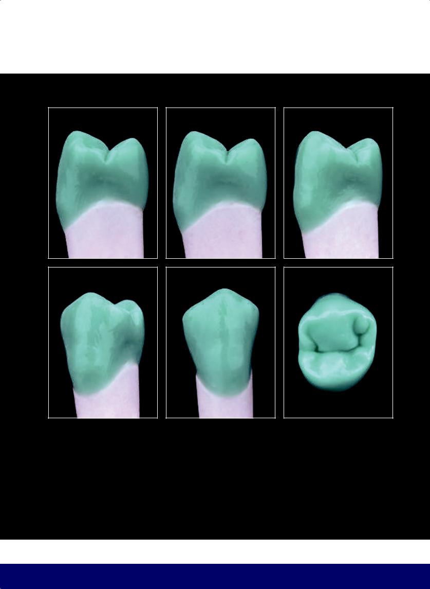
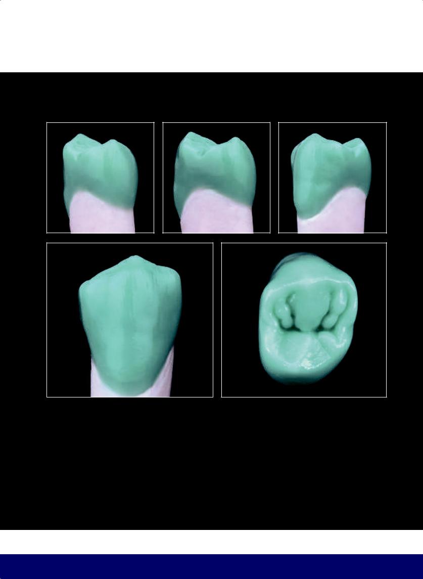
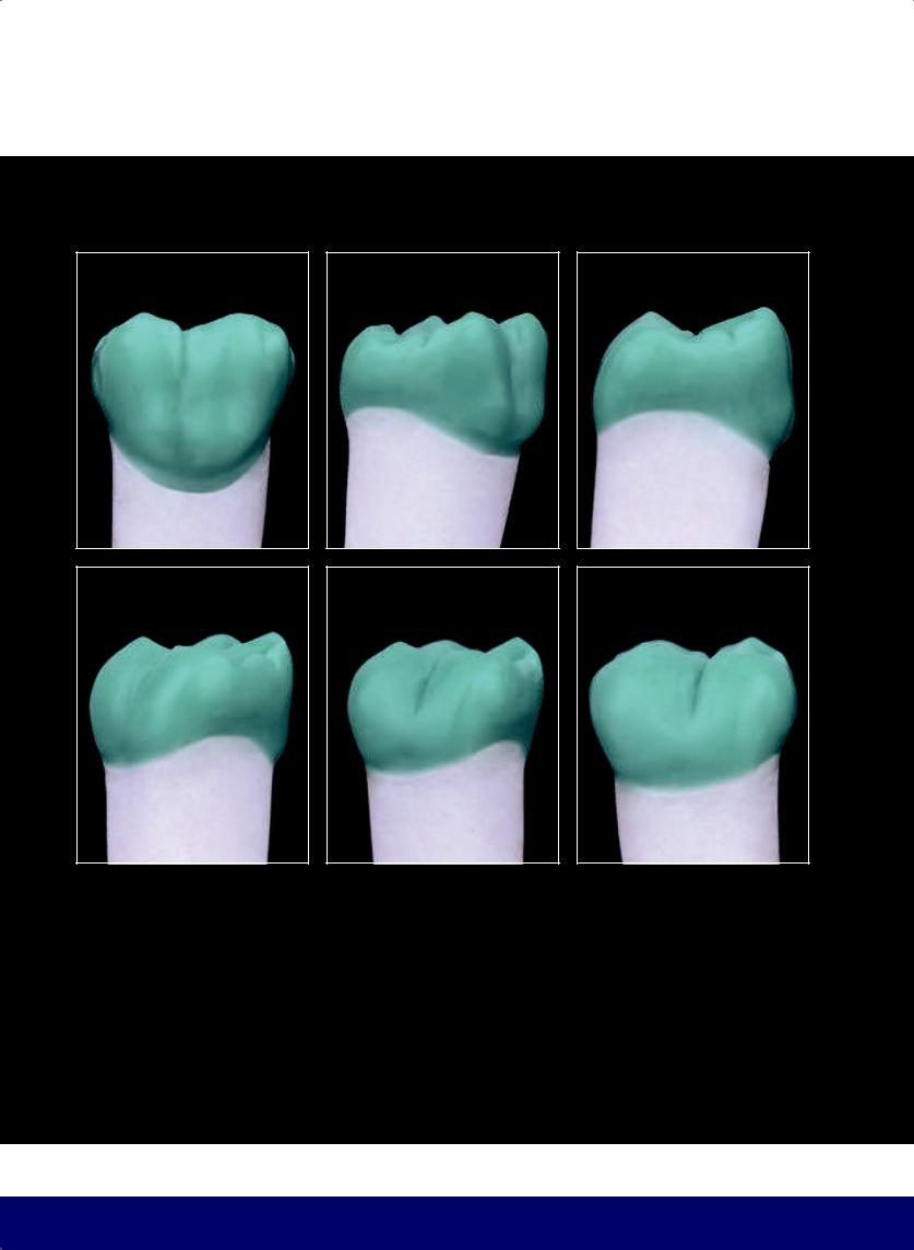
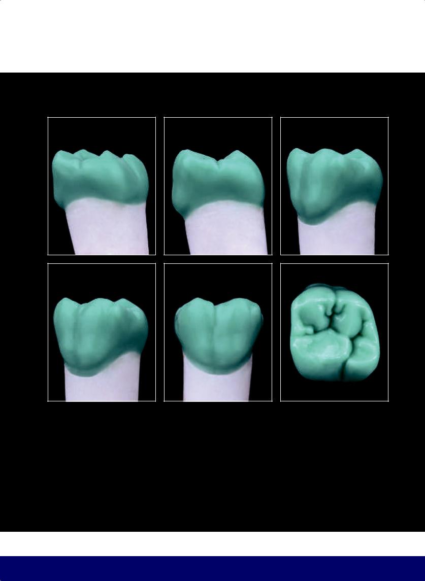
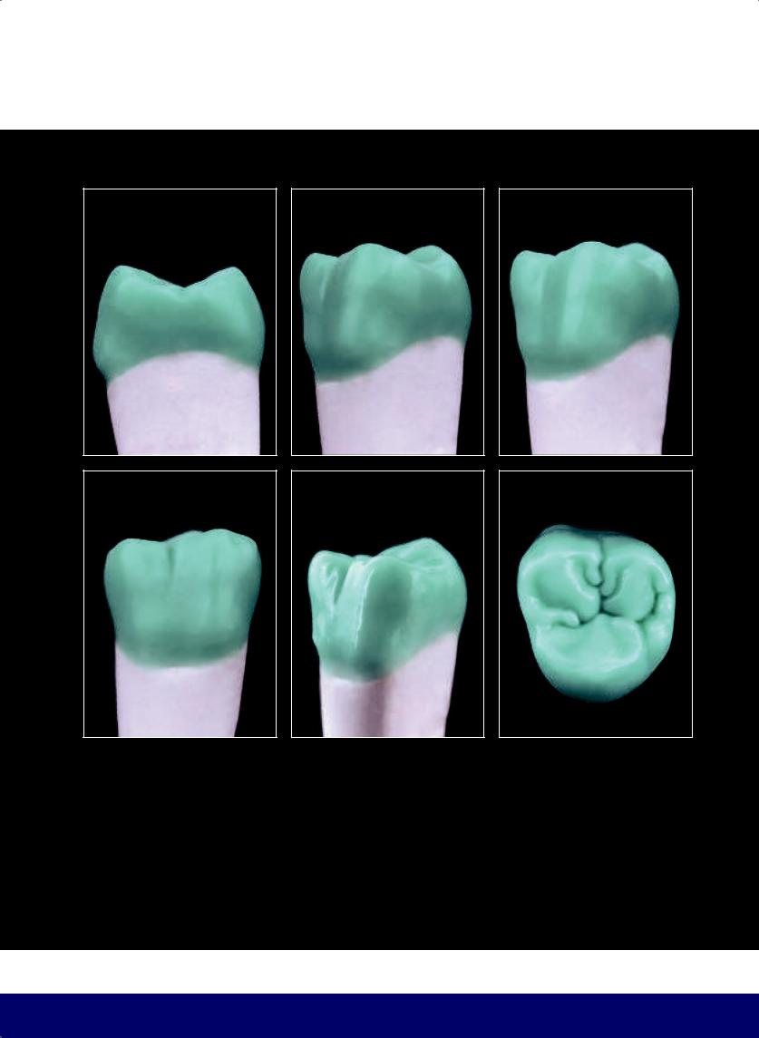
CHAPTER 1
Occlusal
Cervical
Buccal
Mesial |
Distal |
Lingual
2
@dentistinfo стоматологический телеграм канал
THE ESTHETIC AND FUNCTIONAL PARAMETERS OF POSTERIOR TEETH
3
@dentistinfo стоматологический телеграм канал

CHAPTER 1
Ridge |
Cusp tip |
Slope
Overjet |
|
|
|
|
|
3–4 mm |
|
|
|
|
|
|
|
|
|||
|
|
|
|
|
|
|
|
|
|
|
|
|
|
|
|
|
|
|
|
|
|
|
|
|
|
|
|
|
2–3 mm |
|
|
|
|
|
|
|
|
|
||||||
|
|
|
||||||
|
|
|
|
|
|
|
|
|
|
|
|
|
|
|
|
|
|
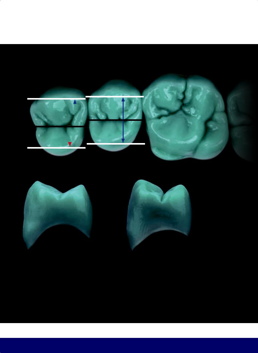
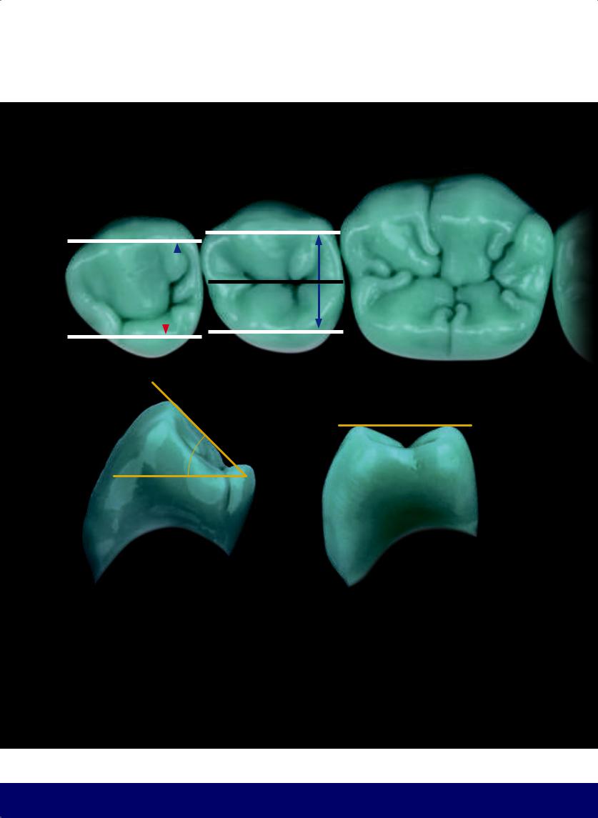
Everycuspispyramidalwithaquadrangularbase,exceptthemesio- |
The OVERJET is the extent of the buccal overlap in the horizontal |
lingual cusp of the maxillary first molar, which is pentagonal. |
plane of the maxillary teeth over the mandibular teeth. The overjet |
|
protects the mouth, lips, and tongue from involuntary bites. The |
|
distance between the lowest point of the bottom of the fossa and |
|
the cusp tip is, on average, between 3 and 4 mm in the HORIZON- |
|
TAL PLANE and between 2 and 3 mm in the VERTICAL PLANE. |
4
@dentistinfo стоматологический телеграм канал
THE ESTHETIC AND FUNCTIONAL PARAMETERS OF POSTERIOR TEETH
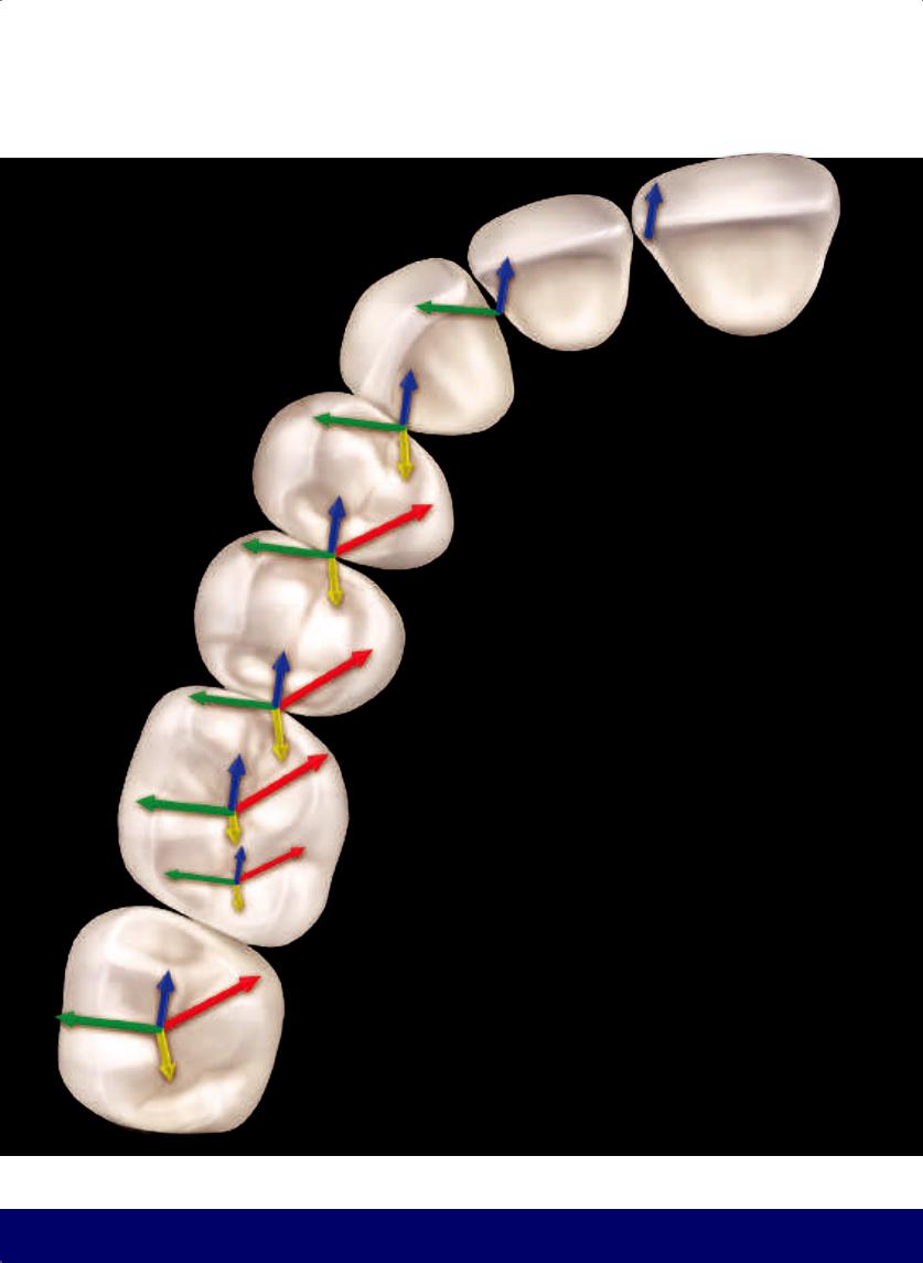
Curve of occlusion, anteroposterior curve, or curve of Spee
Lateral curve or curve of Wilson
5
@dentistinfo стоматологический телеграм канал
CHAPTER 1
CURVE OF MONSON
This is the overlap of the curves of Spee and Wilson, whose radius is in the glabella.
6
@dentistinfo стоматологический телеграм канал
THE ESTHETIC AND FUNCTIONAL PARAMETERS OF POSTERIOR TEETH
ANATOMICAL EQUATOR OF THE TOOTH
 Buccal
Mesial |
Distal |
|
Central |
FOSSAE
7
@dentistinfo стоматологический телеграм канал
CHAPTER 1
AREA OF CONTACT
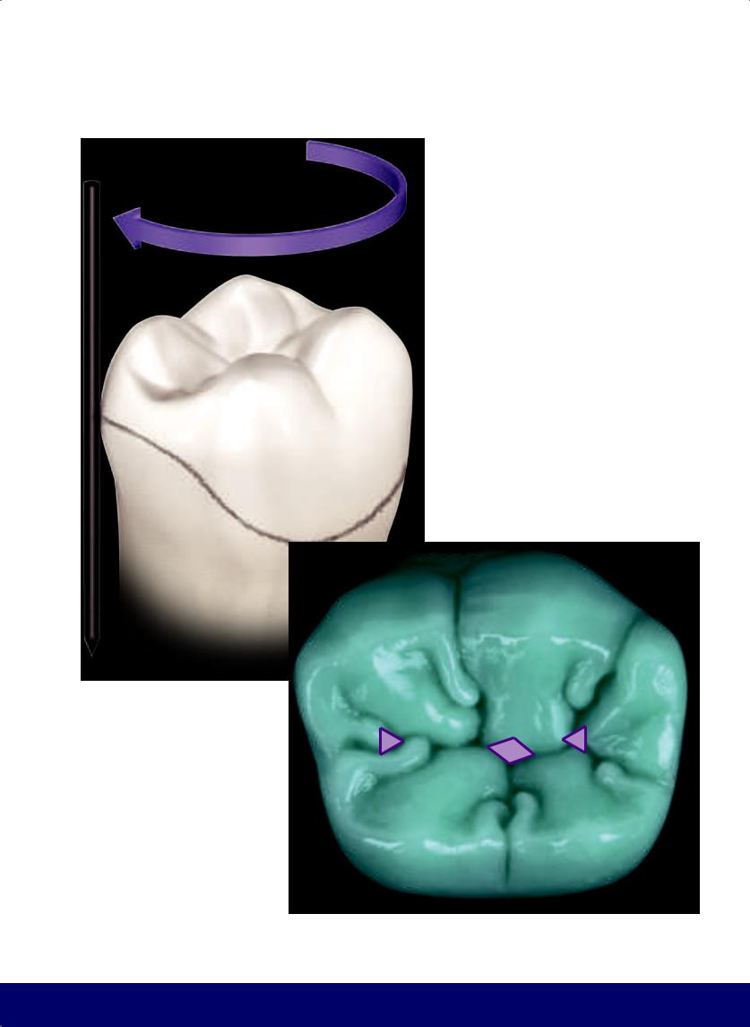
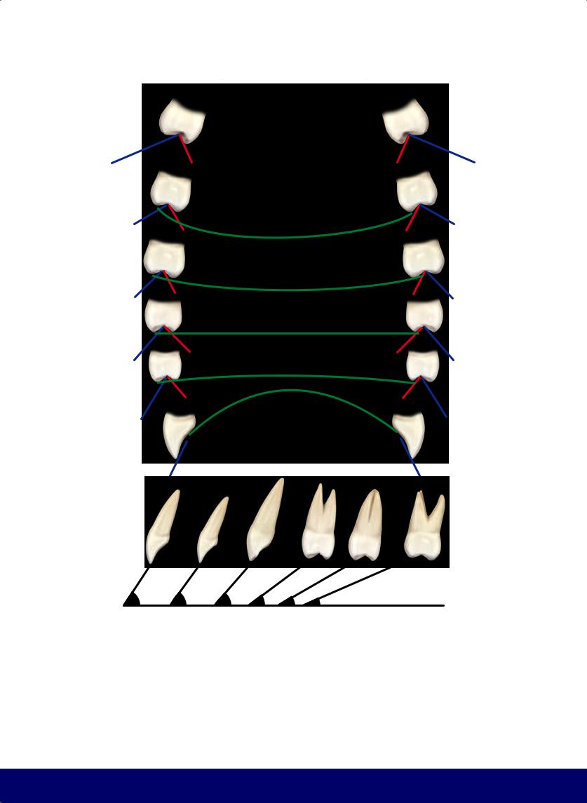
In posterior teeth, the contact areas are horizontal ellipses.
OCCLUSAL SURFACE
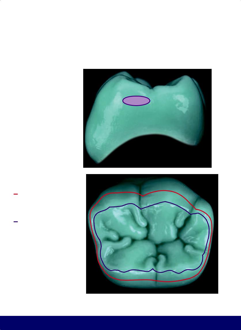
This includes the occlusal table as well as the occlusal third of the buccal and lingual surfaces.
OCCLUSAL TABLE
Anatomical area delineated by marginal ridges and transverse ridges.
8
@dentistinfo стоматологический телеграм канал
THE ESTHETIC AND FUNCTIONAL PARAMETERS OF POSTERIOR TEETH
Longer side
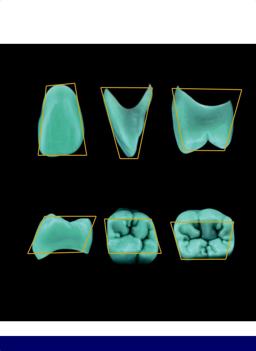
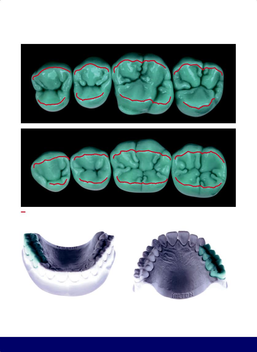
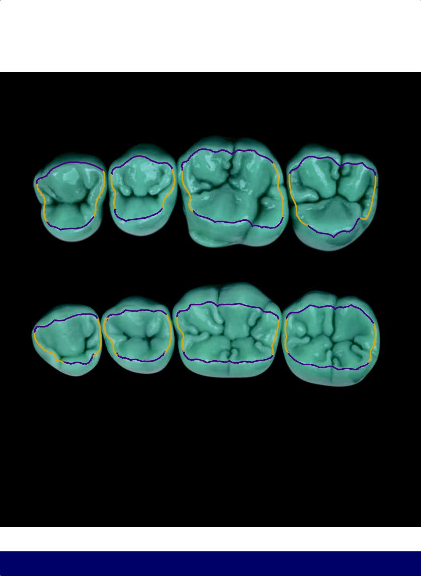
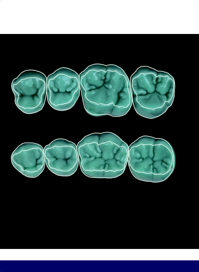
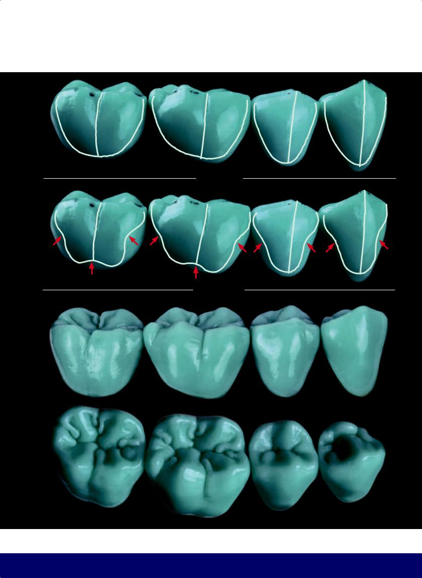
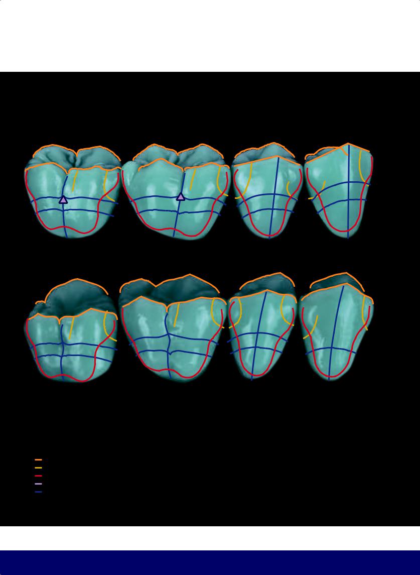
EXTERNAL CONTOUR, OCCLUSAL VIEW
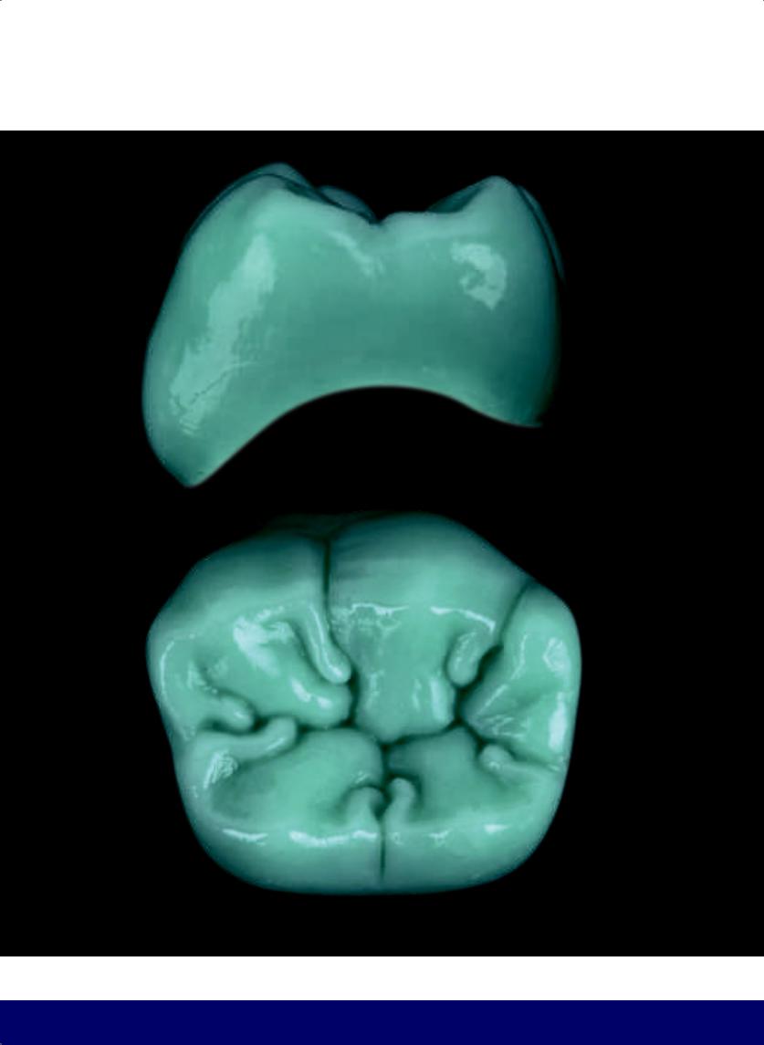
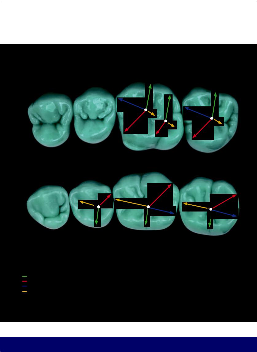
All the teeth are visualized in a trapezoid. Buccolingually, the longer side of the trapezoid is on the buccal aspect, except for the mandibular first molar, where the trapezoid is inverted. In that case, the lingual aspect is longer.
9
@dentistinfo стоматологический телеграм канал
CHAPTER 1
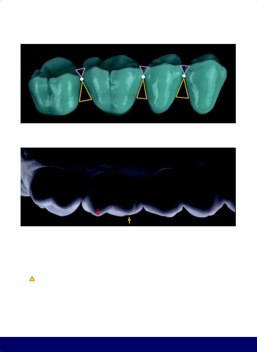
Notice that the contact area between the teeth is more buccal, except between the first and second molars, where it is more lingual.
EMBRASURES
These are triangular spaces between the teeth. The lingual grooves are larger than the buccal grooves, except between the maxillary molars, where an inversion occurs.
10
@dentistinfo стоматологический телеграм канал
THE ESTHETIC AND FUNCTIONAL PARAMETERS OF POSTERIOR TEETH
 SPACE ABOVE THE CONTACT AREA
SPACE BELOW THE CONTACT AREA
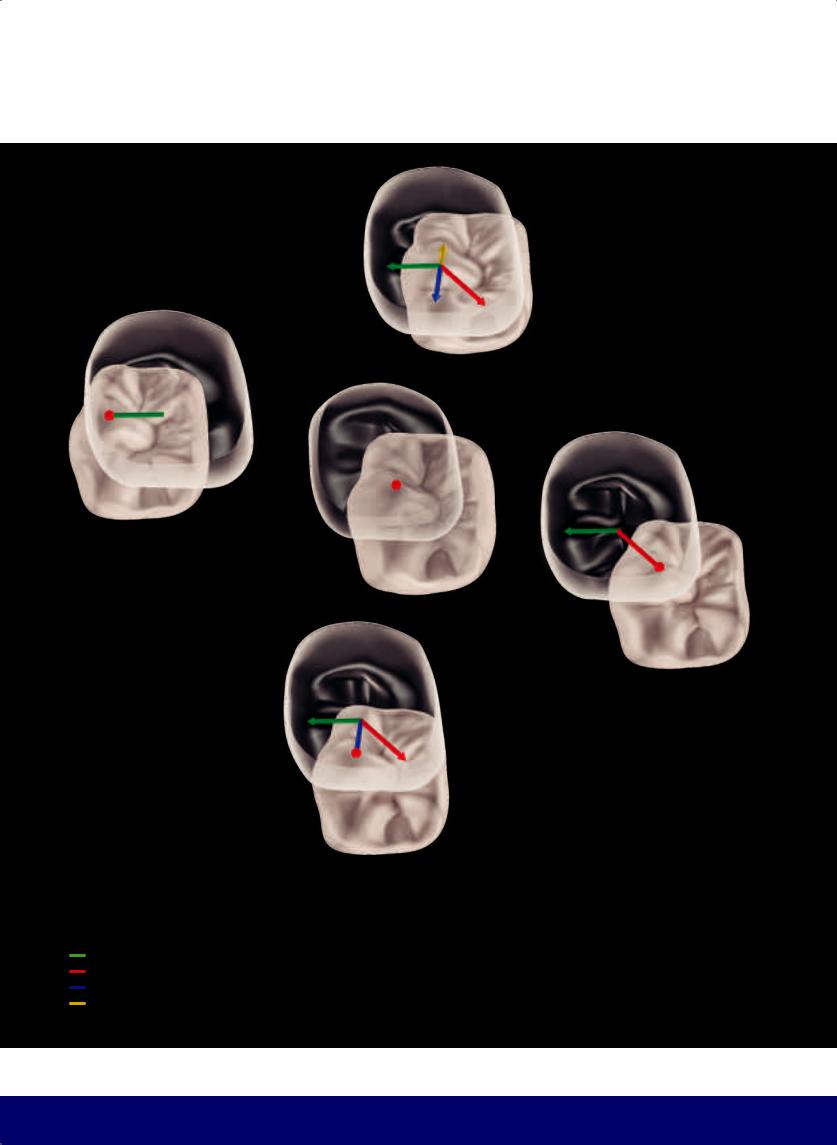
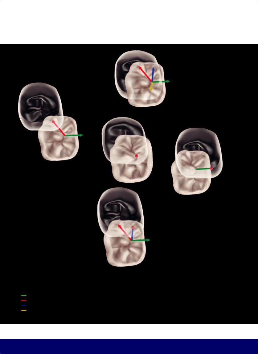
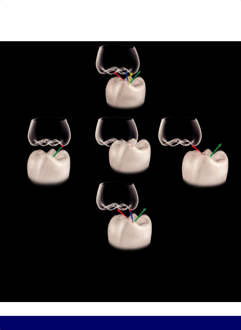
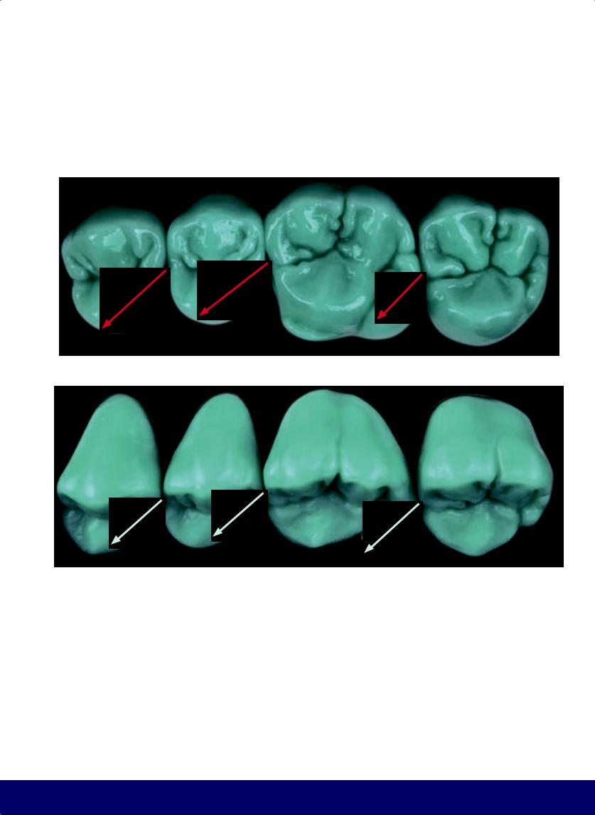
The distobuccal cusp (red arrow) of the maxillary first molar is taller and sharper than the mesiobuccal cusp (yellow arrow), which is smaller and rounder.
11
@dentistinfo стоматологический телеграм канал
CHAPTER 1
BUCCAL AND LINGUAL RIDGES
12
@dentistinfo стоматологический телеграм канал
THE ESTHETIC AND FUNCTIONAL PARAMETERS OF POSTERIOR TEETH
Parallel
Divergent
Parallel
Divergent
ANTEROPOSTERIOR ALIGNMENT OF BUCCAL AND LINGUAL
RIDGES AND OF MESIODISTAL GROOVES
13
@dentistinfo стоматологический телеграм канал
CHAPTER 1
MESIAL AND DISTAL MARGINAL RIDGES
14
@dentistinfo стоматологический телеграм канал
THE ESTHETIC AND FUNCTIONAL PARAMETERS OF POSTERIOR TEETH
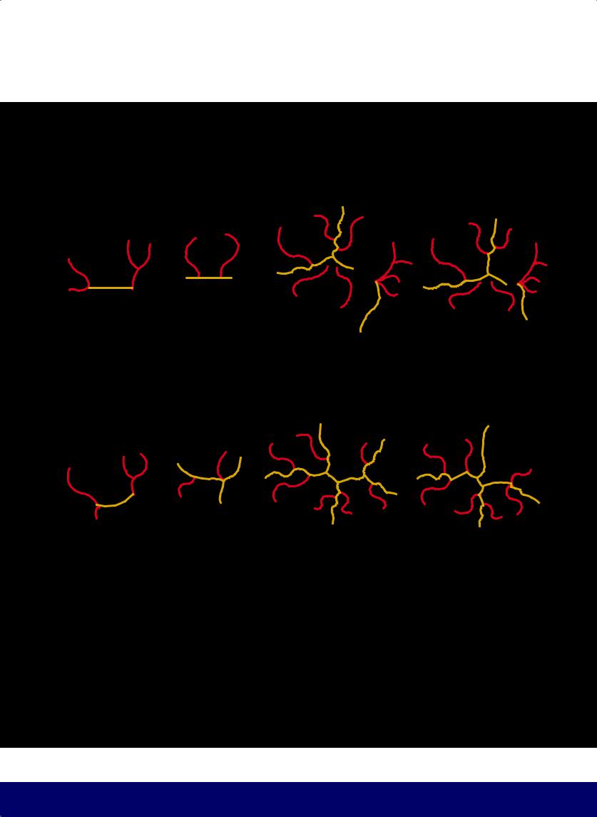
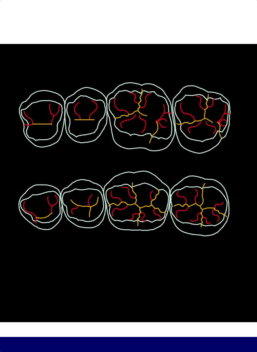
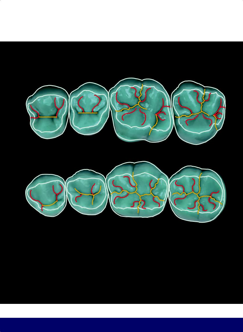
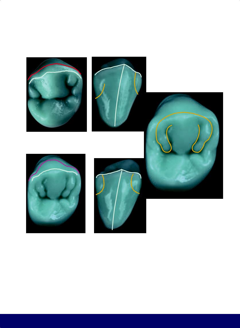
MEMORIZATION BY ANALOGY
The union of all transverse and marginal ridges of the maxillary first molar reveals the number 9 (from Paulo Kano).
15
@dentistinfo стоматологический телеграм канал
CHAPTER 1
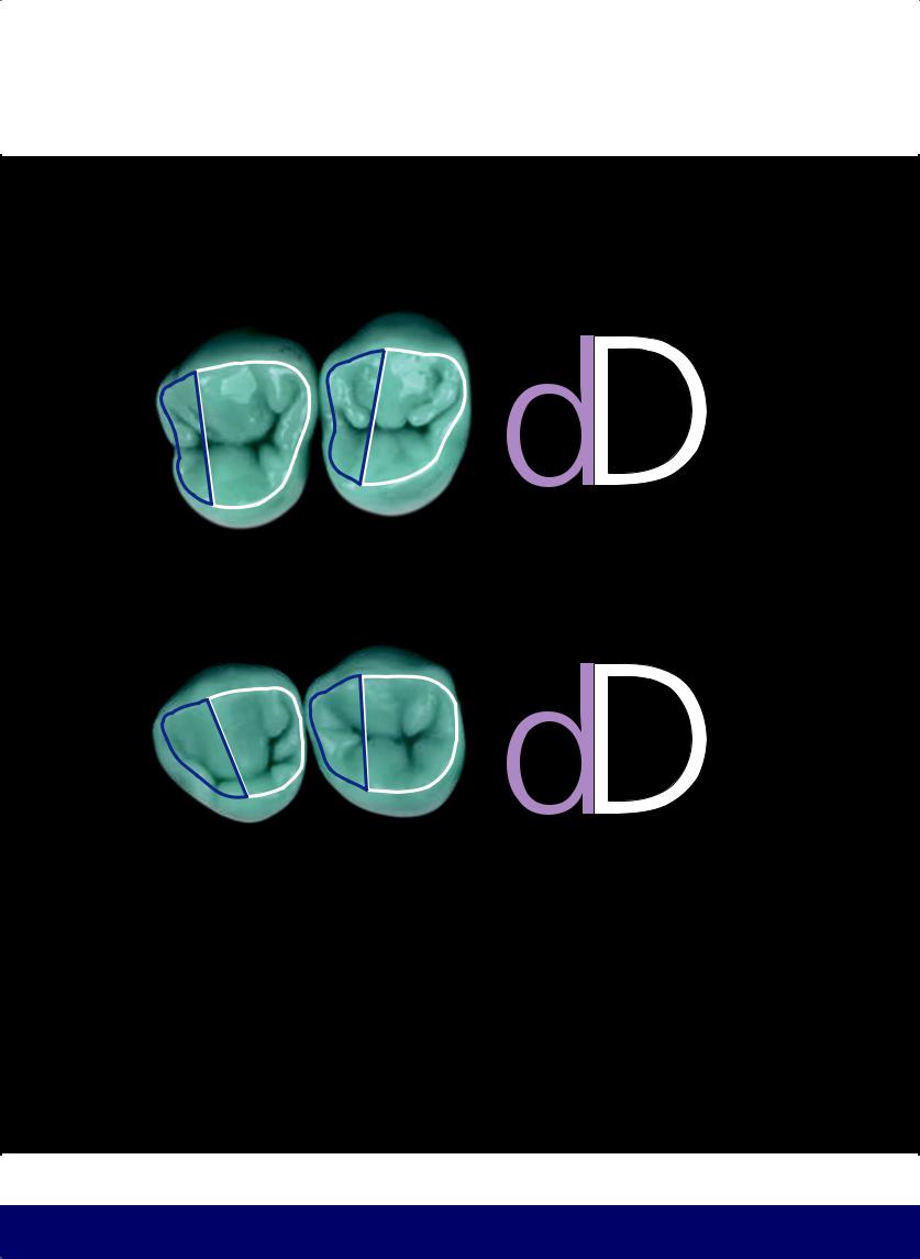
MEMORIZATION BY ANALOGY
By joining all the premolar ridges as shown, we have a larger D on the distal and a smaller, inverted D on the mesial.
16
@dentistinfo стоматологический телеграм канал
THE ESTHETIC AND FUNCTIONAL PARAMETERS OF POSTERIOR TEETH
COMPARISON OF MAXILLARY POSTERIOR TEETH: EXTERNAL CONTOURS AND OCCLUSAL TABLES
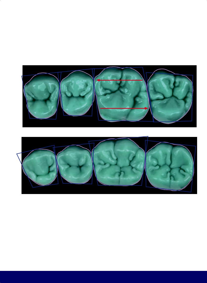
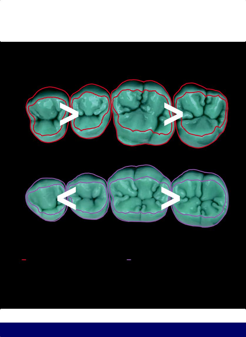
The first premolars are larger than the second premolars and the first molars are larger than the second molars.
COMPARISON OF MANDIBULAR POSTERIOR TEETH: EXTERNAL CONTOURS AND OCCLUSAL TABLES
The first premolars are smaller than the second premolars and the first molars are larger than the second molars.
17
@dentistinfo стоматологический телеграм канал
CHAPTER 1
V |
|
|
V |
|
|
||||
|
|
= |
||
< |
|
|
|
|
|
|
|
L |
|
|
|
|||
|
|
|
||
L |
|
|
|
|
|
|
|
||
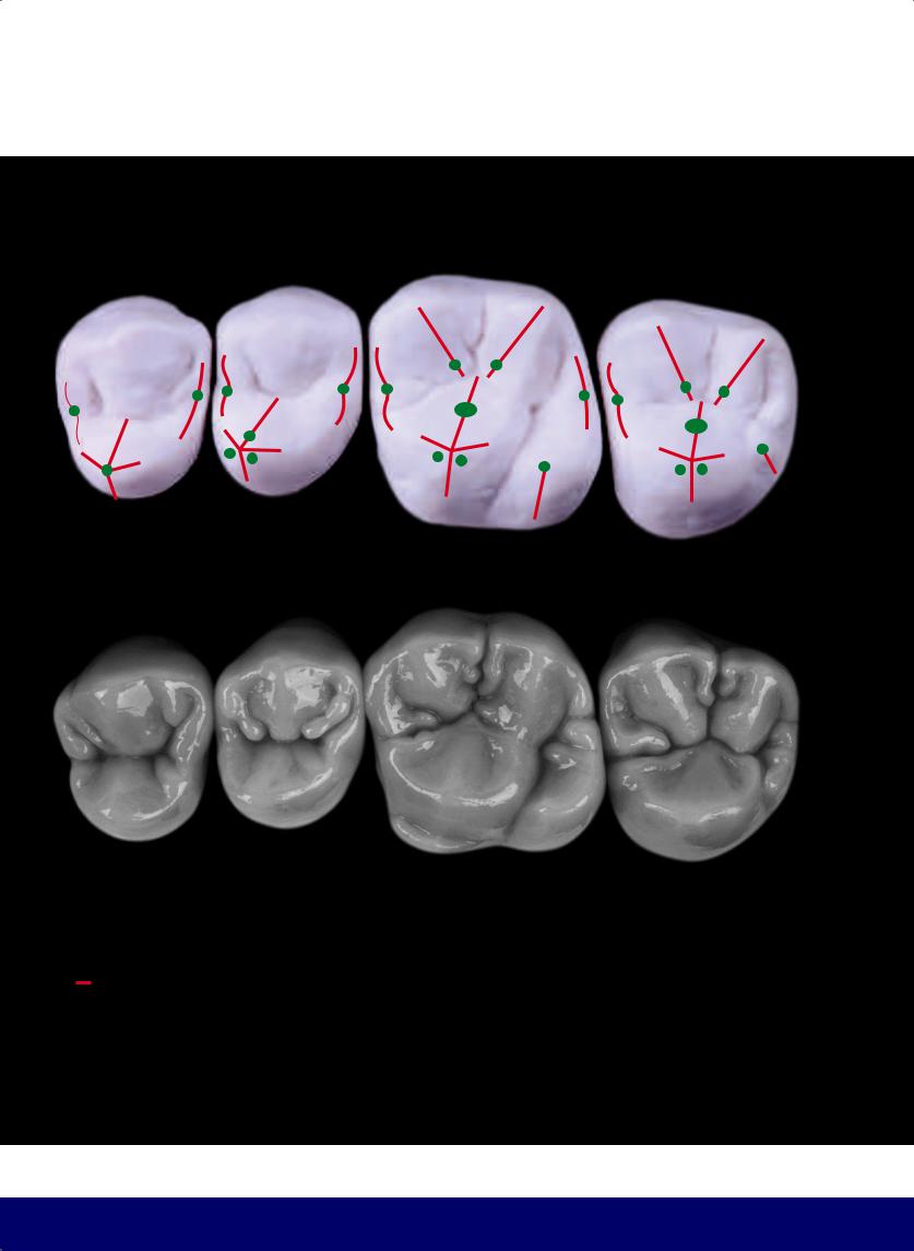
VOLUME OF PREMOLAR CUSPS
The mesiodistal groove of the maxillary first premolar is positioned more toward the lingual, so the buccal cusp is larger than the lingual cusp. In the second premolar, the mesiodistal groove is positioned more at the center of the occlusal surface so that the lingual and buccal cusps have similar volumes.
18
@dentistinfo стоматологический телеграм канал
THE ESTHETIC AND FUNCTIONAL PARAMETERS OF POSTERIOR TEETH
V |
|
|
V |
|
|
||||
|
|
= |
||
> |
|
|
|
|
|
|
|
L |
|
|
|
|
|
|
|
|
|
||
L |
|
|
||
|
|
|
||
30o to 45o
In the mandibular first premolar, the difference in height between the cusps is, on average, between 30 to 45 degrees.
19
@dentistinfo стоматологический телеграм канал
CHAPTER 1
CUSP CRESTS are located inside the occlusal table.
CUSP SLOPES are located outside the occlusal table.
A KIDNEY BEAN PROFILE is visible in mesial marginal ridges of all teeth and also on the distal marginal ridge of the maxillary second premolar.
20
@dentistinfo стоматологический телеграм канал
THE ESTHETIC AND FUNCTIONAL PARAMETERS OF POSTERIOR TEETH
FUNCTIONAL GROOVES
WORKING
BALANCING
PROTRUSIVE
RETRUSIVE
21
@dentistinfo стоматологический телеграм канал
CHAPTER 1
BUCCAL AND OCCLUSAL VIEW OF FUNCTIONAL MOVEMENTS
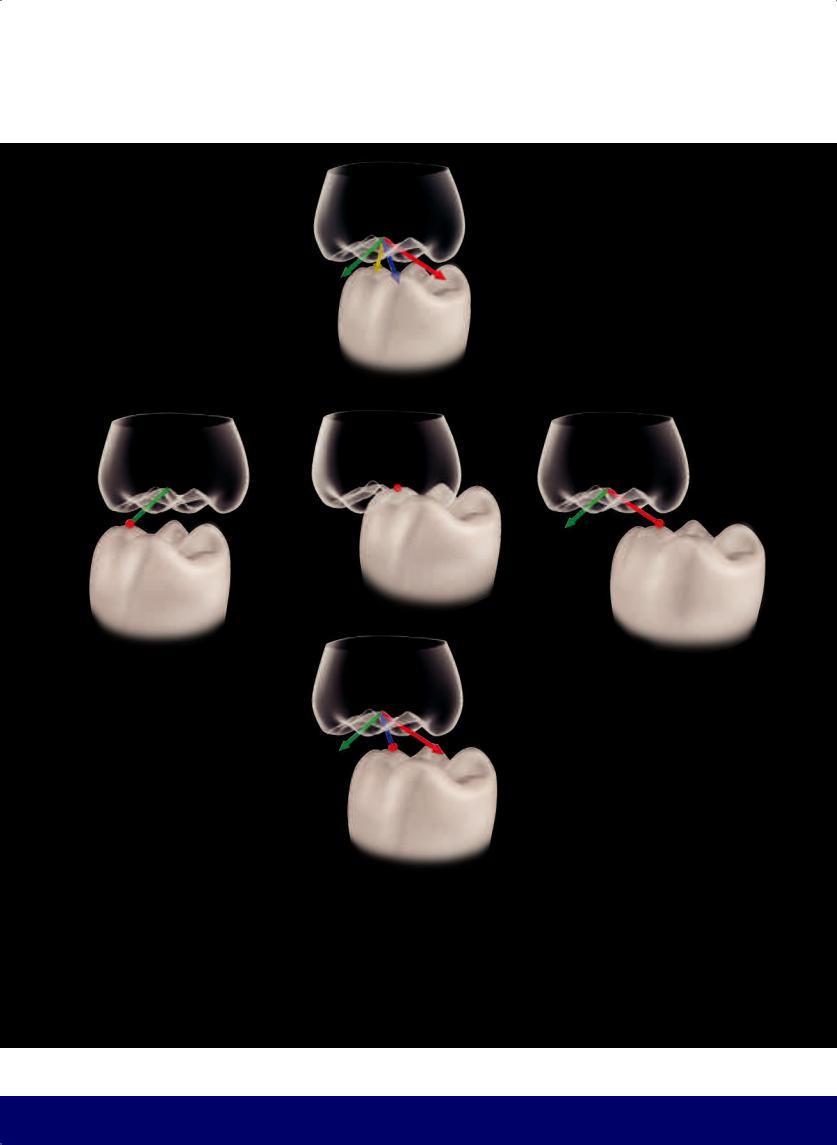
Grooves formed by the medial cusp of the mandibular first molar in the central fossa of the maxillary first molar from the functional movements of:
WORKING
BALANCING
PROTRUSIVE
RETRUSIVE
22
@dentistinfo стоматологический телеграм канал
THE ESTHETIC AND FUNCTIONAL PARAMETERS OF POSTERIOR TEETH
23
@dentistinfo стоматологический телеграм канал
CHAPTER 1
BUCCAL AND OCCLUSAL VIEW OF FUNCTIONAL MOVEMENTS
Grooves formed by the mesiobuccal cusp of the maxillary first molar in the central fossa of the mandibular first molar from the functional movements of:
WORKING
BALANCING
PROTRUSIVE
RETRUSIVE
24
@dentistinfo стоматологический телеграм канал
THE ESTHETIC AND FUNCTIONAL PARAMETERS OF POSTERIOR TEETH
25
@dentistinfo стоматологический телеграм канал
CHAPTER 1
26
@dentistinfo стоматологический телеграм канал
THE ESTHETIC AND FUNCTIONAL PARAMETERS OF POSTERIOR TEETH
FUNCTIONAL GROOVES
WORKING
BALANCING
PROTRUSIVE
RETRUSIVE
27
@dentistinfo стоматологический телеграм канал
CHAPTER 1
INCREASING SLOPE FROM THE INCISAL EDGES IN THE ANTERIOR TEETH TO THE GRINDING RIDGES IN THE POSTERIOR TEETH
(Gustavo Vernazza, 2013)
28
@dentistinfo стоматологический телеграм канал
THE ESTHETIC AND FUNCTIONAL PARAMETERS OF POSTERIOR TEETH
Balance requires that the lingual cusps of the maxillary premolars and distolingual cusps of the maxillary first molars be directed toward the mesial
29
@dentistinfo стоматологический телеграм канал
CHAPTER 1
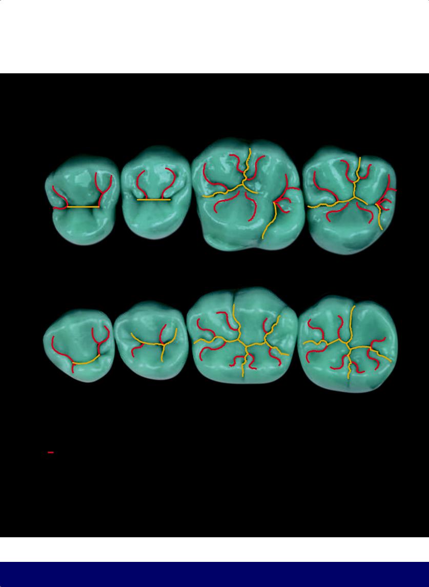
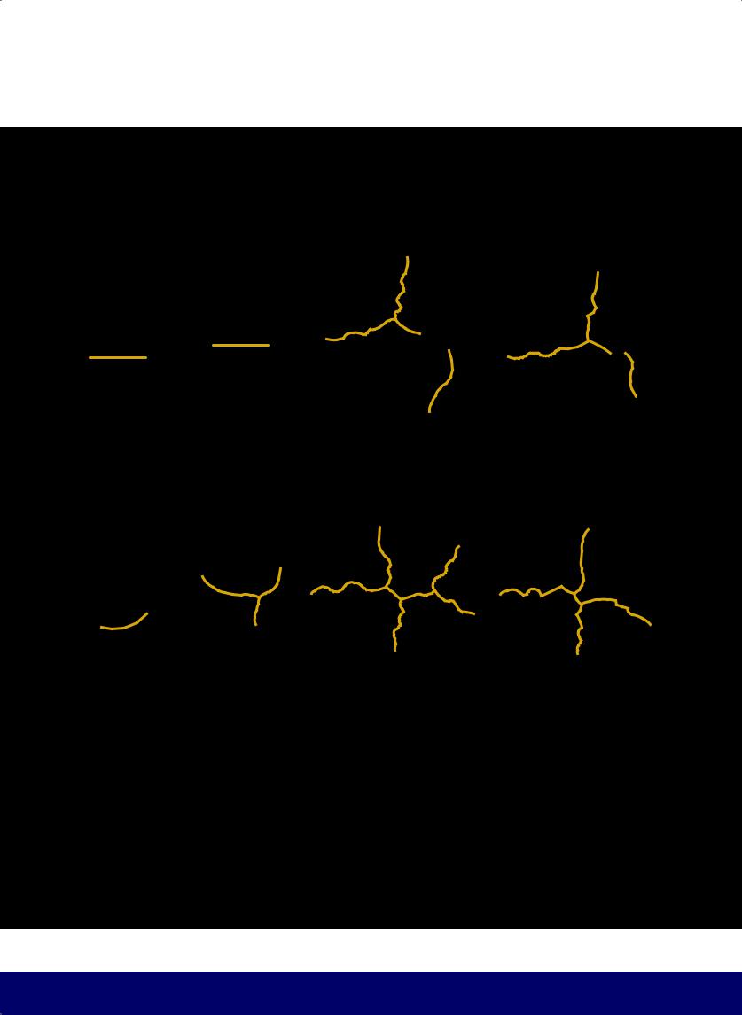
PRIMARY GROOVES separate cusps
30
@dentistinfo стоматологический телеграм канал
THE ESTHETIC AND FUNCTIONAL PARAMETERS OF POSTERIOR TEETH
SECONDARY GROOVES separate the ridges from the cusps
31
@dentistinfo стоматологический телеграм канал
CHAPTER 1
EXTERNAL CONTOUR AND OCLUSAL TABLE
32
@dentistinfo стоматологический телеграм канал

THE ESTHETIC AND FUNCTIONAL PARAMETERS OF POSTERIOR TEETH
DIAGRAM OF EXTERNAL CONTOUR AND OCCLUSAL TABLE
33
@dentistinfo стоматологический телеграм канал
CHAPTER 1
DIAGRAM OF PRIMARY GROOVES
34
@dentistinfo стоматологический телеграм канал
THE ESTHETIC AND FUNCTIONAL PARAMETERS OF POSTERIOR TEETH
DIAGRAM OF PRIMARY AND SECONDARY GROOVES
35
@dentistinfo стоматологический телеграм канал
CHAPTER 1
DIAGRAM OF OCCUSAL MORPHOLOGY
36
@dentistinfo стоматологический телеграм канал
THE ESTHETIC AND FUNCTIONAL PARAMETERS OF POSTERIOR TEETH
DIAGRAM OF OCCLUSAL MORPHOLOGY OVERLAYING WAX-UP
37
@dentistinfo стоматологический телеграм канал
CHAPTER 1
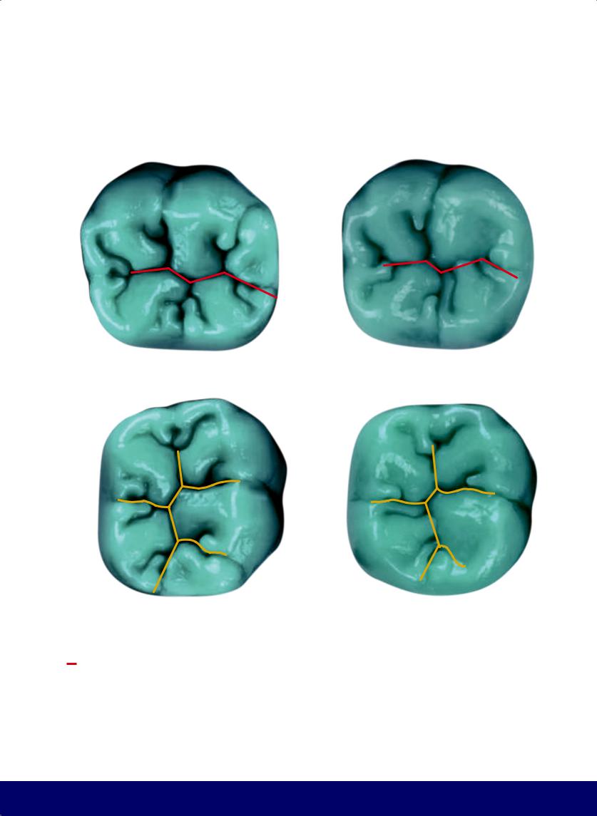
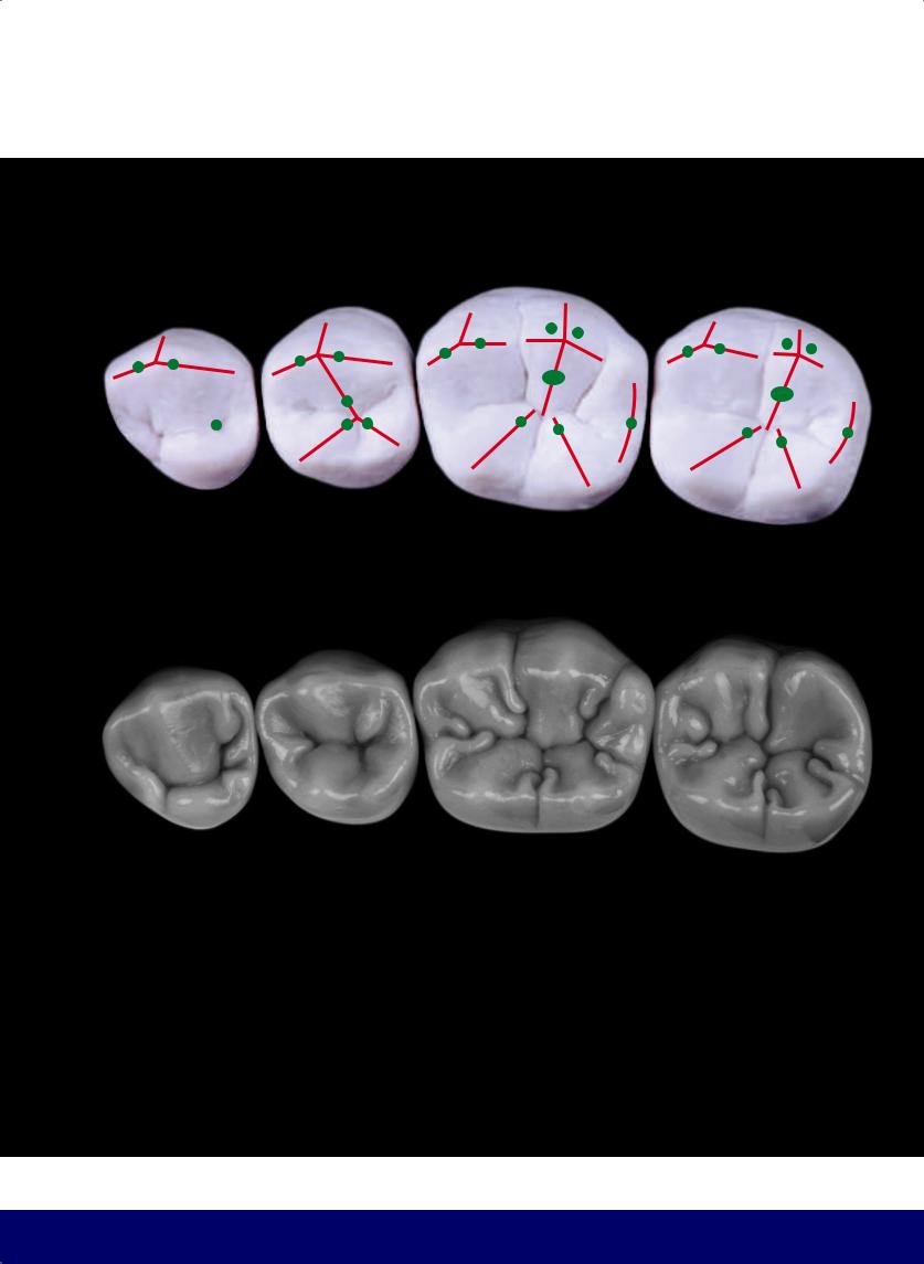
ANALYSIS OF THE OCCLUSAL SURFACE OF THE MAXILLARY PREMOLARS
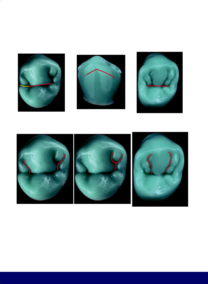
The mesiodistal groove of the maxillary premolar is a line that bends at the apex, and in the first premolar, the mesial marginal ridge is always broken by an extension off of the mesiodistal groove. In maxillary premolars, the mesial and distal secondary grooves resemble the letter S and an inverted S. In the first premolar, they start in the mesial
and distal fossae and proceed toward the corner of the buccal ridge, looking like two outward-spread Zebu horns. From the distal S, another secondary groove forms the shape of a wineglass of varying size. In the second premolar, the secondary grooves begin in the mesiodistal groove, closer to each other, and curve up toward the tip of
38
the buccal cusp, looking like inward-curving Zebu horns. It is common to have two other grooves between these and the marginal ridges, forming two lobes.
@dentistinfo стоматологический телеграм канал
THE ESTHETIC AND FUNCTIONAL PARAMETERS OF POSTERIOR TEETH
Hands on hips
VISUAL GUIDE FOR THE BUCCAL SHAPE OF MAXILLARY PREMOLARS
Developmental grooves
In the maxillary first premolar, the most common developmental groove is the mesial, while in the maxillary second premolar both the mesial and the distal grooves are present. Thus, the buccal aspect of the maxillary second premolar is more curved than that of the maxillary first premolar.
First premolar
On the occlusal surface of the maxillary first premolar, a depression is visible on the buccal aspect because of the exit of the mesial developmental groove.
Second premolar
On the occlusal surface of the second premolar, two depressions are visible on the buccal aspect from the exits of the mesial and distal developmental grooves. The secondary grooves divide the buccal cusp crest into a central lobe and two accessory lobes, resembling a figure with hands on hips.
39
@dentistinfo стоматологический телеграм канал
CHAPTER 1
|
|
|
|
|
|
|
|
|
|
|
|
|
|
|
|
|
|
|
|
|
|
|
|
|
|
|
|
|
|
|
|
|
|
|
|
|
|
|
|
|
|
|
|
|
|
|
|
VISUAL GUIDE FOR THE OCCLUSAL MORPHOLOGY OF |
VISUAL GUIDE FOR THE OCCLUSAL MORPHOLOGY OF |
|
|||||
MAXILLARY MOLARS |
MANDIBULAR PREMOLARS |
|
|||||
|
Mercedes emblem |
|
Kidney bean |
|
|||
|
|
|
|||||
|
Seagull wingspan |
|
Wineglass/outward-spread Zebu horns |
|
|||
|
|
|
|||||
|
Hands on hips |
|
Smile of the mesiodistal groove |
|
|||
|
|
|
|||||
|
Water droplet |
|
Water droplet |
|
|||
|
|
|
|||||
|
|
|
|
|
Letter Y |
|
|
|
|
|
|
|
|
||
40
@dentistinfo стоматологический телеграм канал
THE ESTHETIC AND FUNCTIONAL PARAMETERS OF POSTERIOR TEETH
VISUAL GUIDE FOR THE OCCLUSAL MORPHOLOGY OF
MANDIBULAR MOLARS
Letter M
 Running stick figure (from Paulo Kano)
41
@dentistinfo стоматологический телеграм канал
CHAPTER 1
VISUAL GUIDE FOR THE BUCCAL SURFACES OF MANDIBULAR POSTERIOR TEETH
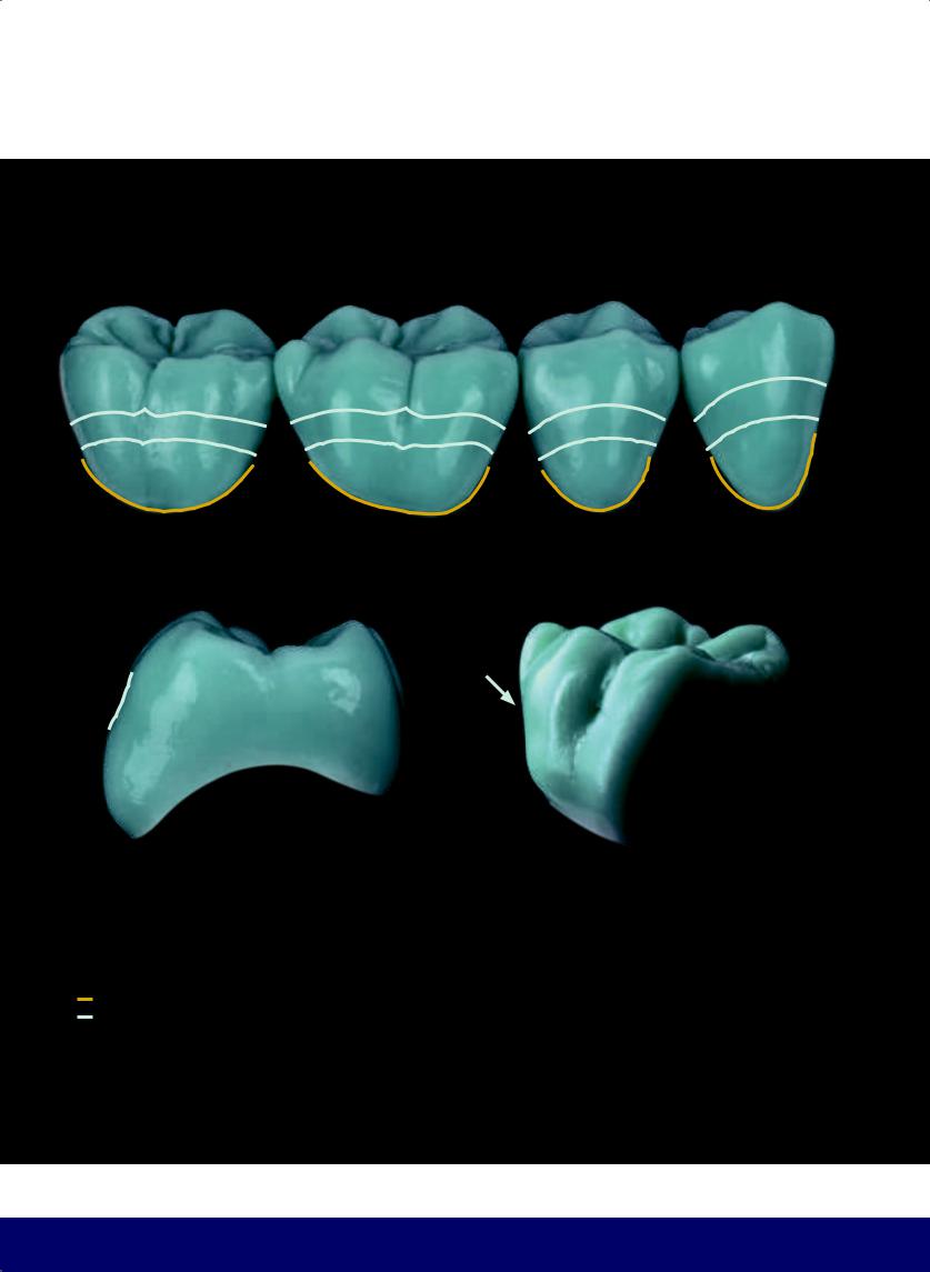
Cementoenamel junction (CEJ).
The buccal central concavity follows a course opposite that of the CEJ.
42
@dentistinfo стоматологический телеграм канал
THE ESTHETIC AND FUNCTIONAL PARAMETERS OF POSTERIOR TEETH
VISUAL GUIDE FOR MANDIBULAR POSTERIOR TEETH
The profile of the transverse ridges resembles curly brackets. The buccal aspect exhibits a narrowed waist, and the junction of the mesiobuccal groove with the horizontal concavity forms the buccal fossa, which resembles a double hook.
43
@dentistinfo стоматологический телеграм канал
CHAPTER 1
Barrel chested
Narrow waisted
44
@dentistinfo стоматологический телеграм канал
THE ESTHETIC AND FUNCTIONAL PARAMETERS OF POSTERIOR TEETH
Barrel chested
Narrow waisted
45
@dentistinfo стоматологический телеграм канал
CHAPTER 1
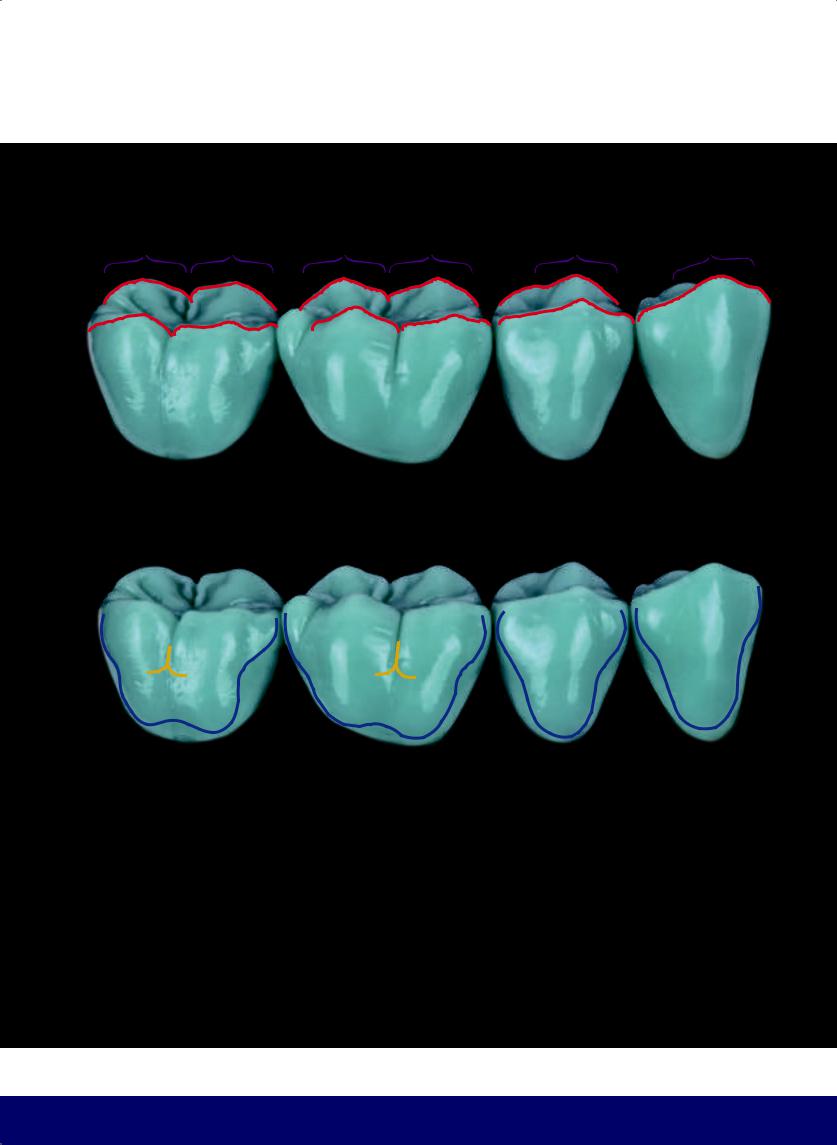
VISUAL GUIDE FOR THE BUCCAL ASPECT OF POSTERIOR TEETH
Curly bracket profile
Developmental grooves
Corseted waist
Buccal fossa
Horizontal texturization on the surface
46
@dentistinfo стоматологический телеграм канал
THE ESTHETIC AND FUNCTIONAL PARAMETERS OF POSTERIOR TEETH
47
@dentistinfo стоматологический телеграм канал
CHAPTER 1
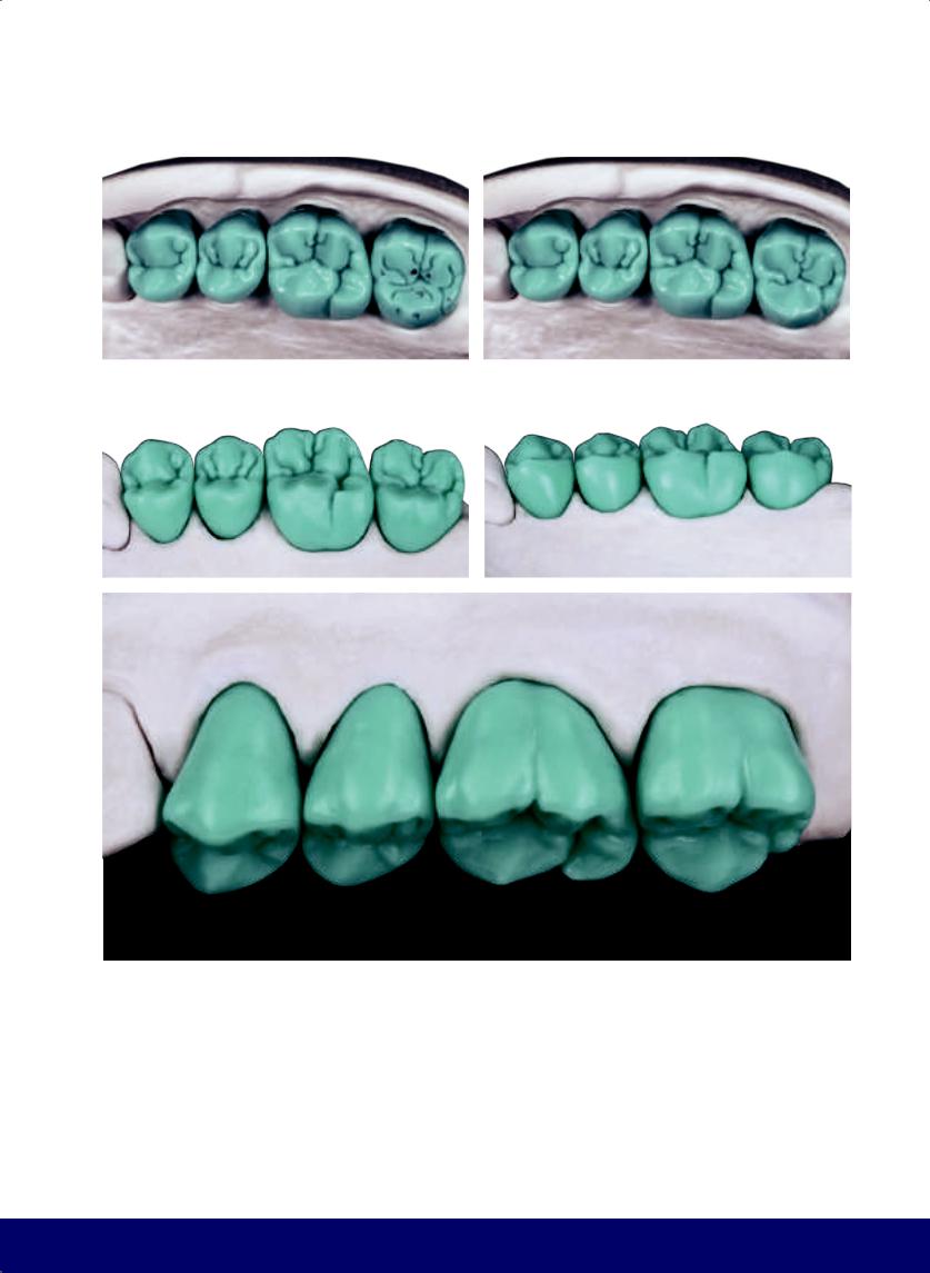
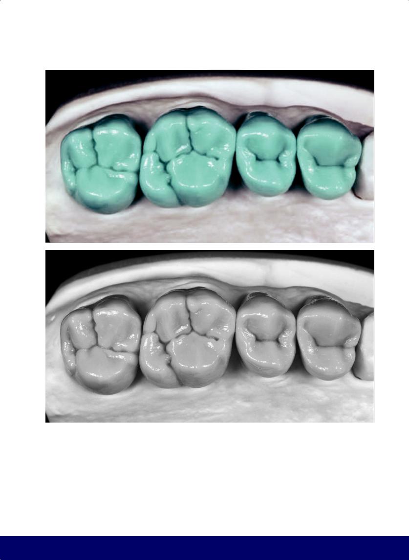
 CONTACT POINTS IN CENTRIC OCCLUSION
ONTRANSVERSERIDGES,CUSPCRESTS,ANDCUSPSLOPES
48
@dentistinfo стоматологический телеграм канал
THE ESTHETIC AND FUNCTIONAL PARAMETERS OF POSTERIOR TEETH
49
@dentistinfo стоматологический телеграм канал
CHAPTER 1
|
|
|
|
11A |
12A |
|
|
6A |
|
|
|
2A |
3A |
7A |
|
|
|
|
9A |
|
|
||
4A |
|
10A |
|
||
|
|
|
|||
|
5A |
|
|
||
|
|
|
|
13A |
|
1A |
|
8A |
|
|
|
|
|
|
|
|
2B |
|
3B |
5B |
7B |
6B |
8B |
9B |
11B |
10B |
12B |
1B |
4B |
|
|||||||||
|
|
|
|
|
1A |
|
|
|
|
|
|
|
13A |
2A |
3A |
|
8A |
|
9A |
|
|
|
|
4A |
|
10A |
12A |
||||
|
|
|
|
|
|
|||
|
|
|
5A |
|
7A |
|
11A |
|
|
|
|
6A |
|
|
|||
|
|
|
|
|
|
|
||
|
|
|
|
|
|
|
|
50
@dentistinfo стоматологический телеграм канал

THE ESTHETIC AND FUNCTIONAL PARAMETERS OF POSTERIOR TEETH
Contact points generated by mandibular teeth on maxillary teeth (Angle Class I)
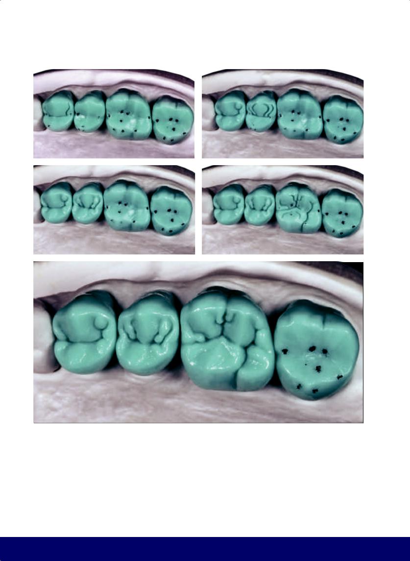
1A: Points of contact on the distal transverse ridge of the buccal cusp of the mandibular first premolar and the mesial marginal ridge of the maxillary first premolar.
2A: Point of contact where the mesial transvers ridge of the buccal cusp of the mandibular second premolar touches the distal marginal ridge of the maxillary first premolar.
3A: Point of contact where the distal transverse ridge of the buccal cusp of the mandibular second premolar touches the distal marginal ridge of the maxillary second premolar.
4A: Point of contact where the mesial transvers ridge of the mesiodistal cusp of the mandibular first molar touches the distal marginal ridge of the maxillary second premolar.
5A: Point of contact where the distal segment of the transvers ridge of the mesiobuccal cusp of the mandibular first molar touches the mesial marginal ridge of the maxillary first molar.
6A: Point of contact where the mesial slope of the medial cusp of the mandibular first molar touches the crest ridge of the mesiobuccal cusp, near the central fossa, of the maxillary first molar.
7A: Point of contact where the distal slope of the medial cusp of the mandibular first molar touches the crest ridge of the distobuccal cusp, near the central fossa, of the maxillary first molar.
8A: Point of contact where the middle of the crest ridge of the medial cusp of the mandibular first molar touches the middle of the crest ridge of the mesiolingual cusp of the maxillary first molar.
9A: Point of contact where the mesial transverse ridge of the mesiobuccal cusp of the mandibular second molar touches the distal marginal ridge of the maxillary first molar.
10A: Point of contact where the distal transverse ridge of the mesiobuccal cusp of the mandibular second molar touches the mesial marginal ridge of the maxilllary second molar.
11A: Point of contact where the mesial slope of the distobuccal cusp of the mandibular second molar touches the crest ridge of the mesiobuccal cusp, near the central fossa, of the maxillary second molar.
12A: Point of contact where the distal slope of the distobuccal cusp of the mandibular second molar touches the crest ridge of the distobuccal cusp, near the central fossa, of the maxillary second molar.
13A: Point of contact where the middle of the crest ridge of the distobuccal cusp of the mandibular second molar touches the middle of the crest ridge of the mesiolingual cusp of the maxillary second molar.
Contact points generated by maxillary teeth on mandibular teeth (Angle Class I)
1B: Point of contact where the tip of the lingual cusp of the maxillary first premolar touches the distal fossa of the mandibular first premolar.
2B: Point of contact where the mesial slope of the lingual cusp of the maxillary second premolar touches the crest ridge of the mesiolingual cusp, near the distal fossa, of the mandibular second premolar.
3B: Point of contact where the distal slope of the lingual cusp of the maxillary second premolar touches the crest ridge of the distolingual cusp, near the distal fossa, of the mandibular second premolar.
4B: Point of contact wher the crest ridge of the lingual cusp of the maxillary second premolar touches the crest ridge of the distolingual cusp, near the distal fossa, of the mandibular second premolar.
5B: Point of contact where the mesial slope of the mesiolingual cusp of the maxillary first molar touches the crest ridge of the mesiolingual cusp, near the distal fossa, of the mandibular first molar.
6B: Point of contact where the distal slope of the mesiolingual cusp of the maxillary first molar touches the crest ridge of the distolingual cusp, near the distal fossa, of the mandibular first molar.
7B: Point of contact where the middle of the crest ridge of the mesiolingual cusp of the maxillary first molar touches the middle of the crest ridge of the medial cusp of the mandibular first molar.
8B: Point of contact where the tip of the distolingual cusp of the maxillary first molar touches the distal marginal ridge of the mandibular first molar.
9B: Point of contact where the mesial slope of the mesiolingual cusp of the maxillary second molar touches the crest ridge of the mesiolingual cusp, near the distal fossa, of the mandibular second molar.
10B: Point of contact where the distal slope of the mesiolingual cusp of the maxillary second molar touches the crest ridge of the distolingual cusp, near the distal fossa, of the mandibular second molar.
11B: Point of contact where the middle of the crest ridge of the mesiolingual cusp of the maxillary second molar touches the middle of the crest ridge of the distobuccal cusp of the mandibular second molar.
12B: Point of contact where the tip of the distolingual cusp of the maxillary second molar touches the distal marginal ridge of the mandibular second molar.
51
@dentistinfo стоматологический телеграм канал

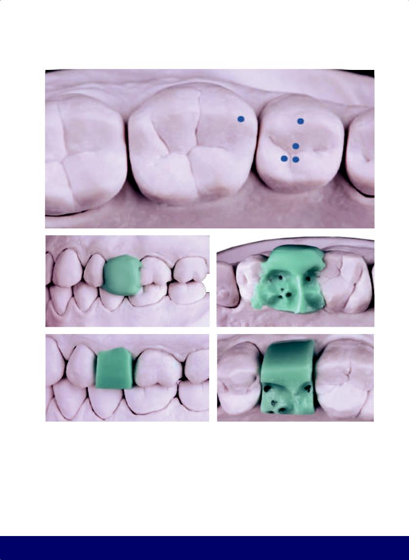
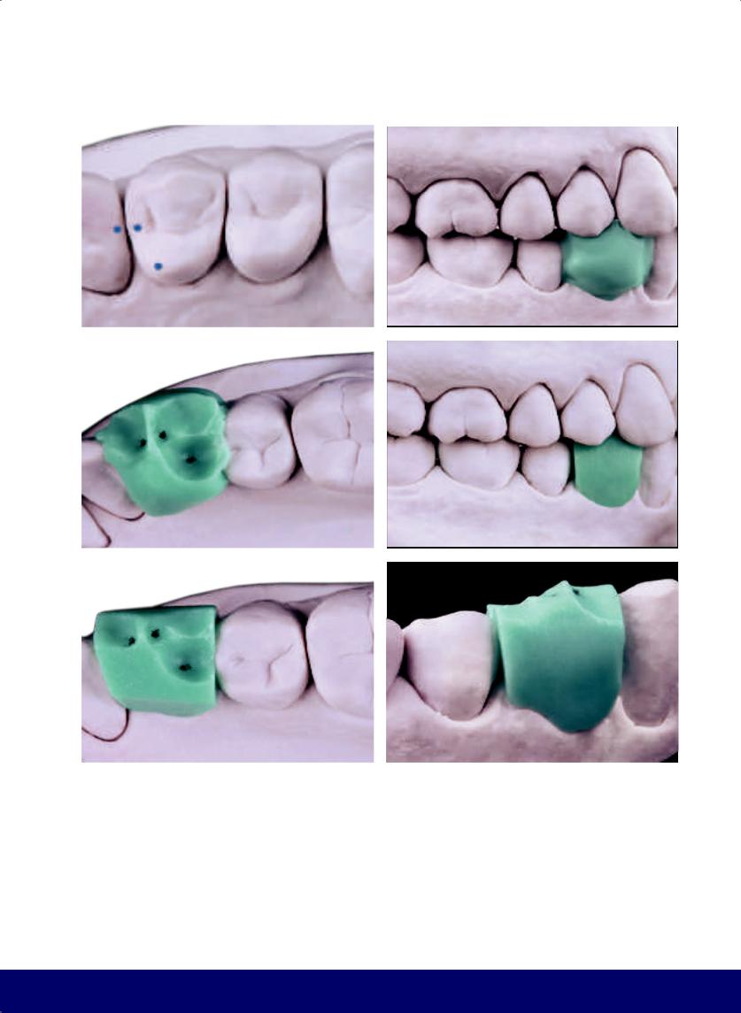
First Premolar
Maxillary
02
52
@dentistinfo стоматологический телеграм канал
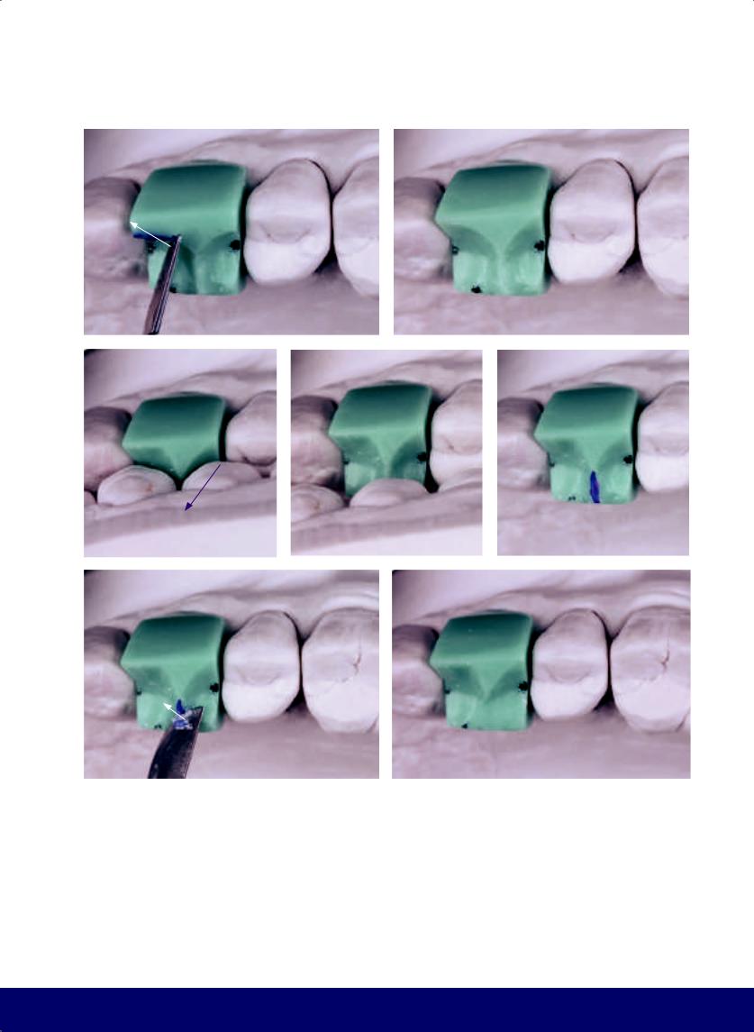
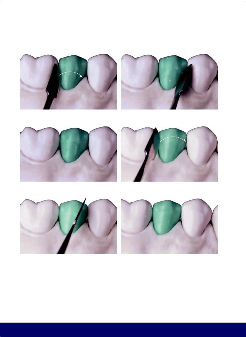
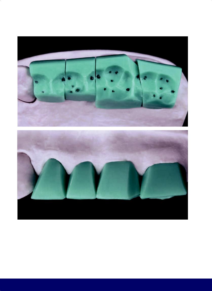
MAXILLARY FIRST PREMOLAR
a
b |
|
c |
|
d |
|
|
|
|
|
e |
|
f |
|
g |
|
|
|
|
|
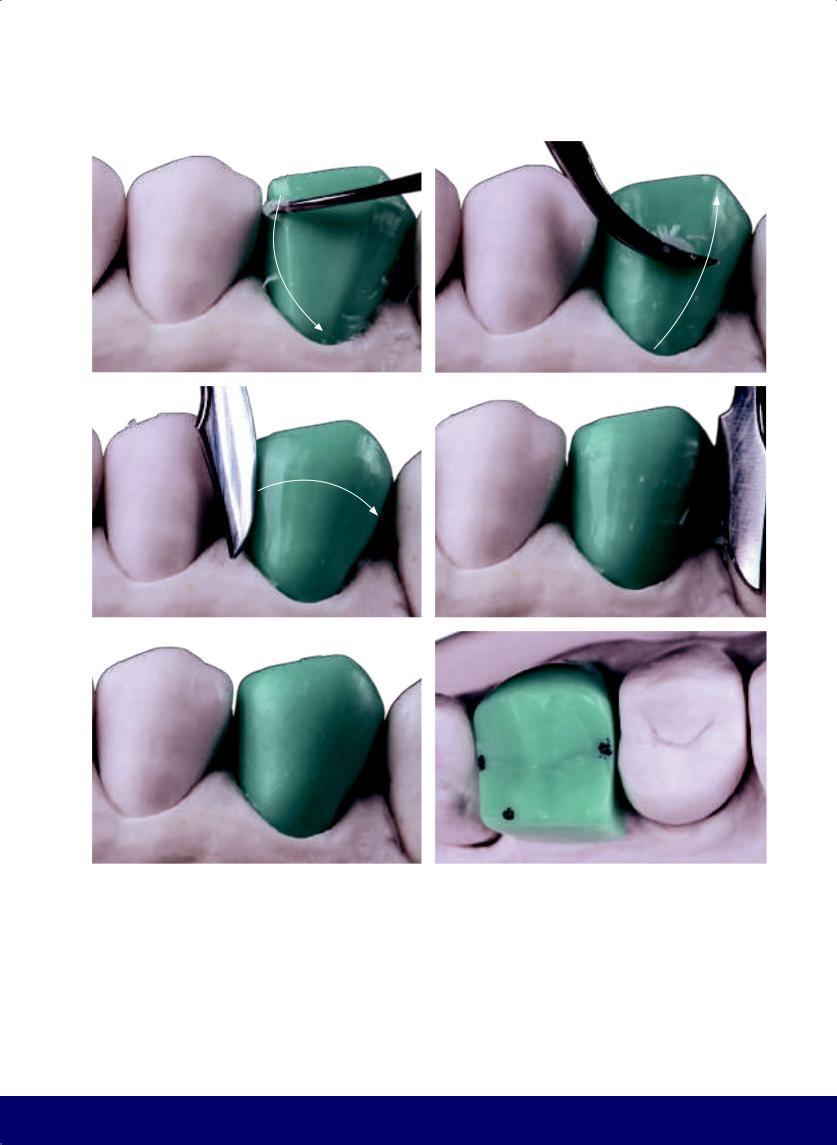
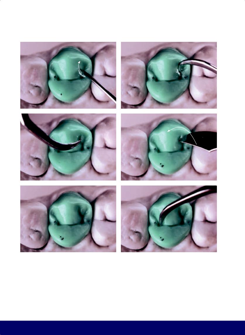
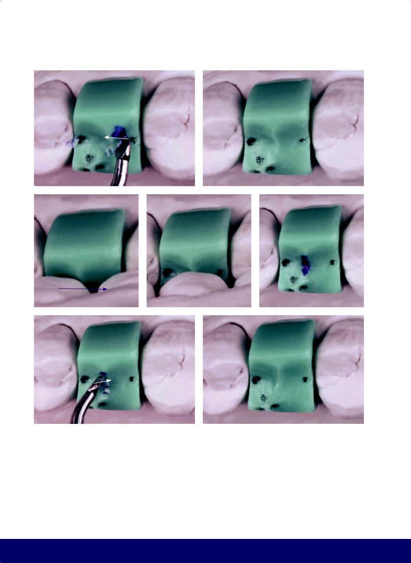
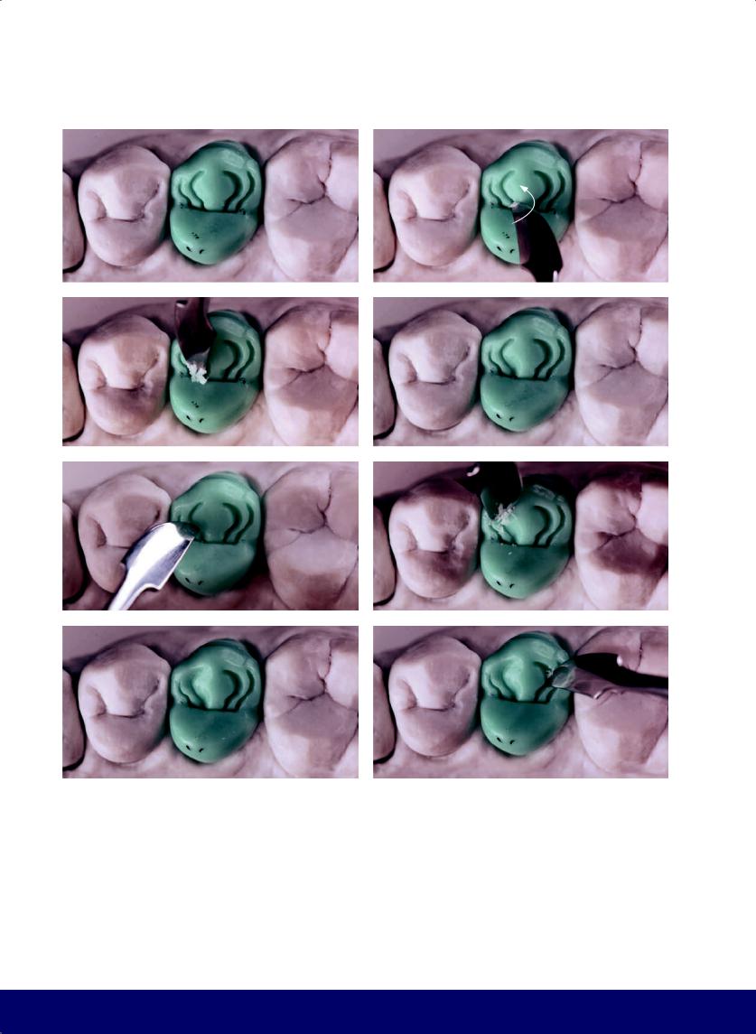
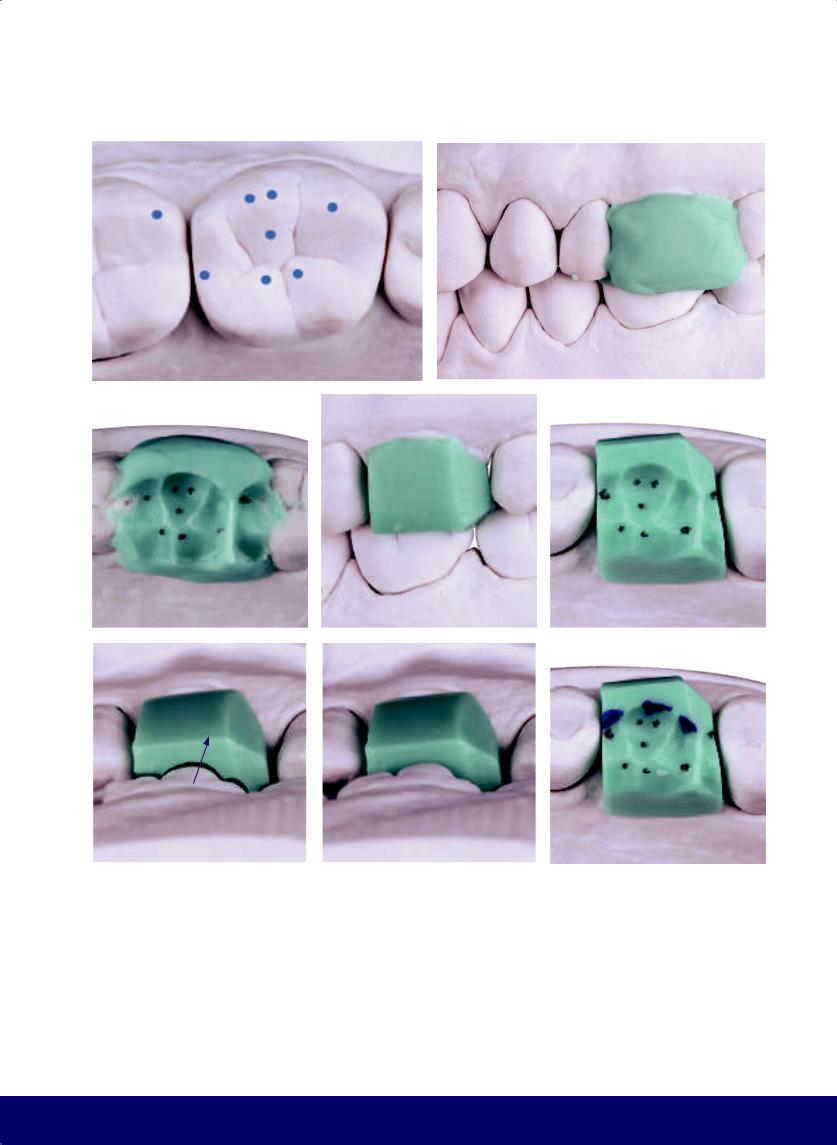
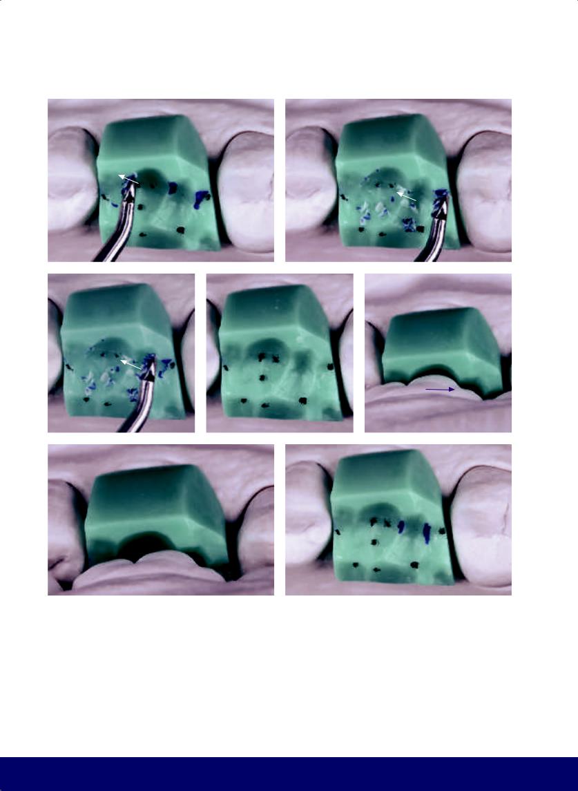
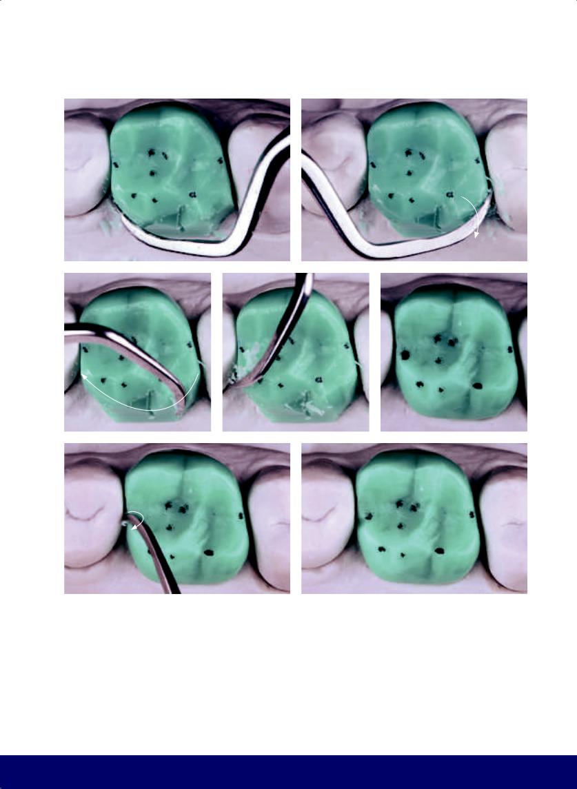
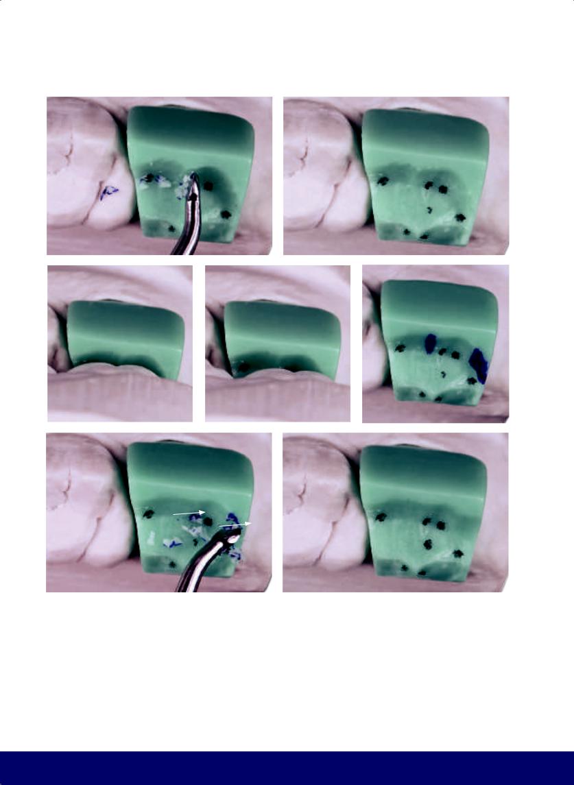
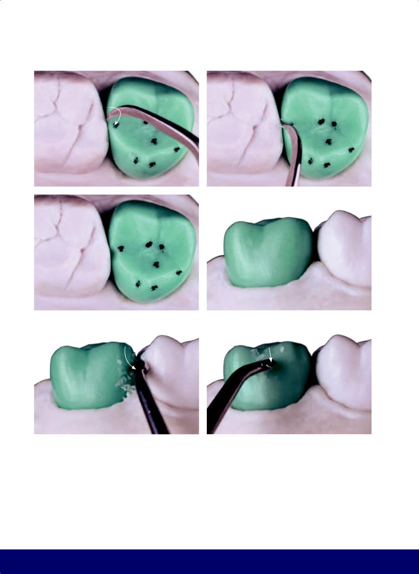
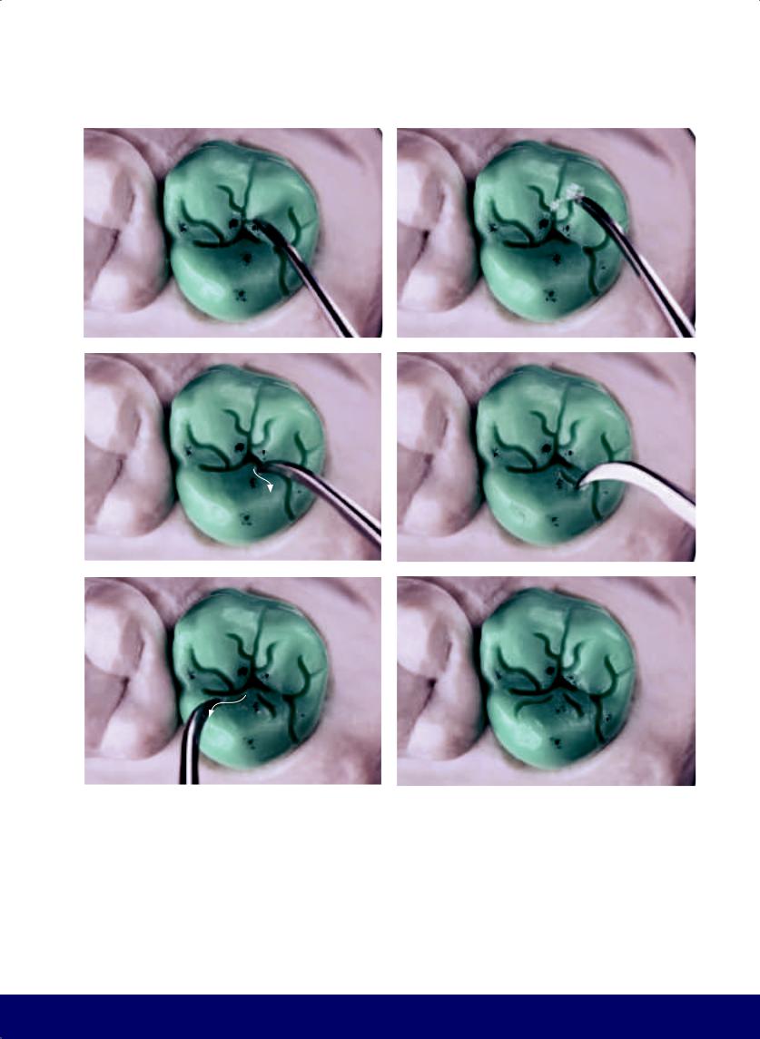
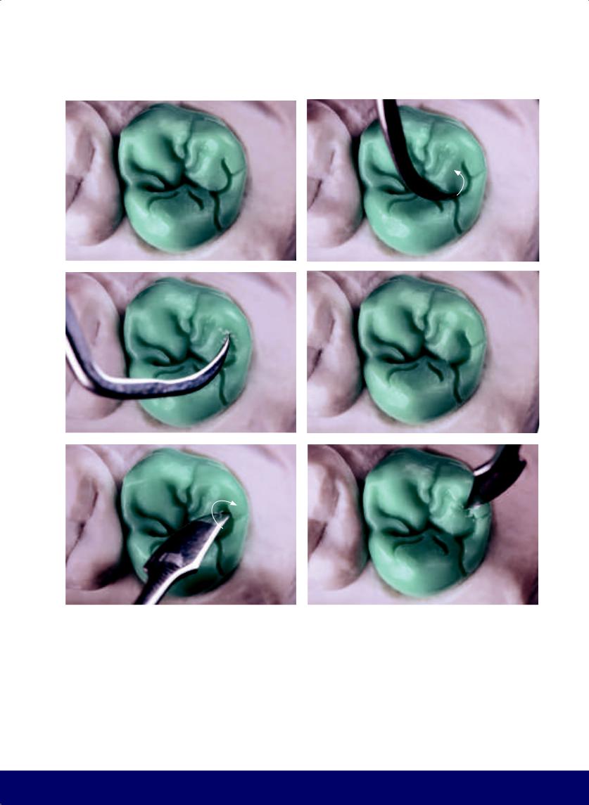
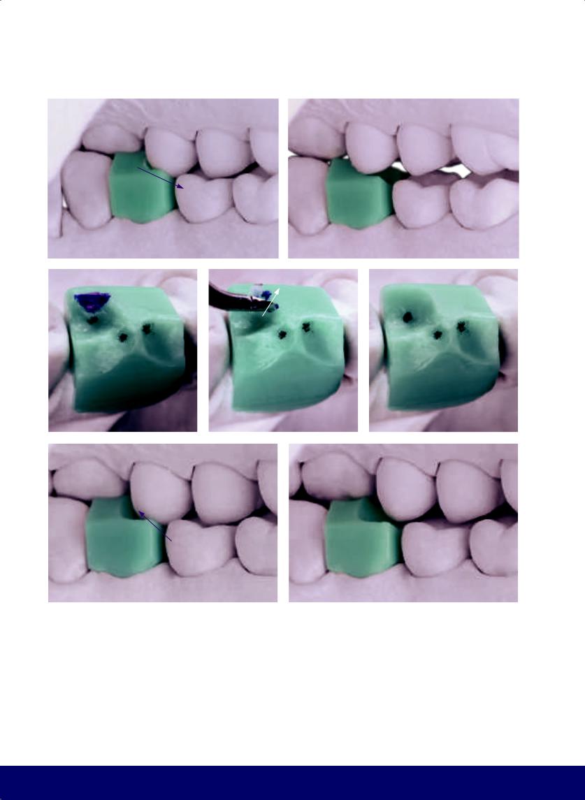
Fig 2-1 | (a) Contact points in the mandibular arch to be reproduced on the wax-up. (b to d) With the heated instrument, the tooth peg is waxed. (e to g) After excess wax is placed over the buccal, lingual, and occlusal surfaces, the most superficial layer of wax is heated. Note the loss of brightness that indicates the ideal point for shaping.
53
@dentistinfo стоматологический телеграм канал
CHAPTER 2
a |
b |
|
|
c |
d |
|
|
e |
f |
g |
|
|
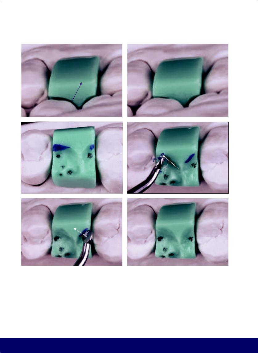
|
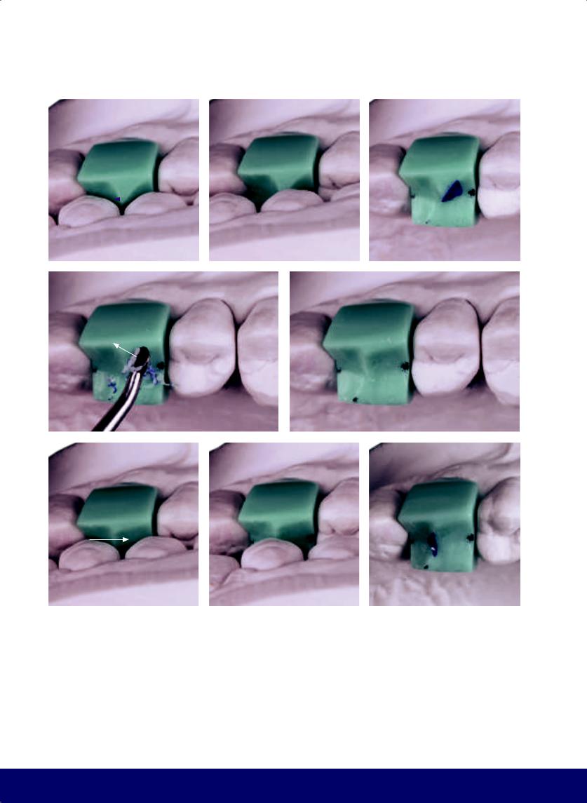
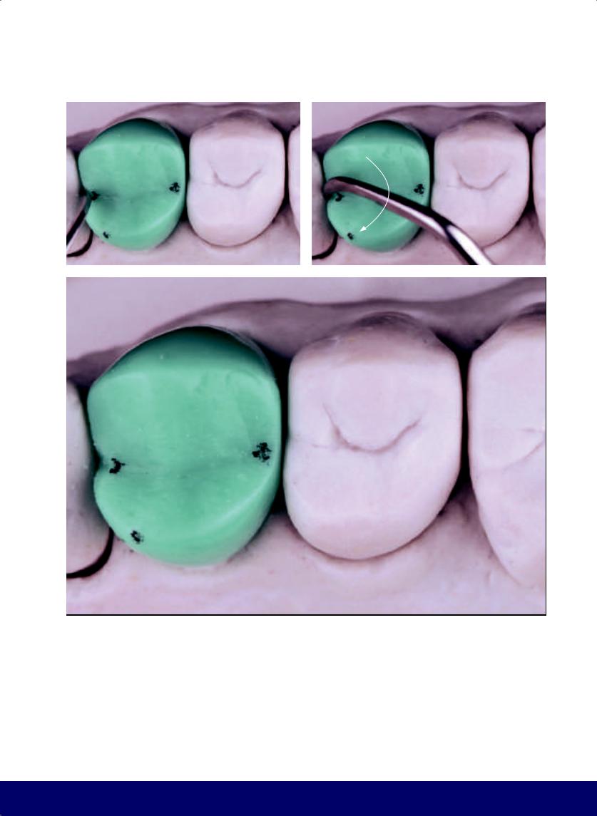
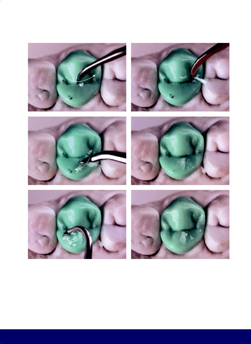
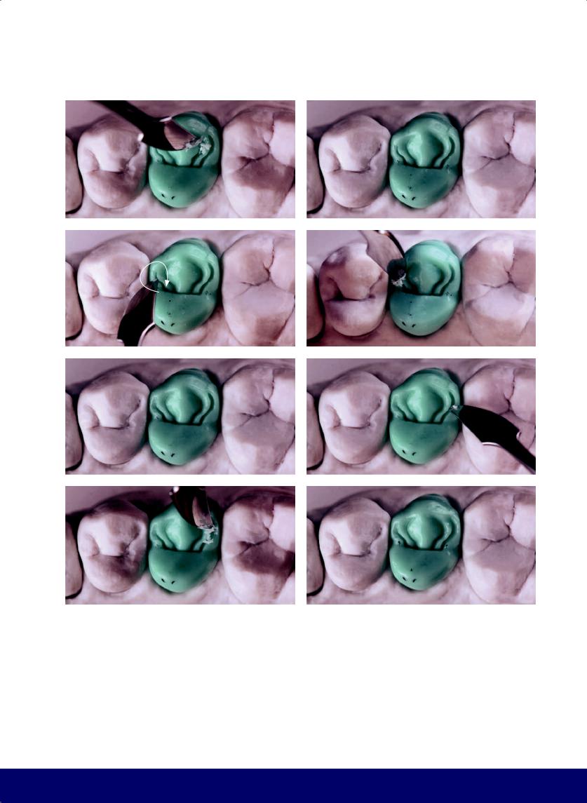
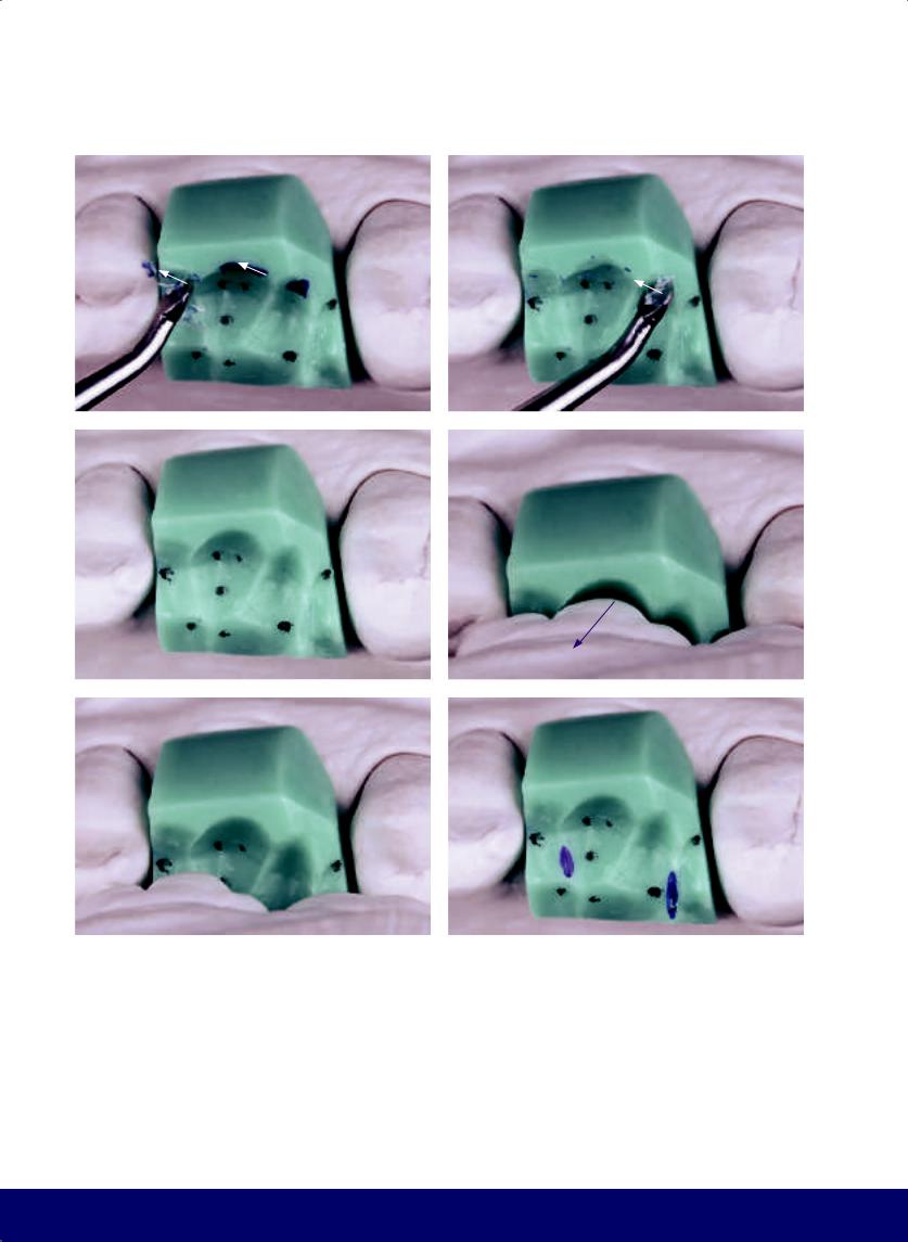
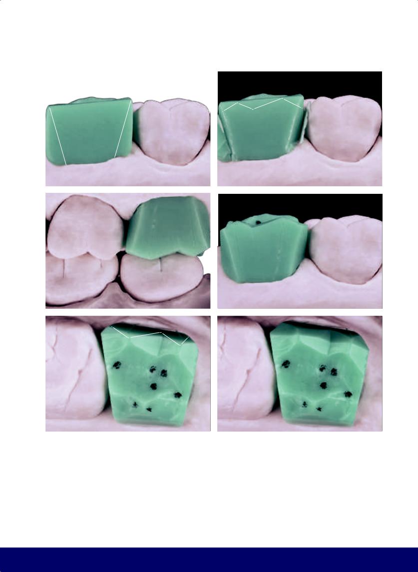
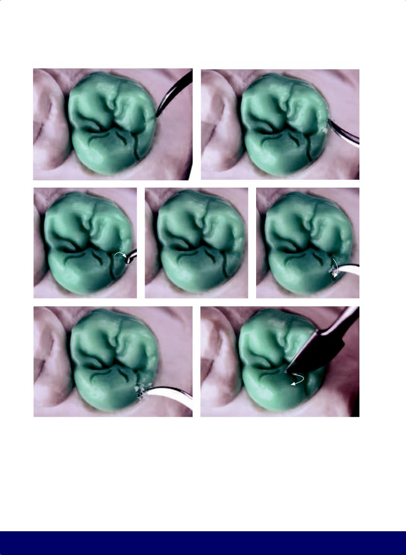
Fig 2-2 | (a) Buccal view showing the cast in occlusion. The softened wax records the occlusal morphology of the opposing mandibular teeth. (b) In the occlusal view, note that the contact points that were marked in pencil on the opposing teeth have also been reproduced on the wax. (c and d) Occlusal and buccal views after removing excess wax. (e to g) Buccal interference in working movement. (Purple arrows indicate the movement of the mandible.)
54
@dentistinfo стоматологический телеграм канал
a |
b |
c |
d |
f |
g |
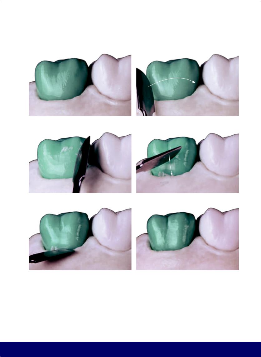
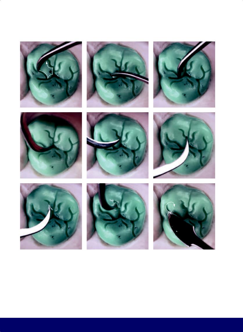
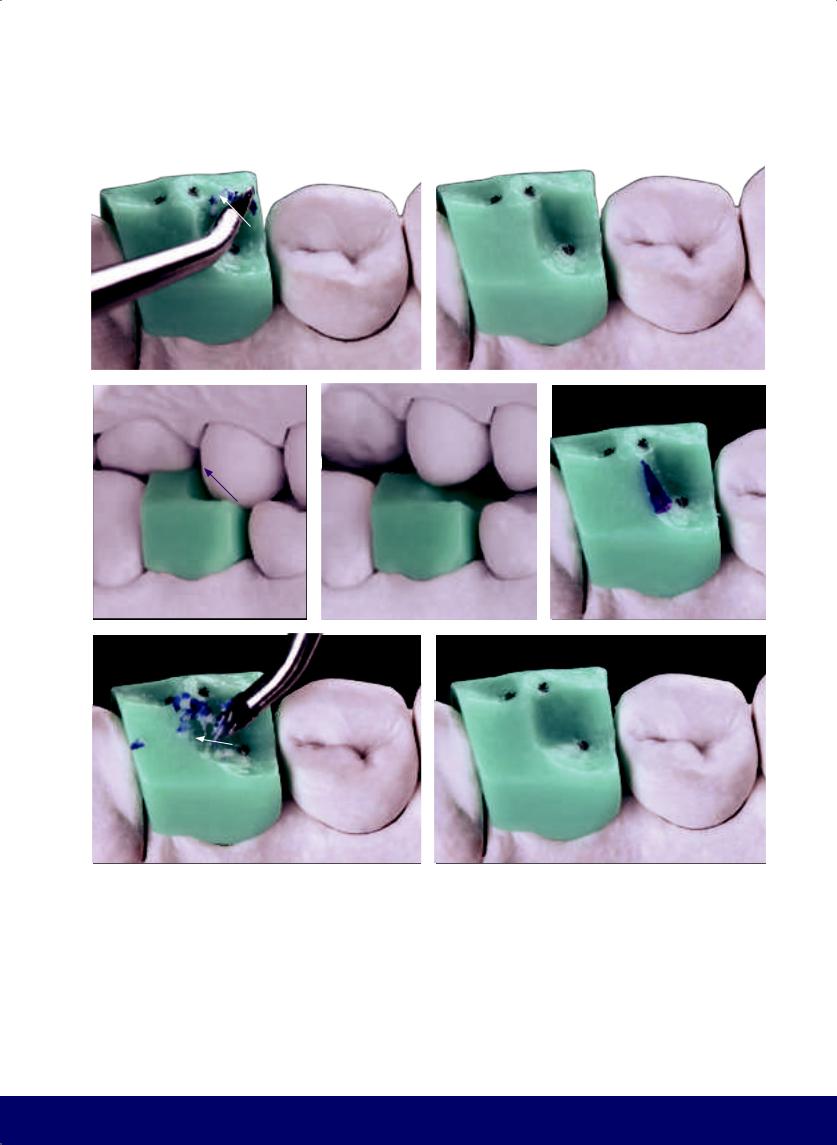
Fig 2-3 | (a and b) Carve away the buccal interference. (c to e) Lingual interference in balancing movement. (f and g) Carve away the lingual interference. (White arrows indicate the direction of instrumentation.)
MAXILLARY FIRST PREMOLAR
e
55
@dentistinfo стоматологический телеграм канал
CHAPTER 2
|
|
|
|
|
a |
|
b |
|
c |
|
|
|
|
|
d |
|
e |
|
|
|
f |
|
g |
|
h |
|
|
|
|
|
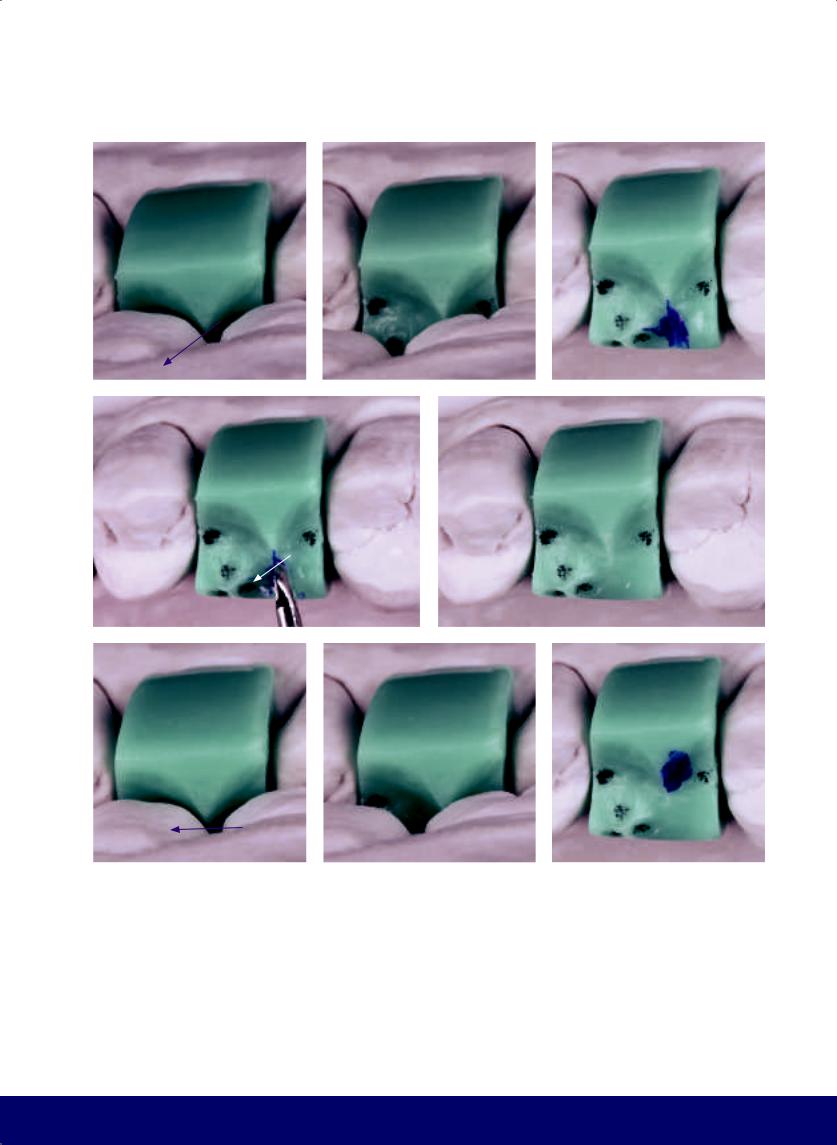
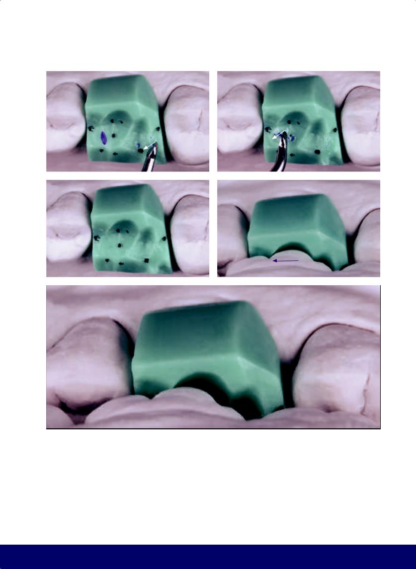
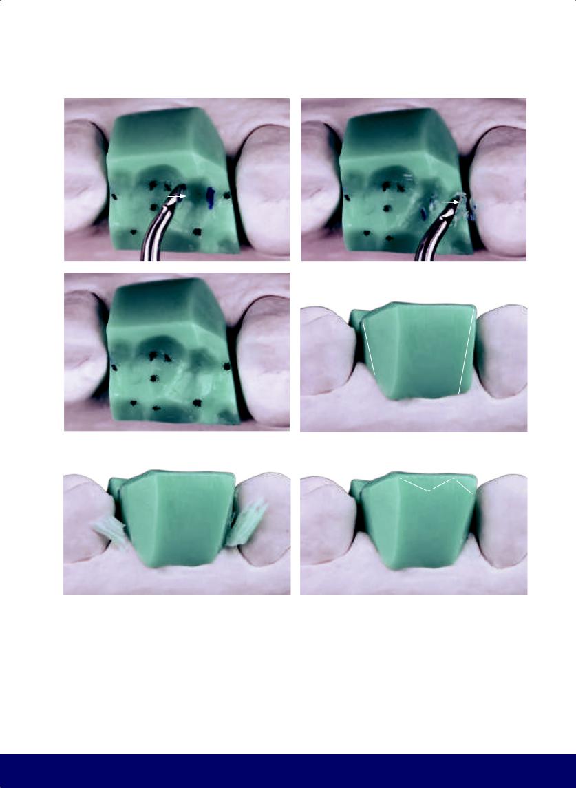
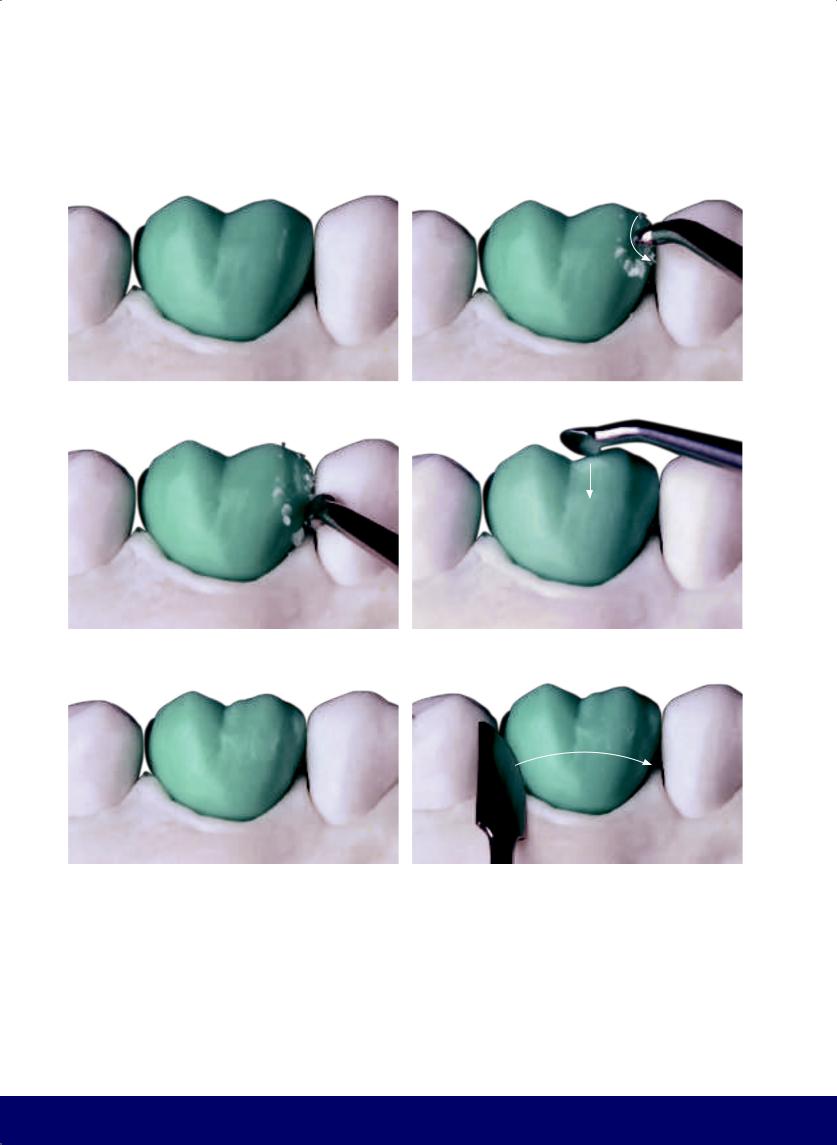
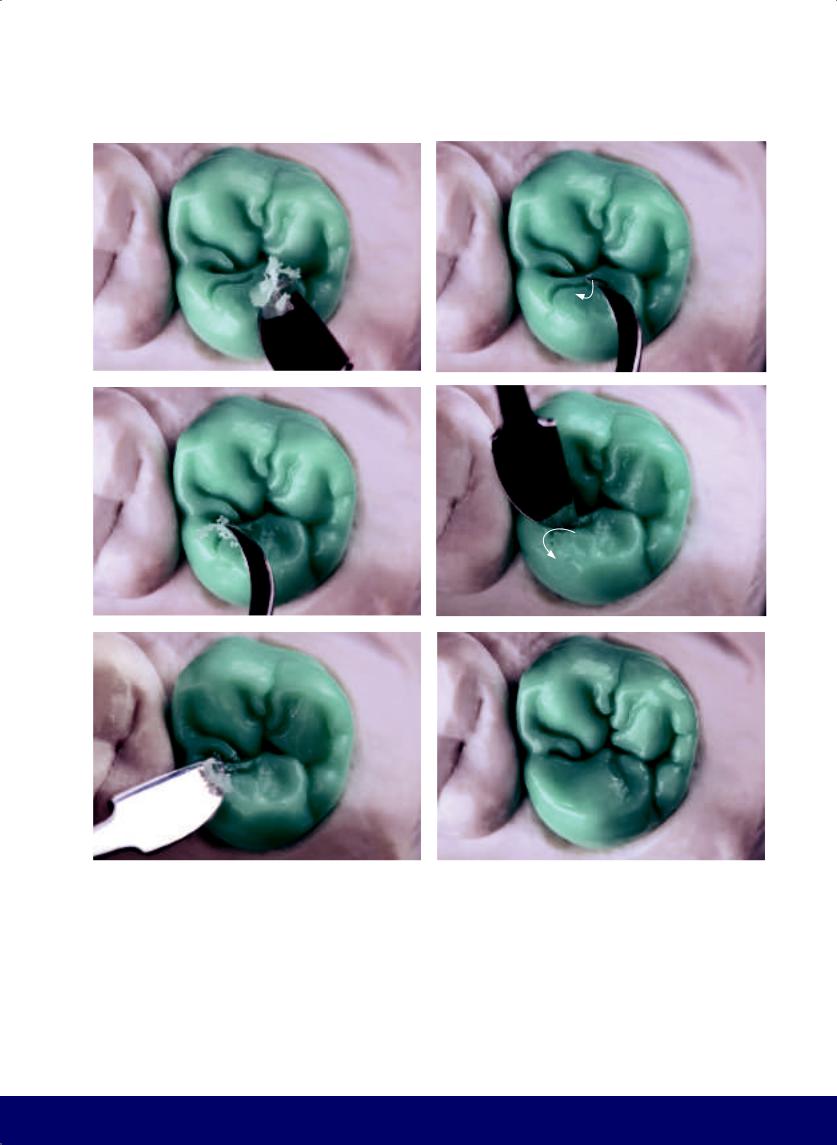
Fig 2-4 | (a to c) Interference in protrusive movement. (d and e) Remove the interference on protrusive movement. (f to h) Interference in retrusive movement.
56
@dentistinfo стоматологический телеграм канал
MAXILLARY FIRST PREMOLAR
a |
|
b |
|
|
|
c |
|
d |
|
e |
|
|
|
|
|
f
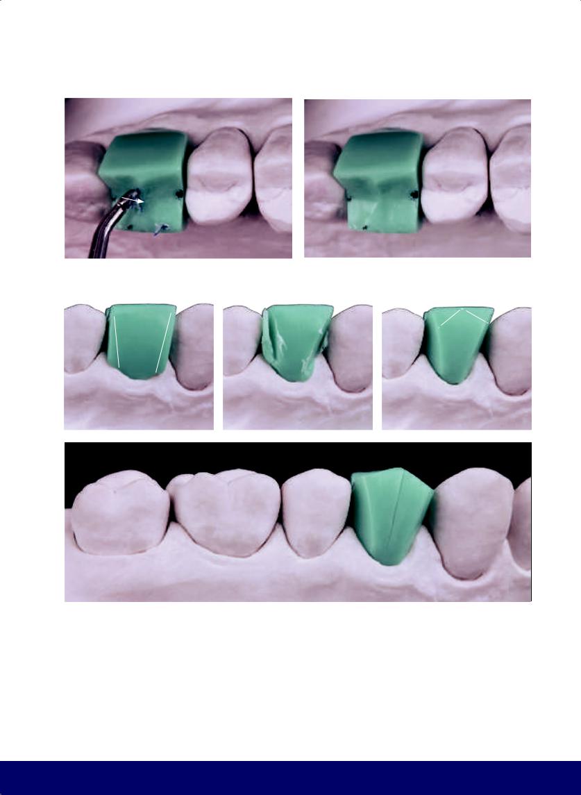
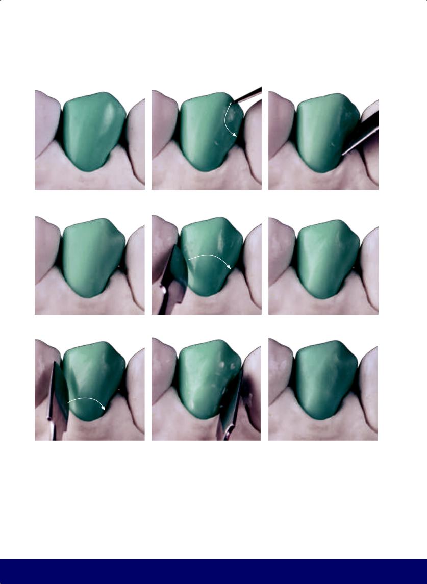
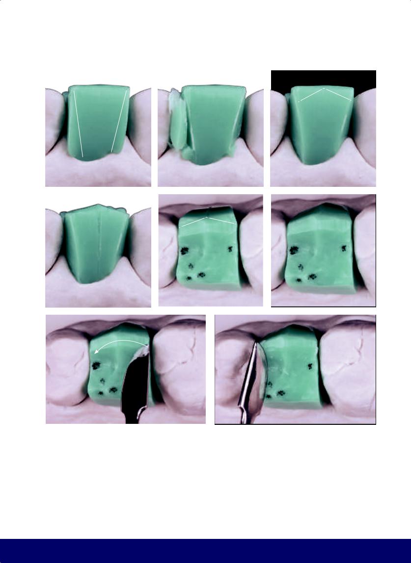
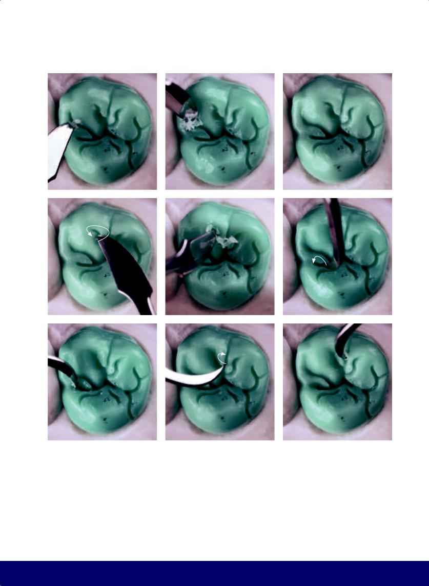
Fig 2-5 | (a and b) Carve away the interference on retrusive movement. (c and d) Cut back wax to open up the buccal embrasures. (e) Form the buccal cusp tip by carving the mesial slope from the distal marginal ridge of the canine to the buccal midline and the distal slop from the midline to the mesial marginal crest of the second premolar. (f) Divide the buccal face into two segments.
57
@dentistinfo стоматологический телеграм канал
CHAPTER 2
a |
|
b |
|
|
|
c |
|
d |
|
e |
|
|
|
|
|
f |
|
g |
|
h |
|
|
|
|
|
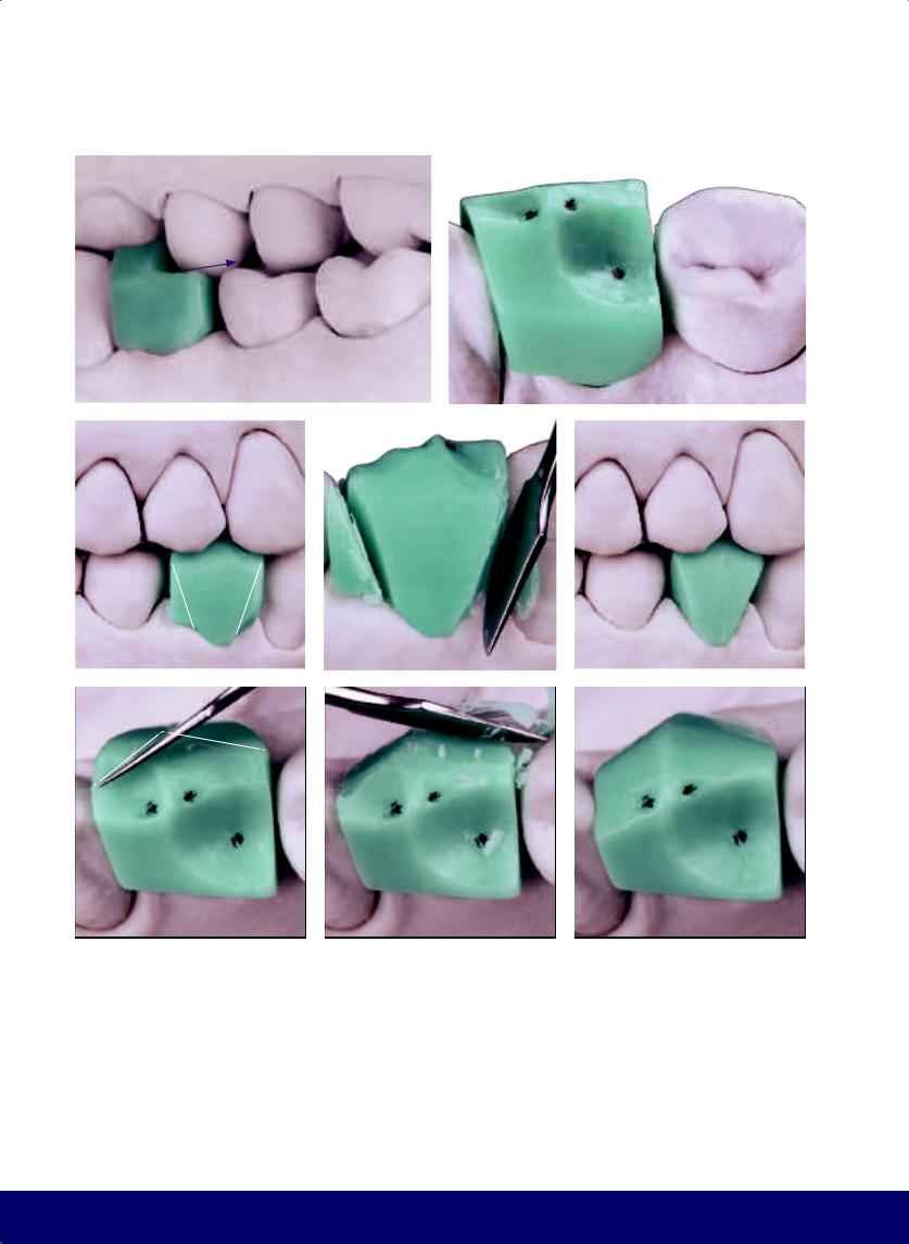
Fig 2-6 | (a and b) Once the mesial plane is carved, the buccal face will have two horizontal slopes: a smaller one mesially and a larger one distally. (c to e) Smooth the angles that resulted from the formation of the occlusal slopes of the buccal cusp tip. (f to m) Finalize the buccal face by rounding it out. (n) Occlusal view.
58
@dentistinfo стоматологический телеграм канал
MAXILLARY FIRST PREMOLAR
i |
|
j |
|
|
|
k |
|
l |
|
|
|
m |
|
n |
|
|
|
59
@dentistinfo стоматологический телеграм канал
CHAPTER 2
a |
|
b |
|
|
|
c |
|
d |
|
|
|
e |
|
f |
|
|
|
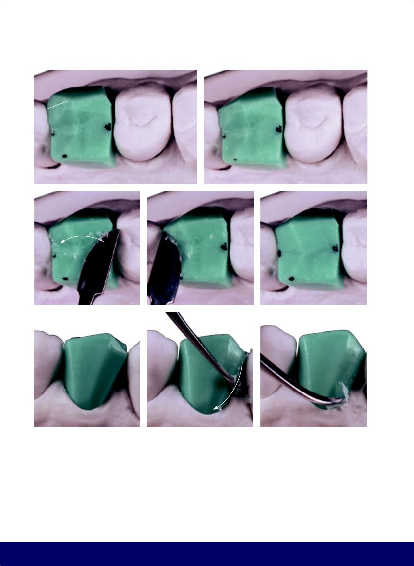
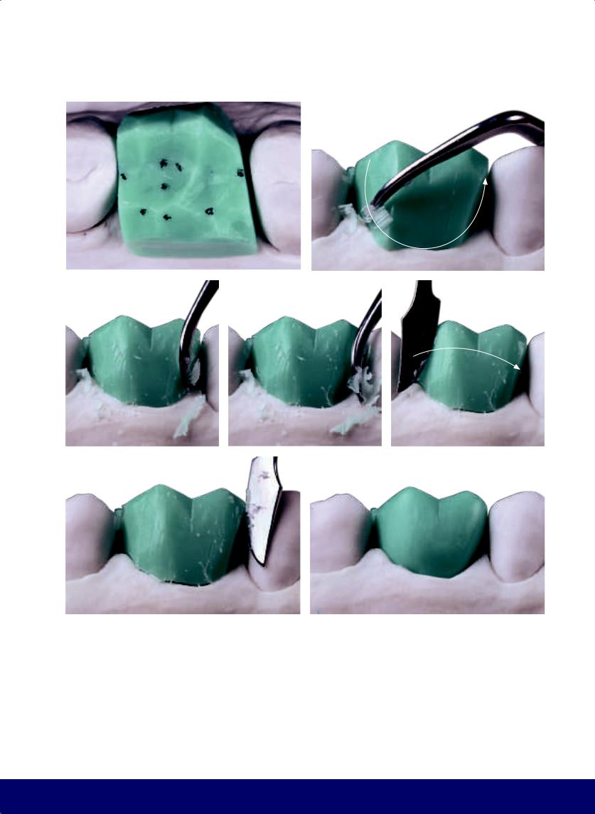
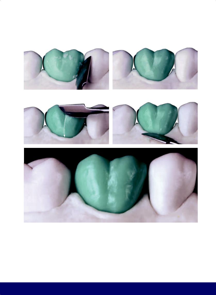
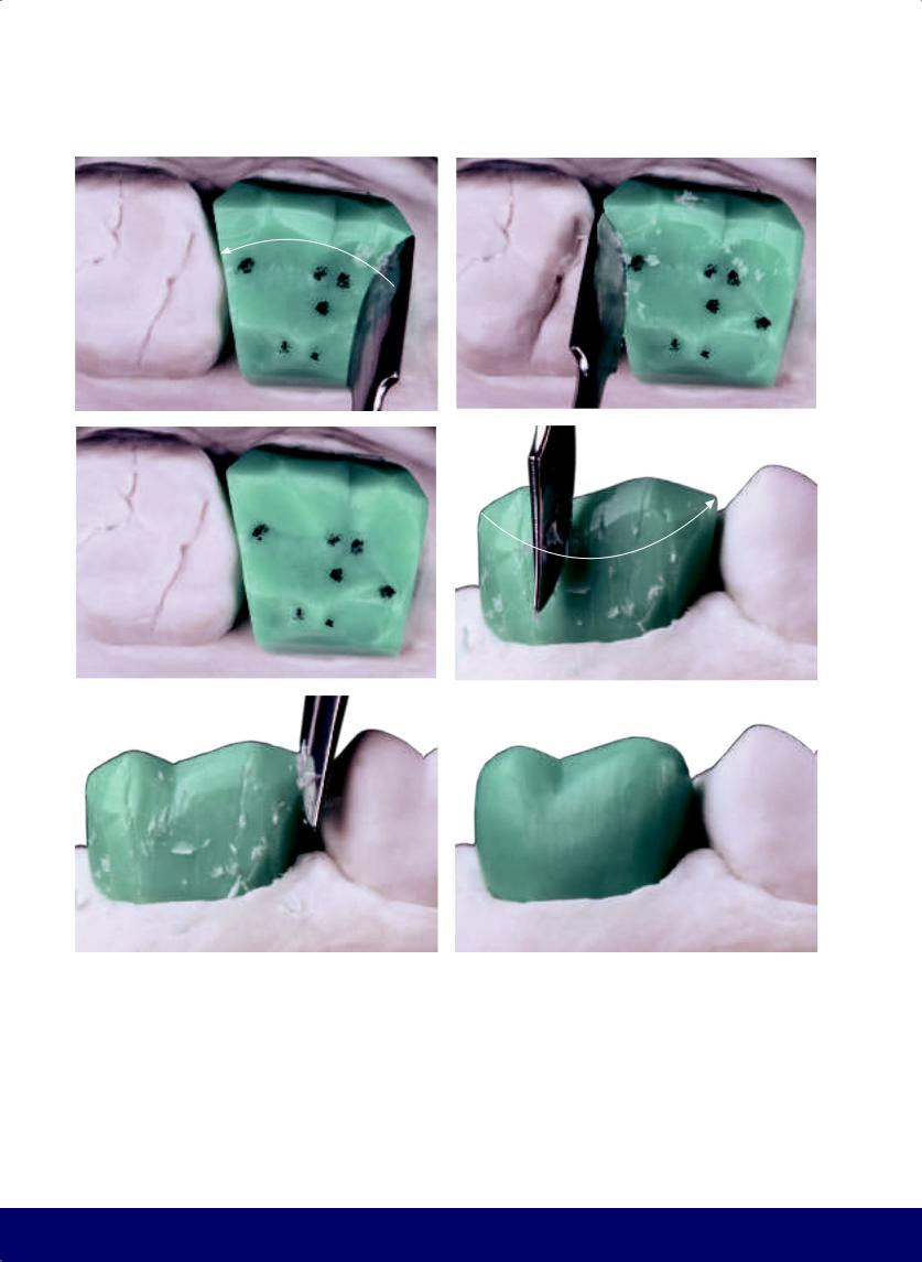
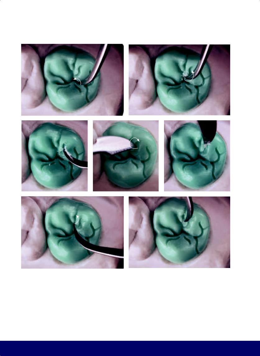
Fig 2-7 | (a and b) Opening the lingual embrasures. (c and d) Finalize the lingual face by breaking the angles and rounding it out. Note the tip of the lingual cusp is directed toward the mesial. (e and f) Remove the die and finish the cervical area.
60
@dentistinfo стоматологический телеграм канал
MAXILLARY FIRST PREMOLAR
a |
|
b |
|
|
|
c
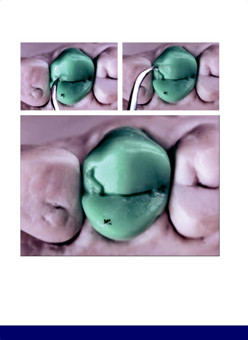
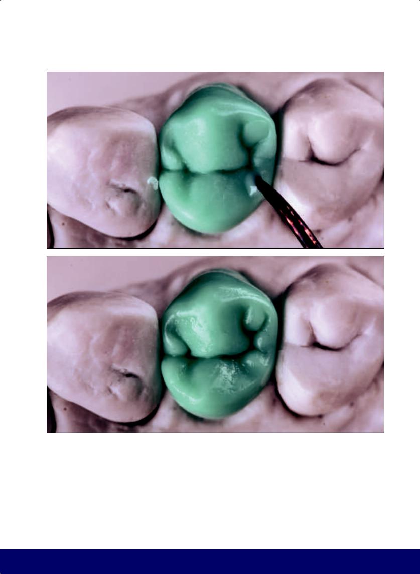
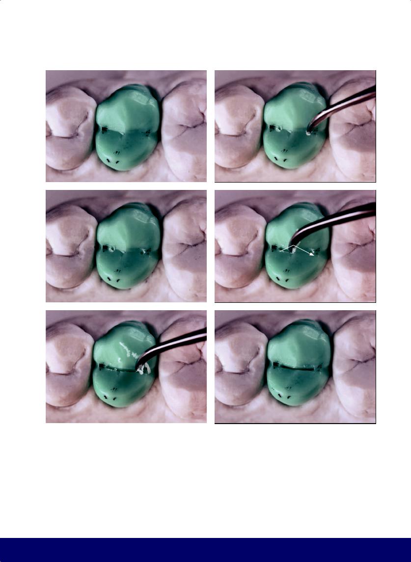
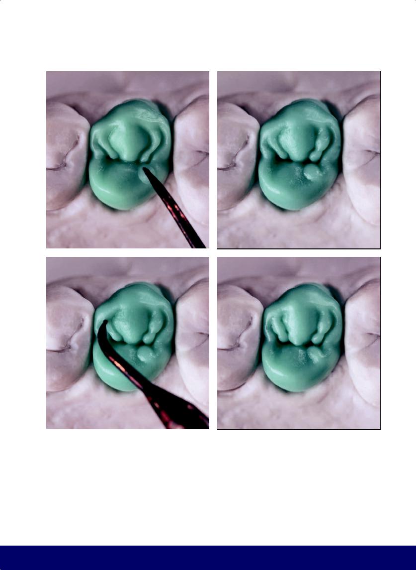
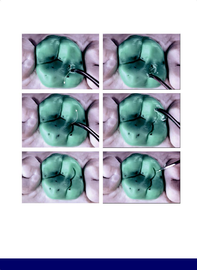
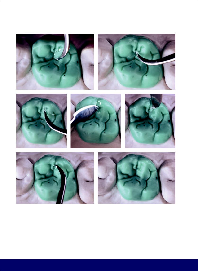
Fig 2-8 | (a to c) Create the kidney bean profile (courtesy of Ivan
Ronald Huanca) on the mesial marginal ridge.
61
@dentistinfo стоматологический телеграм канал
CHAPTER 2
a |
|
b |
|
c |
|
|
|
|
|
d |
|
e |
|
f |
|
|
|
|
|
g |
|
h |
|
i |
|
|
|
|
|
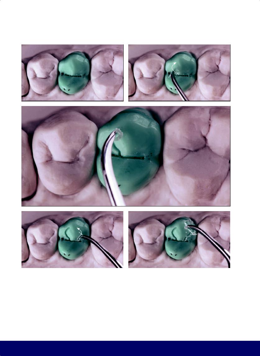
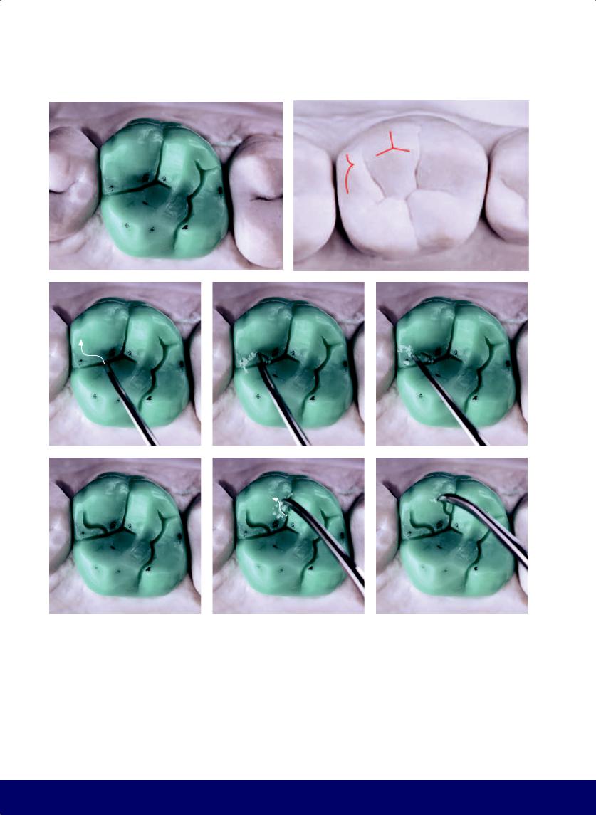
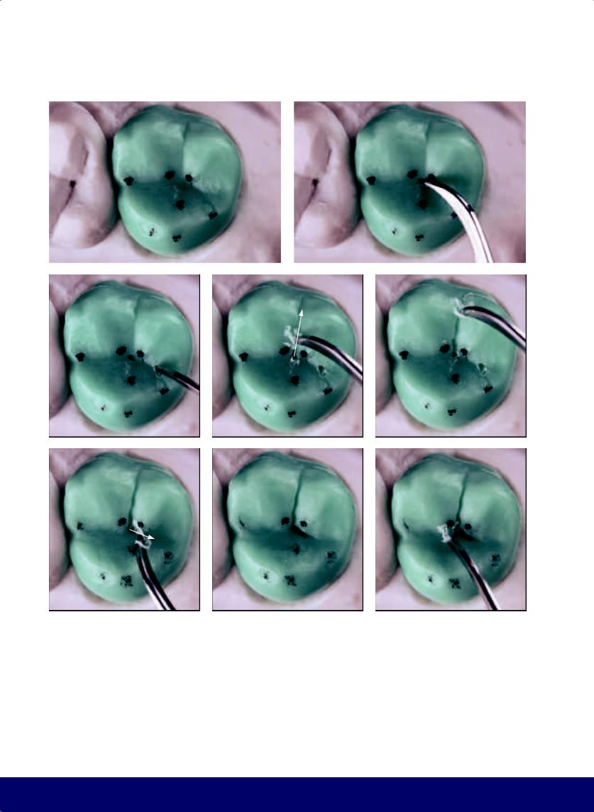
Fig 2-9 | Buccal face. (a) Because the mesial slope of the marginal ridge of the buccal cusp is shorter than the distal slope, the tip of the buccal cusp is slightly toward the mesial. (b to e) The vertical macro texture consists of two developmental grooves. The mesial developmental groove has a beginning, middle, and end that follows from the area of the mesial papilla to the occlusal third. The distal developmental groove, however, goes only to the middle third. (f) The term groove is used metaphorically; they are better described as wide depressions or low reliefs. (g to i)
62
The horizontal macro texture is composed of three or four subtle horizontal depressions, the predominant one being in the middle third and following a trajectory opposite the crown/root line. The macro texture will be repeated in all the teeth, to a greater or lesser degree, depending on the external contour pattern of each person’s teeth. The concrete result of horizontal macro texture is a narrow waist, which improves the silhouette of the buccal face.
@dentistinfo стоматологический телеграм канал
a |
b |
|
|
c |
|
d |
|
|
|
f |
g |
|
|
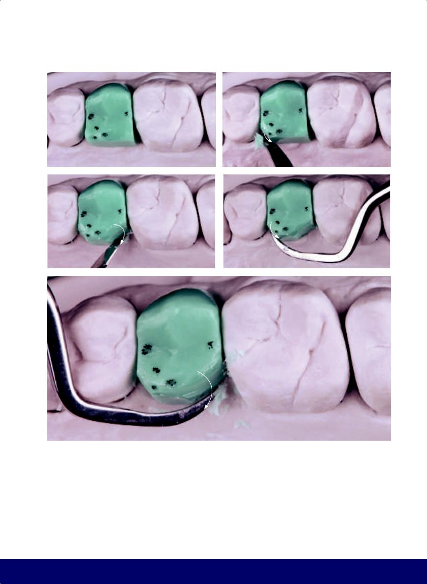
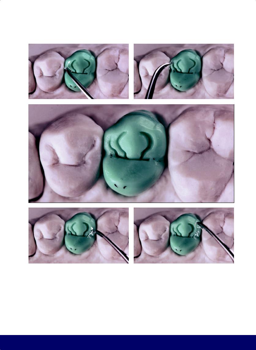
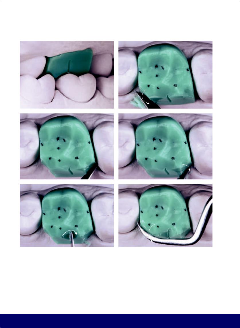
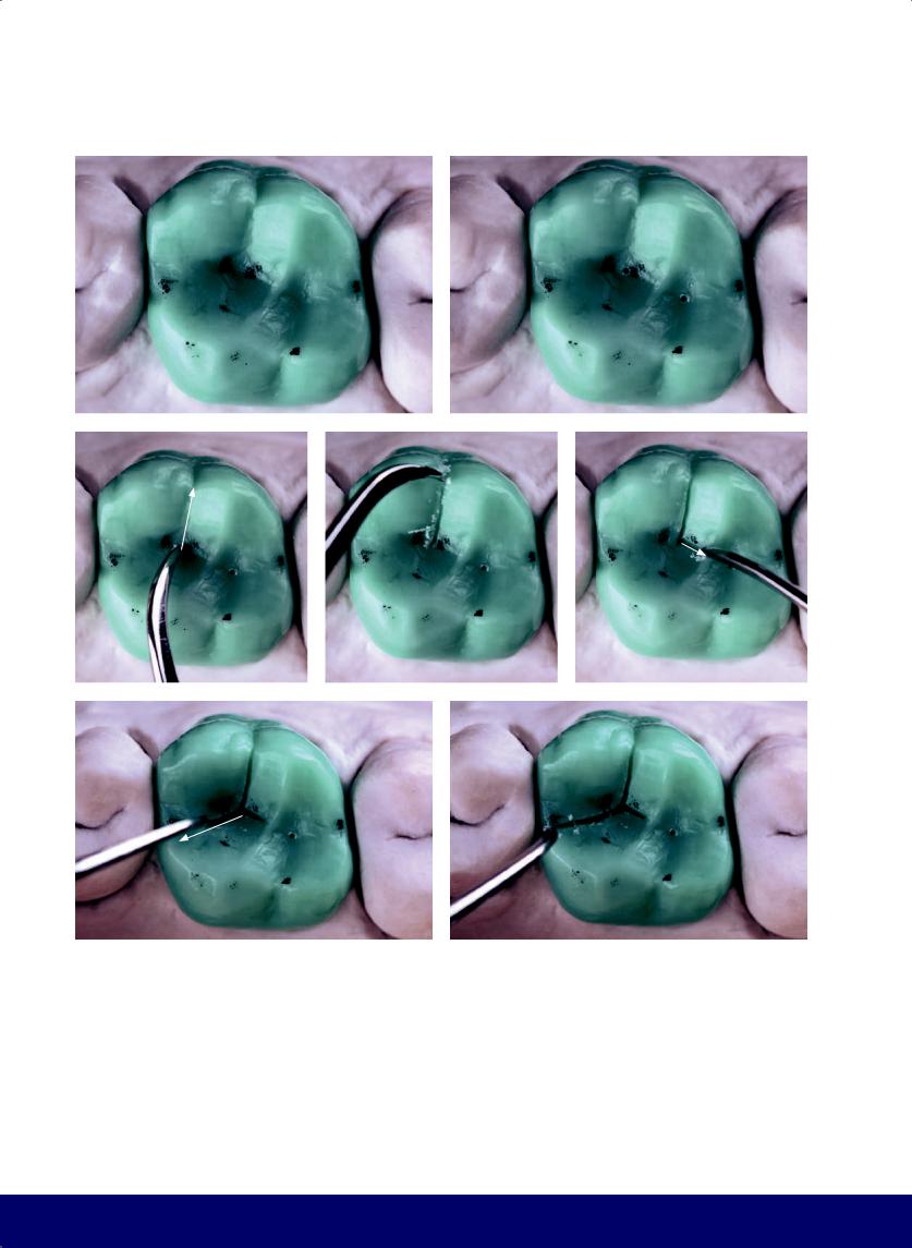
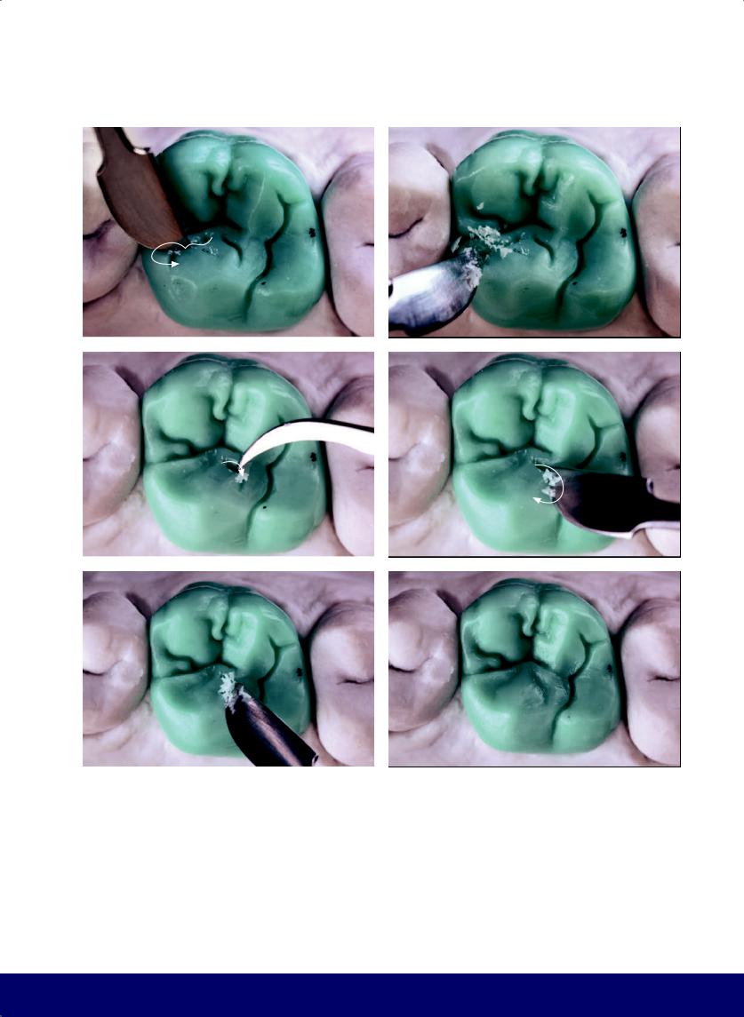
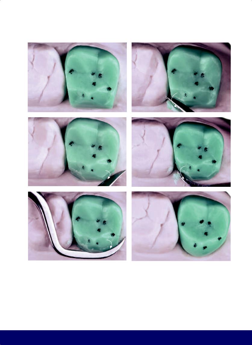
Fig 2-10 | External contour. (a) Observe the vertical and horizontal macro texture from the occlusal view. The occlusal surface is divided into two parts: a smaller mesial side (a small inverted D) and a larger distal side (a larger D). (b to d) Marking the mesial and distal fossae. (e to g) The mesiodistal groove connects the mesial and distal fossae and passes over the apex where the transverse ridges meet.
MAXILLARY FIRST PREMOLAR
e
63
@dentistinfo стоматологический телеграм канал
CHAPTER 2
a |
|
b |
|
|
|
c
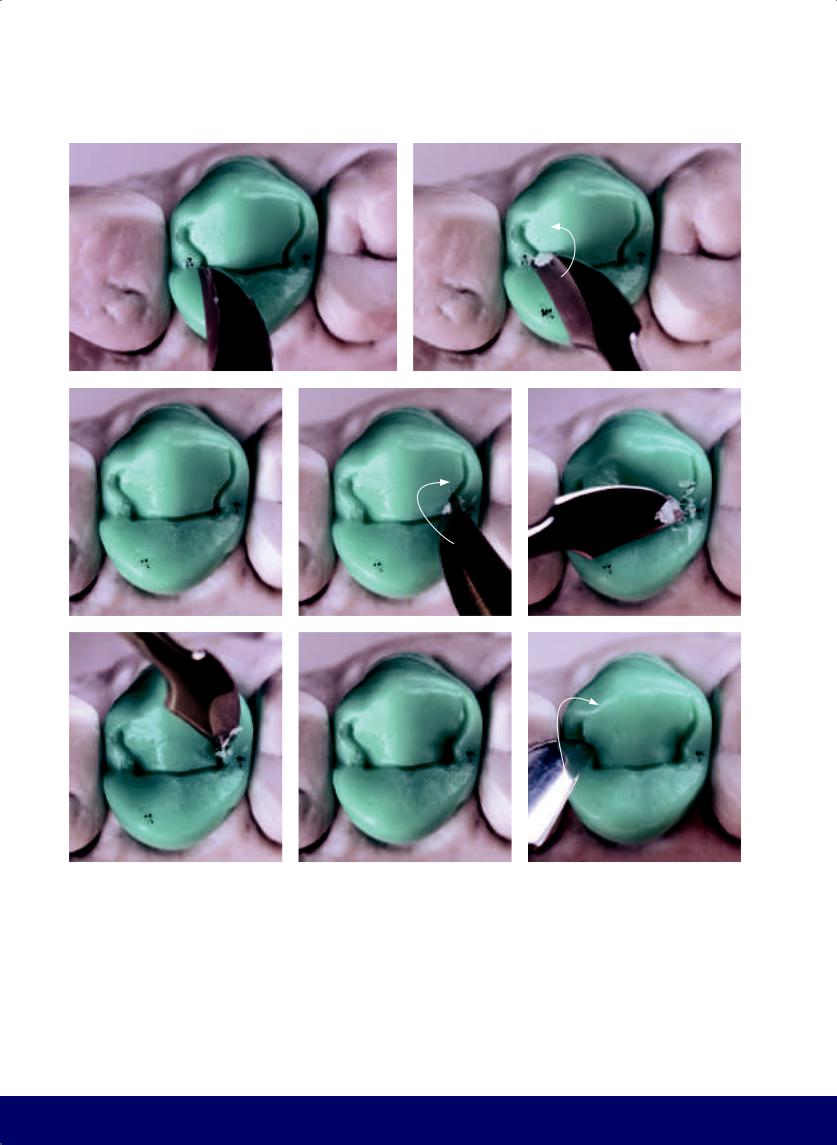
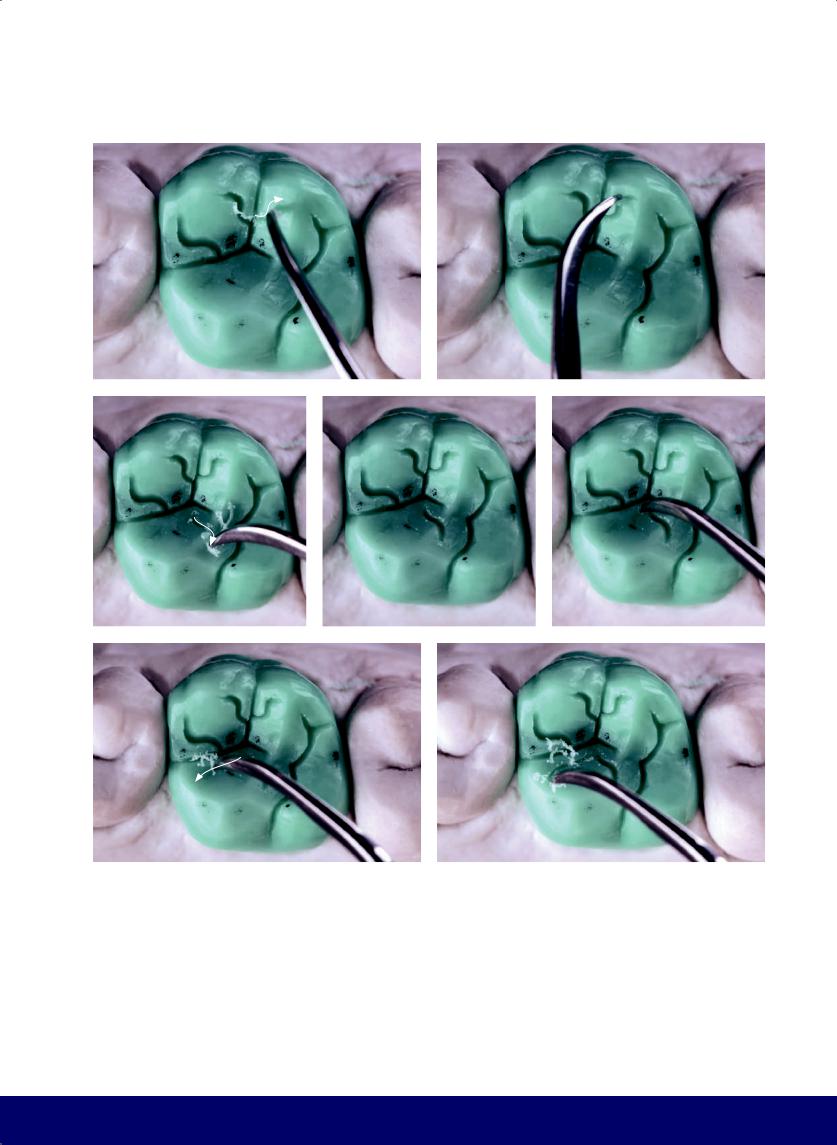
Fig 2-11 | (a to f) The mesial secondary groove is a short S. The distal secondary groove is a longer, inverted S. Both begin in their respective fossae and follow the outward spread of Zebu horns.
64
@dentistinfo стоматологический телеграм канал
MAXILLARY FIRST PREMOLAR
d |
|
e |
|
|
|
f
65
@dentistinfo стоматологический телеграм канал
CHAPTER 2
a |
|
b |
|
|
|
c |
|
d |
|
e |
|
|
|
|
|
f |
|
g |
|
h |
|
|
|
|
|
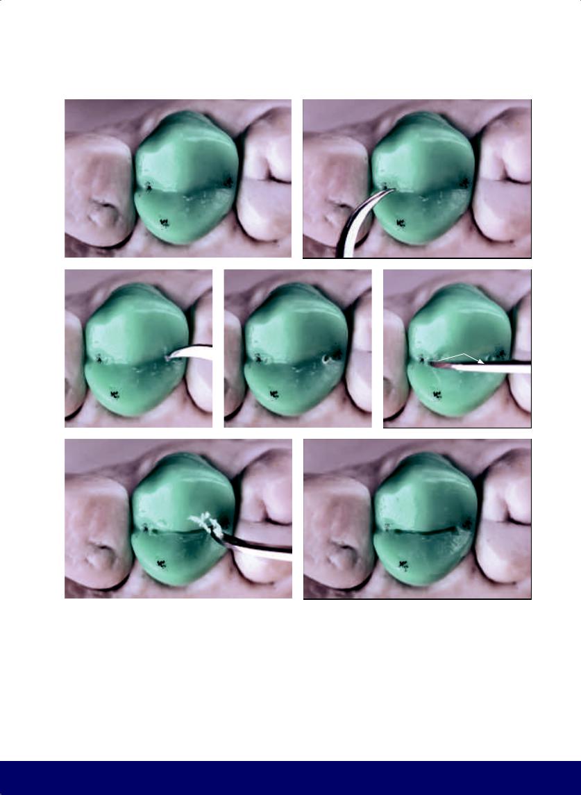
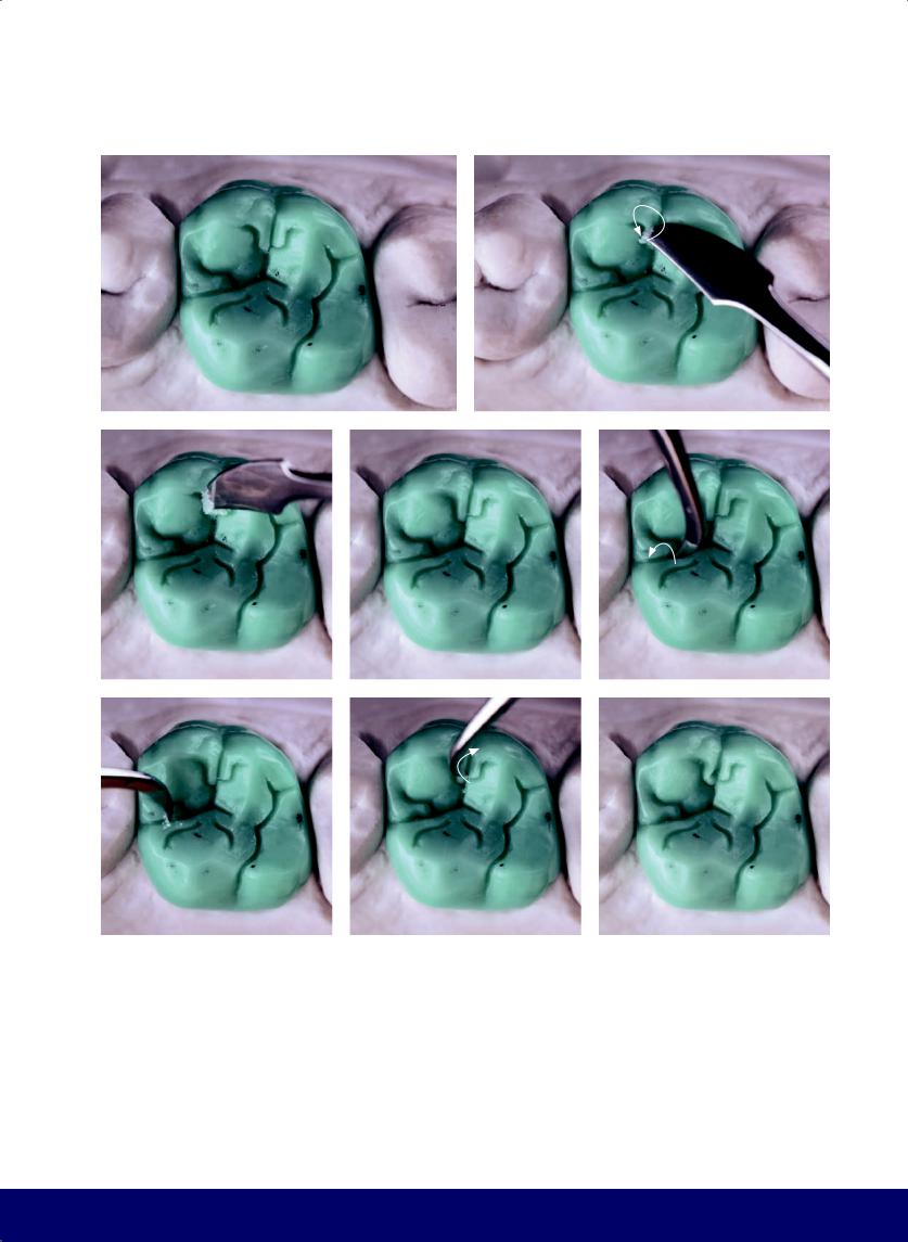
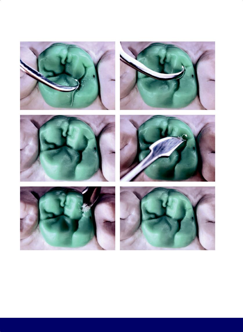
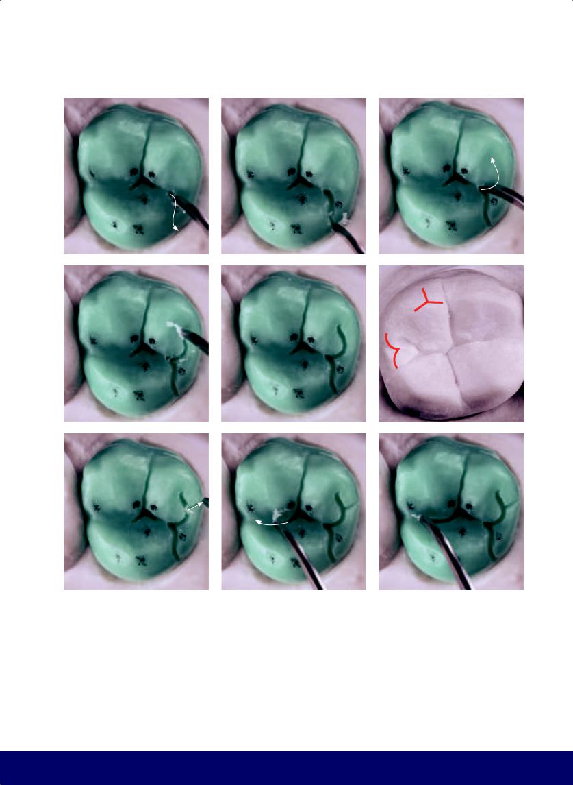
Fig 2-12 | (a to n) Wax is removed to open the grooves and round the fossae. The secondary grooves terminate in an opening that is enlarged and rounded with the turn of the carving instrument.
66
@dentistinfo стоматологический телеграм канал
MAXILLARY FIRST PREMOLAR
i |
|
j |
|
|
|
k |
|
l |
|
|
|
m |
|
n |
|
|
|
67
@dentistinfo стоматологический телеграм канал
CHAPTER 2
a |
|
b |
|
|
|
c |
|
d |
|
|
|
e |
|
f |
|
|
|
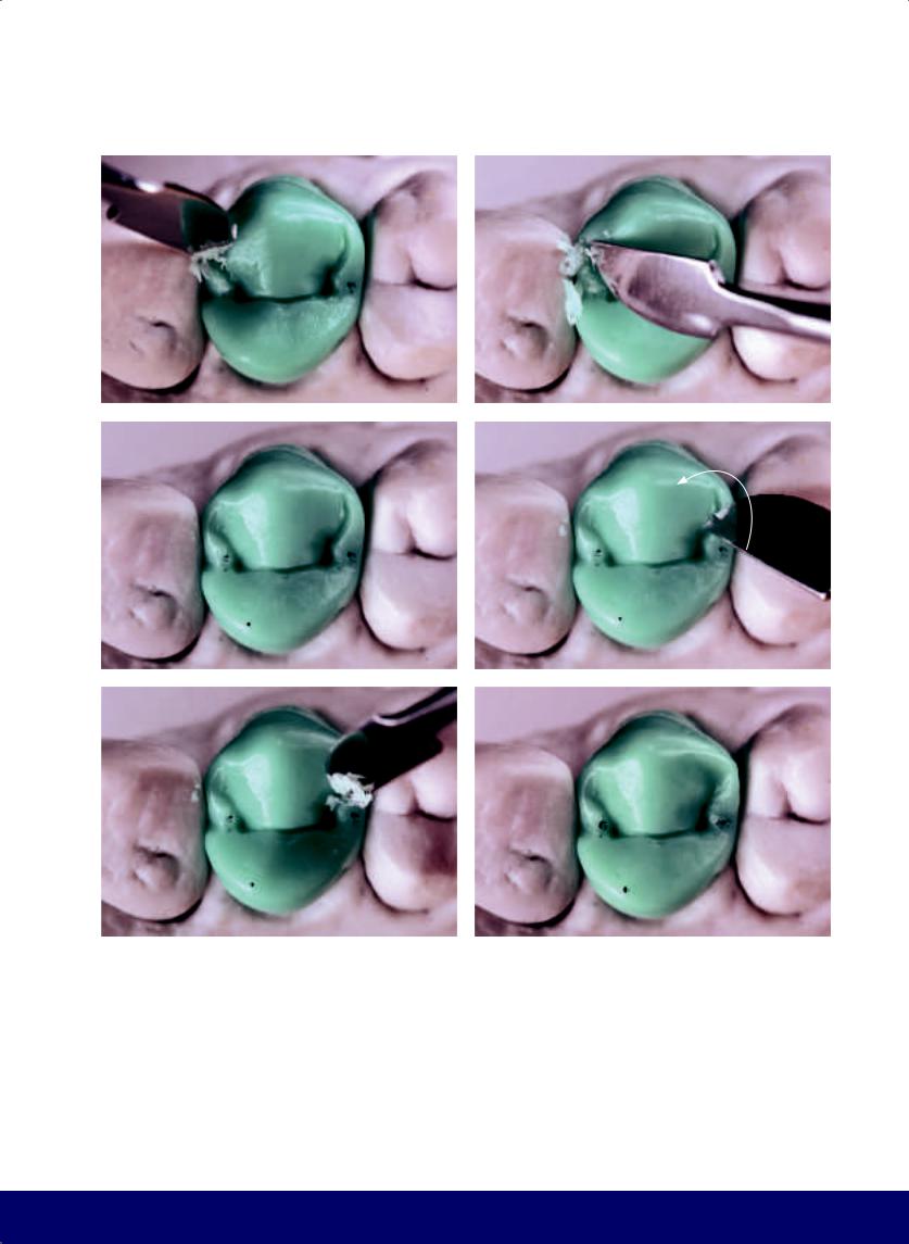
Fig 2-13 | (a to e) A tributary groove starting from the middle of the distal secondary groove completes a wineglass shape. The glass shape can be for red wine (ie, shorter and wider) or white wine (ie, thinner and longer). (f to h) The carving instrument is turned to round the shape and open the mesiodistal groove. (i to l) Slight deepening of the mesial and distal grinding ridges on the lingual cusp.
68
@dentistinfo стоматологический телеграм канал
MAXILLARY FIRST PREMOLAR
g |
|
h |
|
|
|
i |
|
j |
|
|
|
k |
|
l |
|
|
|
69
@dentistinfo стоматологический телеграм канал
CHAPTER 2
a |
|
b |
|
|
|
c
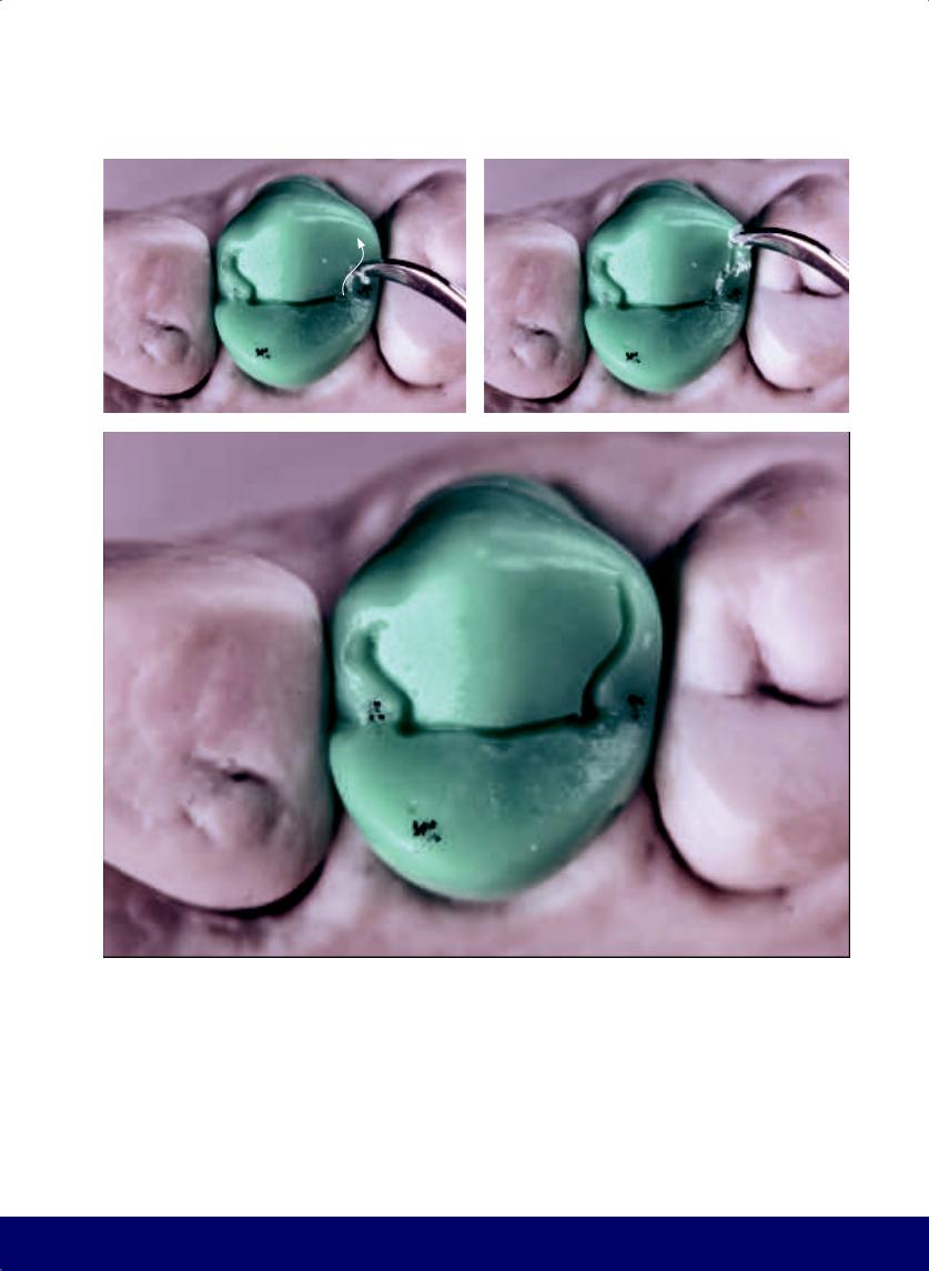
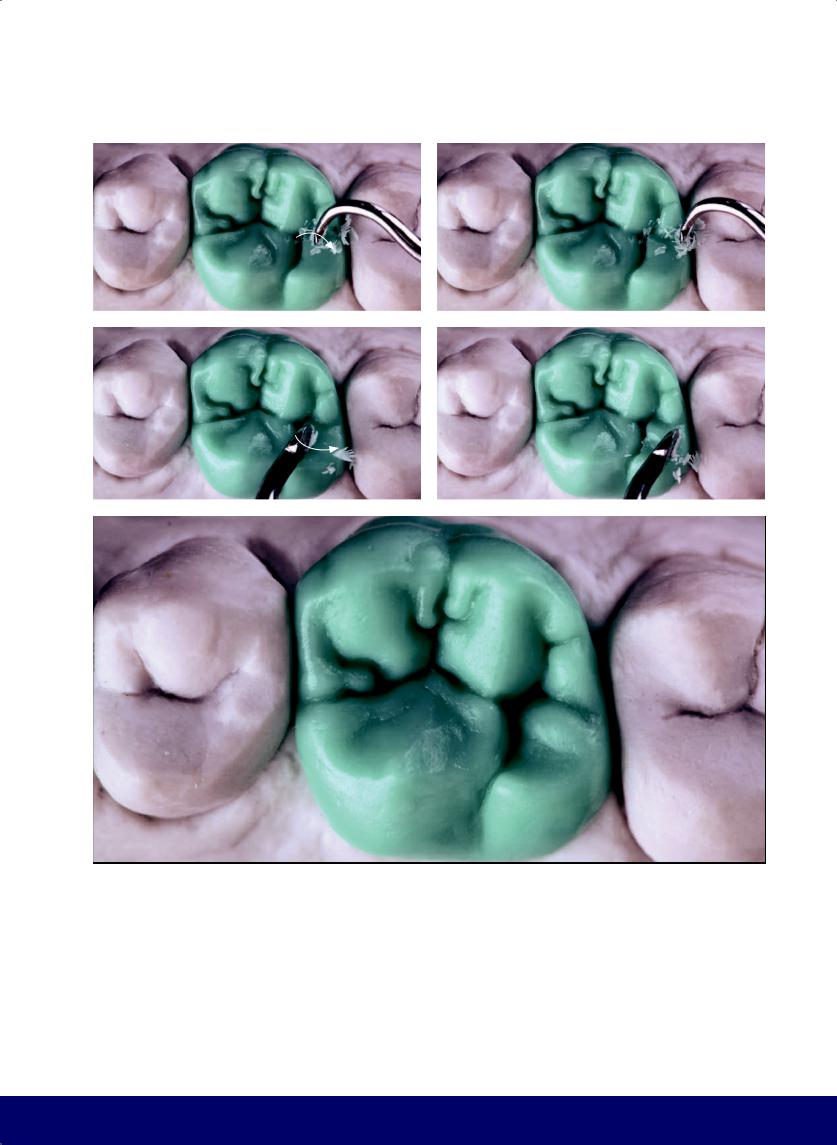
Fig 2-14 | (a and b) The mesiodistal groove is extended across the mesial marginal ridge. This feature is present in all maxillary first premolars. (c) Use wax to fill the cup shape in the distal grinding ridge of the buccal cusp to form a lobe.
70
@dentistinfo стоматологический телеграм канал
MAXILLARY FIRST PREMOLAR
a
b
Fig 2-15 | (a) Create a lobe on the distal marginal crest for occlusal relief. Note the lingual positioning of the mesiodistal groove, which makes the buccal cusp more bulky than the lingual cusp. Also note the secondary grooves (ie, S and inverted S) that start at the mesial and distal fossae; together they mimic the outward spread of Zebu horns. (b) Note the kidney bean profile in the mesial and distal marginal ridges; the mesial inverted D and the larger distal D, separated by the transverse ridges; the wineglass and the lobes of the distal marginal ridge.
71
@dentistinfo стоматологический телеграм канал
CHAPTER 2
a |
b |
c |
|
|
|
d |
e |
f |
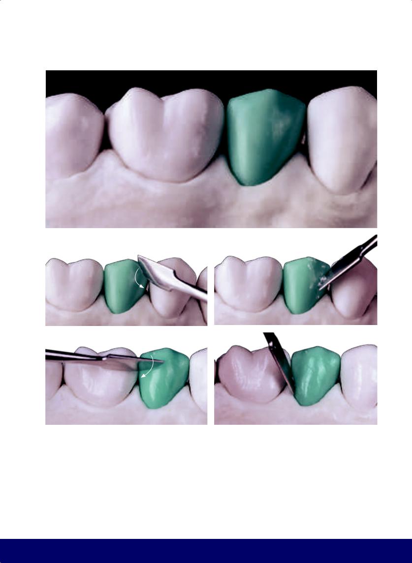
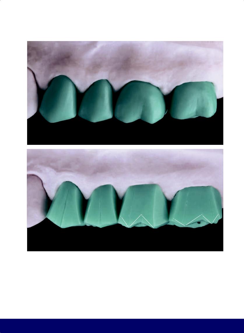
Fig 2-16 | (a to l) Completed contours of the maxillary first premolar.
72
@dentistinfo стоматологический телеграм канал
MAXILLARY FIRST PREMOLAR
g |
h |
i |
|
|
|
j |
k |
l |
73
@dentistinfo стоматологический телеграм канал

Premolar
Maxillary Second
03
74
@dentistinfo стоматологический телеграм канал
MAXILLARY SECOND PREMOLAR
a
b |
|
c |
|
|
|
d |
|
e |
|
|
|
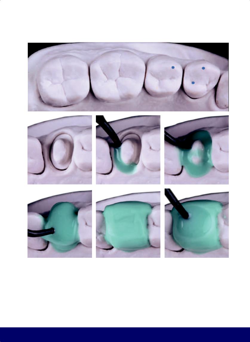
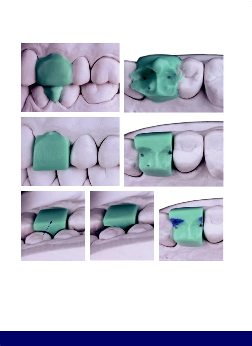
Fig 3-1 | (a) Centric contact points in the mandible to be reproduced on the wax block. (b) With the heated instrument, the wax block is formed with excess material on the buccal, lingual, and occlusal surfaces. The superficial layer of wax is warmed. With the cast in occlusion, the softened wax records the occlusal morphology of the opposing mandibular teeth. (c) Occlusal view of the wax block. Note the points marked in pencil on the opposing teeth are reproduced in the wax. (d and e) Buccal and occlusal views of the wax block with the excess wax removed.
75
@dentistinfo стоматологический телеграм канал
CHAPTER 3
a |
|
b |
|
|
|
c |
d |
|
e |
|
f |
|
|
|
Fig 3-2 | (a to c) Buccal interference in the working movement. (d to f) Carve away the buccal interference. (Purple arrows indicate movement of the mandible. White arrows indicate direction of instrumentation.)
76
@dentistinfo стоматологический телеграм канал
MAXILLARY SECOND PREMOLAR
a |
|
b |
|
c |
|
|
|
|
|
d |
|
e |
|
|
|
f |
|
g |
|
h |
|
|
|
|
|
Fig 3-3 | (a to c) Lingual interference in the balancing movement. (d and e) Carve away the lingual interference. (f to h) Interference in protrusive movement.
77
@dentistinfo стоматологический телеграм канал
CHAPTER 3
a |
|
b |
|
|
|
c |
|
d |
|
e |
|
|
|
|
|
f |
|
g |
|
|
|
Fig 3-4 | (a and b) Carve away the interference on protrusive movement. (c to e) Interference in retrusive movment. (f and g) Carve away interference on retrusive movement.
78
@dentistinfo стоматологический телеграм канал
a |
|
b |
|
|
|
d |
|
e |
|
|
|
g |
h |
|
|
Fig 3-5 | (a to c) Cut back wax to open up the buccal embrasures.
(d) Form the buccal cusp tip by carving the mesial slope from the distal marginal ridge of the first premolar to the buccal midline and the distal slope from the midline to the marginal mesial ridge of the first molar. Divide the buccal aspect into two equal parts. (e and f). Carve the buccal face in two planes. (g and h) Round the angles resulting from the formation of the buccal cusp tip.
MAXILLARY SECOND PREMOLAR
c
f
79
@dentistinfo стоматологический телеграм канал
CHAPTER 3
a |
|
b |
|
c |
|
|
|
|
|
d |
|
e |
|
|
|
f |
|
g |
|
|
|
Fig 3-6 | (a to g) Round the buccal face.
80
@dentistinfo стоматологический телеграм канал
MAXILLARY SECOND PREMOLAR
a |
|
b |
|
|
|
c |
|
d |
|
|
|
e
Fig 3-7 | (a to e) Open up the lingual embrasures and create the transverse cusp ridge. Note the tip of the lingual cusp is directed toward the mesial.
81
@dentistinfo стоматологический телеграм канал

CHAPTER 3
a |
|
b |
|
c |
|
|
|
|
|
d |
|
e |
|
|
|
f |
|
g |
|
|
|
Fig 3-8 | (a) The occlusal surface is divided into two parts: a smaller mesial side (a small inverted D) and a larger distal side (a larger D). This division is made by the transverse grinding ridges of the buccal and lingual cusps. (b to g) The kidney bean profile is carved on the mesial and distal marginal ridges.
82
@dentistinfo стоматологический телеграм канал
MAXILLARY SECOND PREMOLAR
a
b |
|
c |
|
|
|
d |
|
e |
|
|
|
Fig 3-9 | (a) Buccal view. Note that the mesial slope of the marginal ridge of the buccal cusp is smaller than the distal slope and, consequently, the buccal cusp tip turns slightly toward the mesial. (b to e) The vertical macro texture consists of the mesial and distal developmental grooves. These grooves create two small accessory “lobes” on the buccal face.
83
@dentistinfo стоматологический телеграм канал
CHAPTER 3
a |
|
b |
|
|
|
c |
|
d |
|
|
|
e |
|
f |
|
|
|
Fig 3-10 | (a to f) The horizontal macro texture is composed of subtle depressions that are more predominant in the middle third and follow a trajectory opposite to the crown/root line. The result of the horizontal macro texture is the narrow waist that creates the shape of the buccal face and improves its silhouette.
84
@dentistinfo стоматологический телеграм канал
a |
b |
|
|
c |
d |
|
|
e |
f |
|
|
Fig 3-11 | (a) Observe the vertical and horizontal macro textures from an occlusal view. (b to f) The mesiodistal groove connects the mesial and distal fossae and passes over the apex where transverse ridges meet.
MAXILLARY SECOND PREMOLAR
85
@dentistinfo стоматологический телеграм канал
CHAPTER 3
a |
b |
|
c
d |
|
e |
|
|
|
Fig 3-12 | (a to j) The secondary grooves form a letter S and an inverted letter S and together look like inward-curving Zebu horns. They both begin in the mesiodistal groove on either side of the buccal grinding ridge. From the mesial and distal fossae, there are two accessory secondary grooves that, together with each S, form an accessory lobe in each grinding slope of the buccal cusp.
86
@dentistinfo стоматологический телеграм канал
MAXILLARY SECOND PREMOLAR
f |
|
g |
|
|
|
h
i |
|
j |
|
|
|
87
@dentistinfo стоматологический телеграм канал
CHAPTER 3
a |
|
b |
|
|
|
c |
|
d |
|
|
|
e |
|
f |
|
|
|
g |
|
h |
|
|
|
Fig 3-13 | (a to p) Wax is removed to open and round the secondary grooves. The larger secondary grooves terminate in an opening that is enlarged and rounded with the turn of the carving instrument. These two depressions converge toward the tip of the cusp. All four secondary grooves result in two lobes on the grinding slopes of the buccal cusp.
88
@dentistinfo стоматологический телеграм канал
MAXILLARY SECOND PREMOLAR
i |
|
j |
|
|
|
k |
|
l |
|
|
|
m |
|
n |
|
|
|
o |
|
p |
|
|
|
89
@dentistinfo стоматологический телеграм канал
CHAPTER 3
a |
|
b |
|
|
|
c |
|
d |
|
|
|
Fig 3-14 | (a to d) Deepen the mesial and distal grooves of the lingual cusp.
90
@dentistinfo стоматологический телеграм канал
a |
b |
|
|
c |
d |
|
|
Fig 3-15 | (a to c) A lobe is created on the distal marginal crest, and the subtle touch of the dripper tip is used to fill out the rest of the lobes. (d) Occlusal view. Note that the mesiodistal groove is positioned in the center of the occlusal surface to divide the buccal and lingual cusps with similar volumes. Also note the S and inverted S coming off the mesiodistal groove; together they form inward-curving Zebu horns whose terminal points go toward the tip of the buccal cusp. Note the kidney bean profile in the mesial and distal marginal ridges as well as the inverted mesial D and the larger distal D separated by the transverse ridges, and the lobes.
MAXILLARY SECOND PREMOLAR
91
@dentistinfo стоматологический телеграм канал
CHAPTER 3
a |
b |
c |
d |
e |
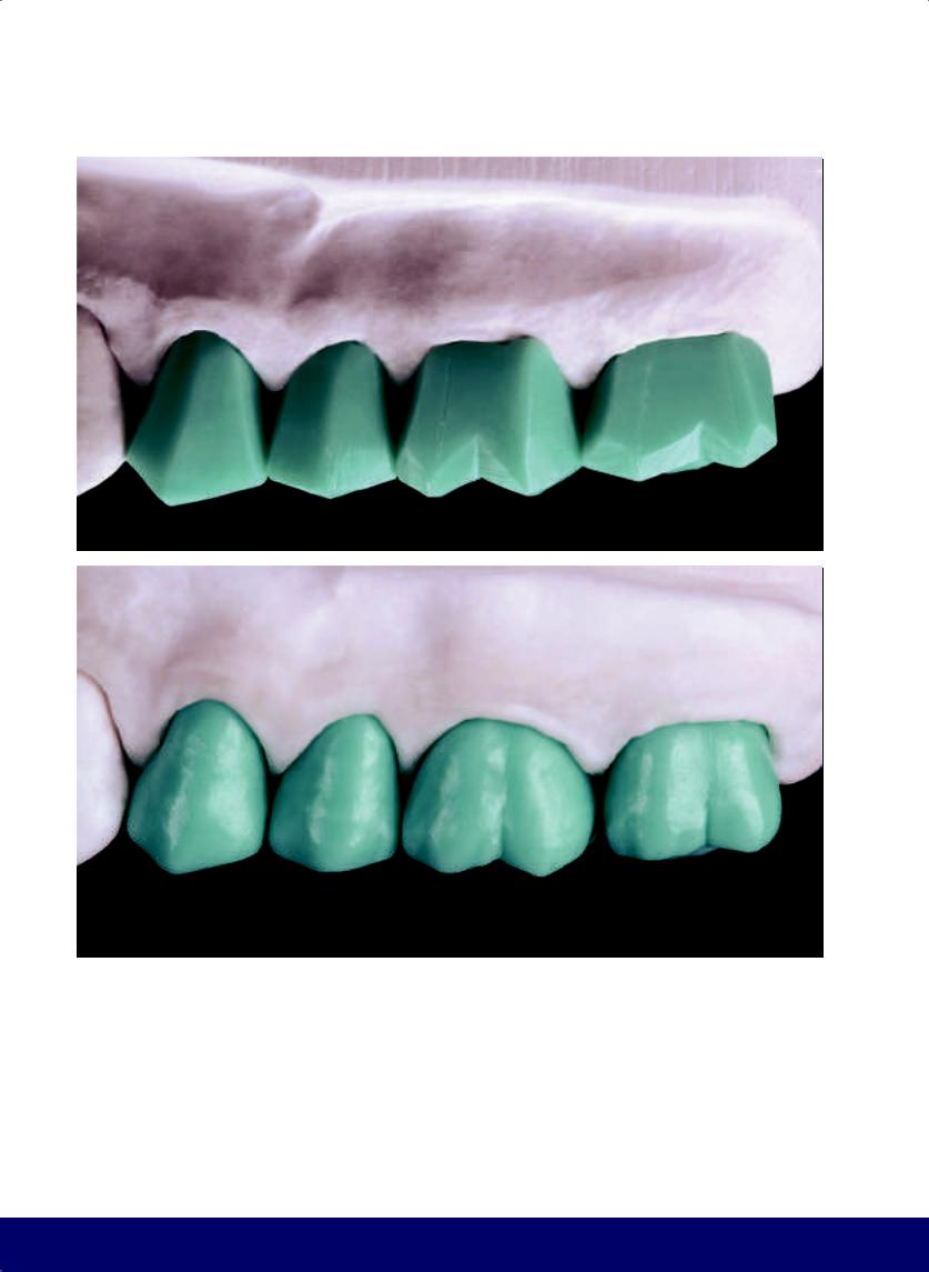
Fig 3-16 | (a to j) Completed contours of the maxillary second premolar.
92
@dentistinfo стоматологический телеграм канал
MAXILLARY SECOND PREMOLAR
f |
g |
h |
i |
j |
93
@dentistinfo стоматологический телеграм канал

First Molar
Maxillary
04
94
@dentistinfo стоматологический телеграм канал
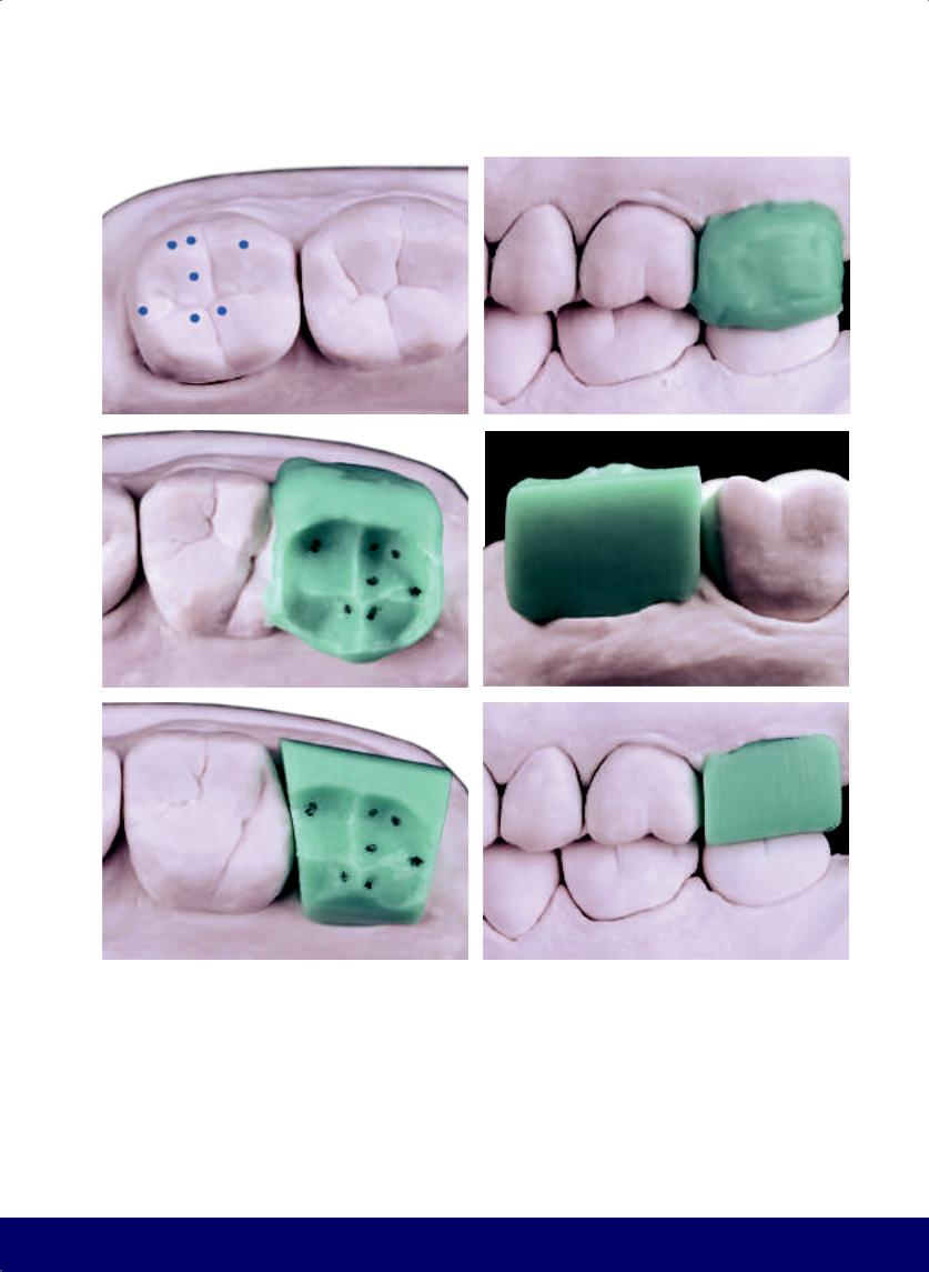
MAXILLARY FIRST MOLAR
a |
|
b |
|
|
|
c |
|
d |
|
e |
|
|
|
|
|
f |
|
g |
|
h |
|
|
|
|
|
Fig 4-1 | (a) Centric contact points in the mandibular molar to be reproduced on the wax-up. (b) Buccal view showing the cast in occlusion. The softened wax records the occlusal morphology of the opposing mandibular teeth. (c) In the occlusal view, note that the contact points marked in pencil on the opposing teeth have been reproduced in the wax. (d) After carving away the lingual excess from the wax-up, the volume and height of the buccal aspect are modeled after the adjacent teeth. (e) The occlusal
surface is divided into two parts by a virtual line created by the distobuccal groove of the mandibular first molar. The mesial side is larger and the distal side is smaller. In addition, note the division of the buccal surface. (f to h) Buccal interference in the working movements. Observe where the buccal cusps of the mandibular first molar touch the wax-up. (Purple arrows indicate the movement of the mandible.)
95
@dentistinfo стоматологический телеграм канал
CHAPTER 4
a |
|
b |
|
|
|
c |
|
d |
|
|
|
e |
|
f |
|
|
|
Fig 4-2 | (a to c) Carve away buccal interference. (d to f) Lingual interference in balancing movement. (White arrows indicate the direction of instrumentation.)
96
@dentistinfo стоматологический телеграм канал
MAXILLARY FIRST MOLAR
a |
|
b |
|
|
|
c |
|
d |
|
|
|
e
Fig 4-3 | (a to c) Carve away lingual interference. (d and e) Interference in protrusive movement.
97
@dentistinfo стоматологический телеграм канал
CHAPTER 4
a |
|
b |
|
|
|
c |
|
d |
|
e |
|
|
|
|
|
f |
|
g |
|
|
|
Fig 4-4 | (a to d) Carve away interference on protrusive movement. (e to g) Interference in retrusive movement.
98
@dentistinfo стоматологический телеграм канал
MAXILLARY FIRST MOLAR
a |
|
b |
|
|
|
c |
|
d |
|
|
|
e |
|
f |
|
|
|
Fig 4-5 | (a to c) Carve away interference on retrusive movement. (d to f) Open up the buccal embrasures.
99
@dentistinfo стоматологический телеграм канал
CHAPTER 4
a |
|
b |
|
|
|
c |
|
d |
|
|
|
e |
|
f |
|
|
|
Fig 4-6 | (a and b) The placement of the buccal cusps of the maxillary first molar relate to the buccal cusps of the mandibular first molar. (c to m) After the cusp tips are formed, the entire buccal surface is rounded. Note that the distobuccal cusp is slightly lower and has a more pointed buccal profile than the mesiobuccal cusp.
100
@dentistinfo стоматологический телеграм канал
MAXILLARY FIRST MOLAR
g |
|
h |
|
|
|
i |
|
j |
|
k |
|
|
|
|
|
l |
|
m |
|
|
|
101
@dentistinfo стоматологический телеграм канал
CHAPTER 4
a |
|
b |
|
|
|
c |
|
d |
|
|
|
e |
|
f |
|
|
|
Fig 4-7 | (a) Mark the position of the tip of the distolingual cusp of the mandibular first molar on the lingual aspect. The lingual groove of the maxillary first molar will emerge from this line. (b to j) Open the lingual embrasures, delineate the longitudinal ridge, and soften the angles by rounding of the lingual aspect. (k) Occlusal view of the completed external contour. Look for the two external references for creating the buccal and lingual grooves. (l and m) Place the kidney bean profile on the mesial marginal ridge.
102
@dentistinfo стоматологический телеграм канал
MAXILLARY FIRST MOLAR
g |
|
h |
|
|
|
i |
|
j |
|
k |
|
|
|
|
|
l |
|
m |
|
|
|
103
@dentistinfo стоматологический телеграм канал
CHAPTER 4
a |
|
b |
|
|
|
c |
|
d |
|
|
|
e |
|
f |
|
|
|
Fig 4-8 | (a to k) The vertical macro texture is defined by the pseudo developmental grooves on the mesiobuccal cusp. The horizontal macro texture (the windshield wiper effect) results in a narrowed waist, which shapes the silhouette of the buccal aspect.
104
@dentistinfo стоматологический телеграм канал
MAXILLARY FIRST MOLAR
g |
|
h |
|
|
|
i |
|
j |
|
|
|
k
105
@dentistinfo стоматологический телеграм канал
CHAPTER 4
a |
|
b |
|
|
|
c |
|
d |
|
e |
|
|
|
|
|
f |
|
g |
|
|
|
Fig 4-9 | (a to c) Define the central and distal fossae. (d) Open the buccal groove that connects the bottom of the central fossa to the vertex of the angle formed between the buccal cusps (working groove). (e to g) Extend two grooves from the bottom of the central fossae, which correspond to the arms of the Mercedes star logo. The mesial arm of the star is connected to the kidney bean profile on the mesial marginal crest.
106
@dentistinfo стоматологический телеграм канал
a |
b |
|
|
c |
d |
|
|
e |
f |
|
|
Fig 4-10 | (a to c) Delineate the lingual wing of the “seagull.” This is a main groove because it separates cusps. (d and e) The other wing of the seagull, a curve toward the buccal, is a secondary groove, as it is on the distal grinding slope of the distobuccal cusp.
(f) The seagull detail with a broken feather.
MAXILLARY FIRST MOLAR
107
@dentistinfo стоматологический телеграм канал
CHAPTER 4
a |
|
b |
|
|
|
c |
|
d |
|
e |
|
|
|
|
|
f |
|
g |
|
h |
|
|
|
|
|
Fig 4-11 | (a and b) Maxillary first molar grooves and their counterparts on the opposing tooth. (c to h) Sketch the two secondary grooves (forming an S and an inverted S) from the mesiodistal groove, like two inward-curving Zebu horns. The protrusive groove is formed from the S groove and the mesiodistal groove from the central fossa.
108
@dentistinfo стоматологический телеграм канал
MAXILLARY FIRST MOLAR
a |
|
b |
|
|
|
c |
|
d |
|
e |
|
|
|
|
|
f |
|
g |
|
|
|
Fig 4-12 | (a and b) Create of a secondary groove off of the labial (working) groove by forming an S in the mesial grinding slope of the distobuccal cusp. (c to g) Sketch inward-curving Zebu horns on the mesiolingual cusp. The S on the mesial is also a functional balancing groove.
109
@dentistinfo стоматологический телеграм канал
CHAPTER 4
a |
|
b |
|
c |
|
|
|
|
|
d |
|
e |
|
f |
|
|
|
|
|
g |
|
h |
|
i |
|
|
|
|
|
Fig 4-13 | (a to q) Round the corners and open the terminal points of the grooves. Preserve the transverse and longitudinal ridges, and highlight the figure with hands on hips on the mesiobuccal cusp.
110
@dentistinfo стоматологический телеграм канал
MAXILLARY FIRST MOLAR
j |
|
k |
|
|
|
l |
|
m |
|
n |
|
|
|
|
|
o |
|
p |
|
q |
|
|
|
|
|
111
@dentistinfo стоматологический телеграм канал
CHAPTER 4
a |
|
b |
|
|
|
c |
|
d |
|
e |
|
|
|
|
|
f |
|
g |
|
|
|
Fig 4-14 | (a to g) Round the corners of the distobuccal cusp, of the inverted S coming off the working groove on the mesial grinding slope, and of the opening at the terminal point of the secondary groove.
112
@dentistinfo стоматологический телеграм канал
a |
b |
|
|
c |
d |
|
|
e |
f |
|
|
Fig 4-15 | (a to f) The middle of the mesiolingual cusp should remain as a high relief because it has a ridge with a centric point of contact. Its high relief is enhanced by deepening the inwardcurving horns.
MAXILLARY FIRST MOLAR
113
@dentistinfo стоматологический телеграм канал
CHAPTER 4
a |
|
b |
|
|
|
c |
|
d |
|
|
|
e |
|
f |
|
|
|
Fig 4-16 | (a to c) Round the corners around the seagull form on the mesiolingual cusp. (d to f) As with every secondary groove, the terminal point of the seagull’s wing is also opened up. (g to k) Make a lobe near the head of the seagull.
114
@dentistinfo стоматологический телеграм канал
MAXILLARY FIRST MOLAR
g |
|
h |
|
|
|
i |
|
j |
|
|
|
k
115
@dentistinfo стоматологический телеграм канал
CHAPTER 4
a |
b |
|
|
d |
e |
Fig 4-17 | (a to l) Completed external contour and occlusal morphology. Note that the secondary grooves (S shapes) start tentatively and open out toward the end. Together, the secondary grooves resemble inward-curving horns. Also note the kidney bean profile in the mesial marginal ridge and the difference between the opening of the grooves. Note the winding and vertical relief of the wing of the seagull with the broken feather.
116
c
f
@dentistinfo стоматологический телеграм канал
MAXILLARY FIRST MOLAR
g |
h |
i |
|
|
|
j |
k |
l |
117
@dentistinfo стоматологический телеграм канал

Molar
Maxillary Second
05
118
@dentistinfo стоматологический телеграм канал
a |
b |
|
|
c |
d |
|
|
e |
f |
|
|
Fig 5-1 | (a) Mandibular contact points to be reproduced in the wax-up. (b and c) Cast in occlusion. Note the points that were marked in pencil on the opposing molar were reproduced in the wax. (d to f) Buccal and occlusal views after removing excess wax from the distal, lingual, and buccal aspects.
MAXILLARY SECOND MOLAR
119
@dentistinfo стоматологический телеграм канал
CHAPTER 5
a |
|
b |
|
|
|
c |
|
d |
|
|
|
e |
f |
Fig 5-2 | (a to c) Buccal interference in working movement. Observe where the buccal cusps of the mandibular first molar touch the wax. (d to f) Carve away buccal interference. (Purple arrows indicate the movement of the mandible, while white arrows indicate the direction of instrumentation.)
120
@dentistinfo стоматологический телеграм канал
a |
b |
|
|
c |
|
d |
|
|
|
f |
g |
Fig 5-3 | (a to c) Lingual interference in balancing movement. (d and e) Carve away lingual interference. (f to h) Interference in protrusive movement.
MAXILLARY SECOND MOLAR
e
h
121
@dentistinfo стоматологический телеграм канал
CHAPTER 5
a |
|
b |
|
|
|
c |
|
d |
|
e |
|
|
|
|
|
f |
|
g |
|
|
|
Fig 5-4 | (a and b) Carve away interference on protrusive movement. (c to e) Interference in retrusion. (f and g) Carve away interference on retrusive movement.
122
@dentistinfo стоматологический телеграм канал
a |
b |
|
|
c |
d |
|
|
e |
f |
Fig 5-5 | (a and b) Open the buccal embrasures. (c to f) The references for locating the mesiobuccal cusp and the path of the buccal grooves of the maxillary second molar are, respectively, the buccal groove and the smooth transverse ridge of the distobuccal of the mandibular second molar.
MAXILLARY SECOND MOLAR
123
@dentistinfo стоматологический телеграм канал
CHAPTER 5
a |
|
b |
|
|
|
c |
|
d |
|
|
|
e |
|
f |
|
|
|
Fig 5-6 | (a to g) Form the cusp tips and shape the entire buccal face. (h to l) Open the lingual embrasures, delineate the longitudinal ridge, and smooth the angles with the final shaping of the lingual face.
124
@dentistinfo стоматологический телеграм канал
MAXILLARY SECOND MOLAR
g |
|
h |
|
|
|
i |
|
j |
|
|
|
k |
|
l |
|
|
|
125
@dentistinfo стоматологический телеграм канал
CHAPTER 5
a |
|
b |
|
|
|
c |
|
d |
|
|
|
e |
|
f |
|
|
|
Fig 5-7 | (a and b) Kidney bean profile on the mesial marginal ridge. (c) Occlusal view of the final external contour. It is important to emphasize the two external references that will serve to create buccal and lingual grooves. (d to l) Define the vertical macro texture and the pseudo developmental grooves on the mesiobuccal cusp. The horizontal macro texture (the windshield wiper effect) results in a narrowed waist, which shapes the silhouette of the buccal aspect.
126
@dentistinfo стоматологический телеграм канал
MAXILLARY SECOND MOLAR
g |
|
h |
|
|
|
i |
|
j |
|
|
|
k |
|
l |
|
|
|
127
@dentistinfo стоматологический телеграм канал
CHAPTER 5
a |
|
b |
|
|
|
c |
d |
e |
f |
g |
h |
Fig 5-8 | (a to c) Define the central and distal fossae. (d and e) Open the buccal groove that connects the bottom of the central fossa to the vertex of the angle formed between the buccal cusps (ie, the working groove). (f to h) Create two groove extensions from the bottom of the central fossa, which correspond to the arms of the Mercedes star logo. The mesial arm of the star is connected to the kidney bean profile on the mesial marginal crest.
128
@dentistinfo стоматологический телеграм канал
MAXILLARY SECOND MOLAR
a |
|
b |
|
c |
|
|
|
|
|
d |
|
e |
|
f |
|
|
|
|
|
g |
|
h |
|
i |
|
|
|
|
|
Fig 5-9 | (a to d) Delineate the lingual wing of the “seagull.” The other wing of the seagull, a curve toward the buccal aspect, is a secondary groove. (e and f) The maxillary first molar grooves correspond with landmarks on the antagonist. (g) The seagull has a broken feather. (h and i) Extend the mesiodistal groove to the mesial marginal ridge.
129
@dentistinfo стоматологический телеграм канал
CHAPTER 5
a |
|
b |
|
|
|
c |
|
d |
|
|
|
e |
|
f |
|
|
|
Fig 5-10 | (a to d) Outline the two secondary grooves that form a letter S. The one from the mesiodistal groove is inverted. Together they are two inward-curving Zebu horns. The first letter S, together with the mesiodistal groove from the central fossa, forms the protrusive groove. (e to h) Create a secondary groove from the labial (working) groove by forming an S in the mesial grinding slope of the distobuccal cusp. (i to l) Place inward-curving Zebu horns on the mesiolingual cusp. The S toward the mesial is also a functional balancing groove.
130
@dentistinfo стоматологический телеграм канал
MAXILLARY SECOND MOLAR
g |
|
h |
|
|
|
i |
|
j |
|
|
|
k |
|
l |
|
|
|
131
@dentistinfo стоматологический телеграм канал
CHAPTER 5
a |
|
b |
|
c |
|
|
|
|
|
d |
|
e |
|
f |
|
|
|
|
|
g |
|
h |
|
i |
|
|
|
|
|
Fig 5-11 | (a to r) Round the corners and open the terminal points of the grooves. Preserve the transverse and longitudinal ridges, and highlight the figure with hands on hips on the mesiobuccal cusp.
132
@dentistinfo стоматологический телеграм канал
MAXILLARY SECOND MOLAR
j |
|
k |
|
l |
|
|
|
|
|
m |
|
n |
|
o |
|
|
|
|
|
p |
|
q |
|
r |
|
|
|
|
|
133
@dentistinfo стоматологический телеграм канал
CHAPTER 5
a |
|
b |
|
|
|
c |
|
d |
|
e |
|
|
|
|
|
f |
|
g |
|
|
|
Fig 5-12 | (a to h) Round the corners of the distobuccal cusp, of the inverted S coming off the working groove on the mesial grinding slope, and of the opening at the terminal point of the secondary groove. (i to k) Round the corners of the seagull by the distobuccal cusps. Note the broken feather. (l and m) As in every secondary groove, the terminal point of the seagull’s wing is also open.
134
@dentistinfo стоматологический телеграм канал
MAXILLARY SECOND MOLAR
h |
|
i |
|
|
|
j |
|
k |
|
|
|
l |
|
m |
|
|
|
135
@dentistinfo стоматологический телеграм канал
CHAPTER 5
a |
|
b |
|
|
|
c |
|
d |
|
e |
|
|
|
|
|
f |
|
g |
|
|
|
Fig 5-13 | (a) Make a lobe near the seagull’s head. (b to m) The middle of the mesiolingual cusp should remain in a high relief because it has a ridge with a centric point of contact. The high relief is enhanced by the inward-curving Zebu horns.
136
@dentistinfo стоматологический телеграм канал
MAXILLARY SECOND MOLAR
h |
|
i |
|
|
|
j |
|
k |
|
|
|
l |
|
m |
|
|
|
137
@dentistinfo стоматологический телеграм канал
CHAPTER 5
a |
b |
|
|
d |
e |
Fig 5-14 | (a to l) Completed external contour and occlusal morphology. Note that the secondary grooves (S shapes) start tentatively and open out toward the end. Together, the secondary grooves resemble inward-curving horns. Also note the kidney bean profile in the mesial marginal ridge and the difference between the openings of the grooves. Note the winding and vertical relief of the wing of the seagull with the broken feather.
138
c
f
@dentistinfo стоматологический телеграм канал
MAXILLARY SECOND MOLAR
g |
h |
i |
|
|
|
j |
k |
l |
139
@dentistinfo стоматологический телеграм канал
Quadrant
Maxillary Posterior
06
140
@dentistinfo стоматологический телеграм канал
MAXILLARY POSTERIOR QUADRANT
a
b
Fig 6-1 | (a) Reference for the ridges and slopes (grinding and smooth) and the centric contact points (Angle Class I) that must be recreated in the wax-up. (b) Height reference for the cusps. Note the line touching the tip of the buccal cusps of the premolars and the distobuccal cusp of the first molar.
141
@dentistinfo стоматологический телеграм канал
CHAPTER 6
a
b
Fig 6-2 | (a) Wax blocks carved individually, following the standard dimensions. (b) Reference for the buccal volume. Note the straight line that touches the buccal face of the premolars and first molar, from the cervical to the occlusal.
142
@dentistinfo стоматологический телеграм канал
MAXILLARY POSTERIOR QUADRANT
a
b
Fig 6-3 | (a) Reference for the lingual volume. Note the line that touches the lingual aspect of the first premolar and molar, from the cervical to the occlusal. The lingual face of the second premolar is 1 mm from the line, opening the dental arch. (b) Centric contact points are referenced to be marked in pencil on the antagonist.
143
@dentistinfo стоматологический телеграм канал
CHAPTER 6
a
b
Fig 6-4 | (a) Buccal and lingual excess has already been removed. Note that the centric contact points have been reproduced in pencil on the wax. (b) The buccal embrasures have been opened.
144
@dentistinfo стоматологический телеграм канал
MAXILLARY POSTERIOR QUADRANT
a
b
Fig 6-5 | (a) Form the cusp tips. (b) The buccal aspects must be divided into different planes. In the first premolar, the mesial plane is smaller than the distal one. In the second premolar, the two planes are equal. In the molars, smaller planes better define the buccal cusp tips.
145
@dentistinfo стоматологический телеграм канал
CHAPTER 6
a
b
Fig 6-6 | (a) Round the buccal aspect. Note the barrel shape. (b) Completed buccal aspect with vertical developmental grooves, a more narrow-waisted silhouette, and macro horizontal depressions in all teeth.
146
@dentistinfo стоматологический телеграм канал
MAXILLARY POSTERIOR QUADRANT
a
b
c
Fig 6-7 | (a) Notice the different buccal planes. (b) The external and occlusal contour of the teeth has been created with the kidney bean profile in all mesial marginal ridges. (c) View of the macro texture of the buccal aspect. Note the final external contour.
147
@dentistinfo стоматологический телеграм канал
CHAPTER 6
a |
|
b |
|
|
|
c |
|
d |
|
|
|
e
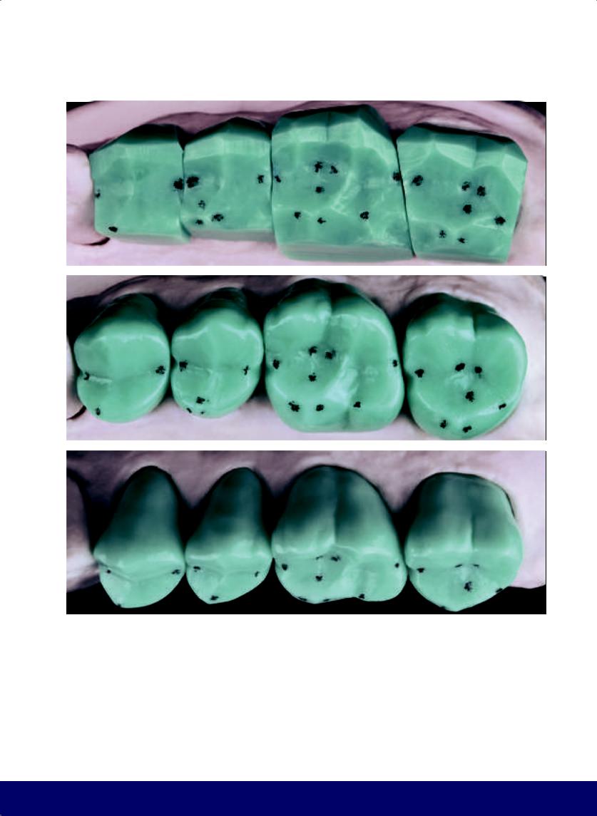
Fig 6-8 | (a to g) Sequence of positioning the main and secondary grooves on the teeth in the maxillary posterior quadrant. (h to j) Completed occlusal morphology for the quadrant.
148
@dentistinfo стоматологический телеграм канал
MAXILLARY POSTERIOR QUADRANT
f |
|
g |
|
|
|
h |
|
i |
|
|
|
j
149
@dentistinfo стоматологический телеграм канал
CHAPTER 6
a
b
Fig 6-9 | (a) Defined external contour. (b) Delineated primary grooves. (c and d) Completed occlusal surface, without secondary grooves. This reveals the three characteristics that define a tooth: the definition of the external contour, the delineation of the ridges, and the positioning of the primary grooves.
150
@dentistinfo стоматологический телеграм канал
MAXILLARY POSTERIOR QUADRANT
c
d
151
@dentistinfo стоматологический телеграм канал

Premolar
Mandibular First
07
152
@dentistinfo стоматологический телеграм канал
a |
b |
|
|
c |
d |
|
|
e |
f |
|
|
Fig 7-1 | (a) Maxillary contact points to be reproduced on the wax-up. (b and c) Buccal and occlusal views of the softened wax recording the occlusal morphology of the opposing maxillary dentition. Note that the contact points that were marked in pencil on the opposing teeth have also been reproduced on the wax. (d to f) Buccal and occlusal perspective of the wax block after removing excess wax.
MANDIBULAR FIRST PREMOLAR
153
@dentistinfo стоматологический телеграм канал
CHAPTER 7
a |
|
b |
|
|
|
c |
|
d |
|
e |
|
|
|
|
|
f |
|
g |
|
|
|
Fig 7-2 | (a to c) Lingual interference in working movement. (d and e) Carve away lingual interference. (f and g) Buccal interference in balancing movement. (Purple arrows indicate the movement of the mandible, while white arrows indicate the direction of instrumentation.)
154
@dentistinfo стоматологический телеграм канал
MANDIBULAR FIRST PREMOLAR
a |
|
b |
|
|
|
c |
d |
e |
f |
g |
Fig 7-3 | (a and b) Carve away buccal interference. (c to e) Interference in protrusive movement. (f and g) Carve away interference on protrusive movement.
155
@dentistinfo стоматологический телеграм канал
CHAPTER 7
a |
|
b |
|
|
|
c |
|
d |
|
e |
|
|
|
|
|
f |
g |
h |
Fig 7-4 | (a and b) There is no interference in retrusive movement. (c to e) Cut back wax to open up the buccal embrasures. (f to h) Divide the buccal face into two planes: a smaller mesial plane and a larger distal plane. Note the shape of the buccal face.
156
@dentistinfo стоматологический телеграм канал
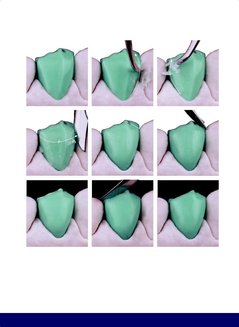
MANDIBULAR FIRST PREMOLAR
a |
|
b |
|
c |
|
|
|
|
|
d |
|
e |
|
f |
|
|
|
|
|
g |
h |
i |
Fig 7-5 | (a to e) Smooth the angles by rounding and finishing the buccal face. (f to i) Form the tip of the buccal cusp by working the mesial and distal segments of the longitudinal ridge of the buccal cusp.
157
@dentistinfo стоматологический телеграм канал
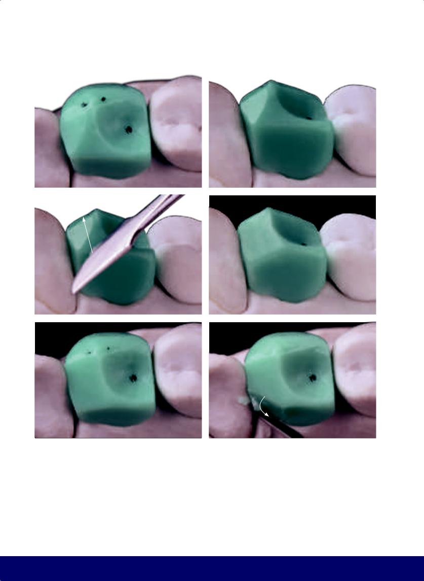
CHAPTER 7
a |
|
b |
|
|
|
c |
d |
|
|
e |
f |
Fig 7-6 | (a to e) Divide the occlusal surface in two parts: a smaller mesial part and a larger distal part. Reducing the inclination of the mesial segment forms the enamel bridge that will join the buccal and lingual cusps. (f to j) Open the lingual embrasures and round the definitive lingual face. (k and l) Create the kidney bean profile on the mesial marginal ridge. (m) In the occlusal view of the external contour, note the shorter inverted D on the mesial and larger D on the distal.
158
@dentistinfo стоматологический телеграм канал
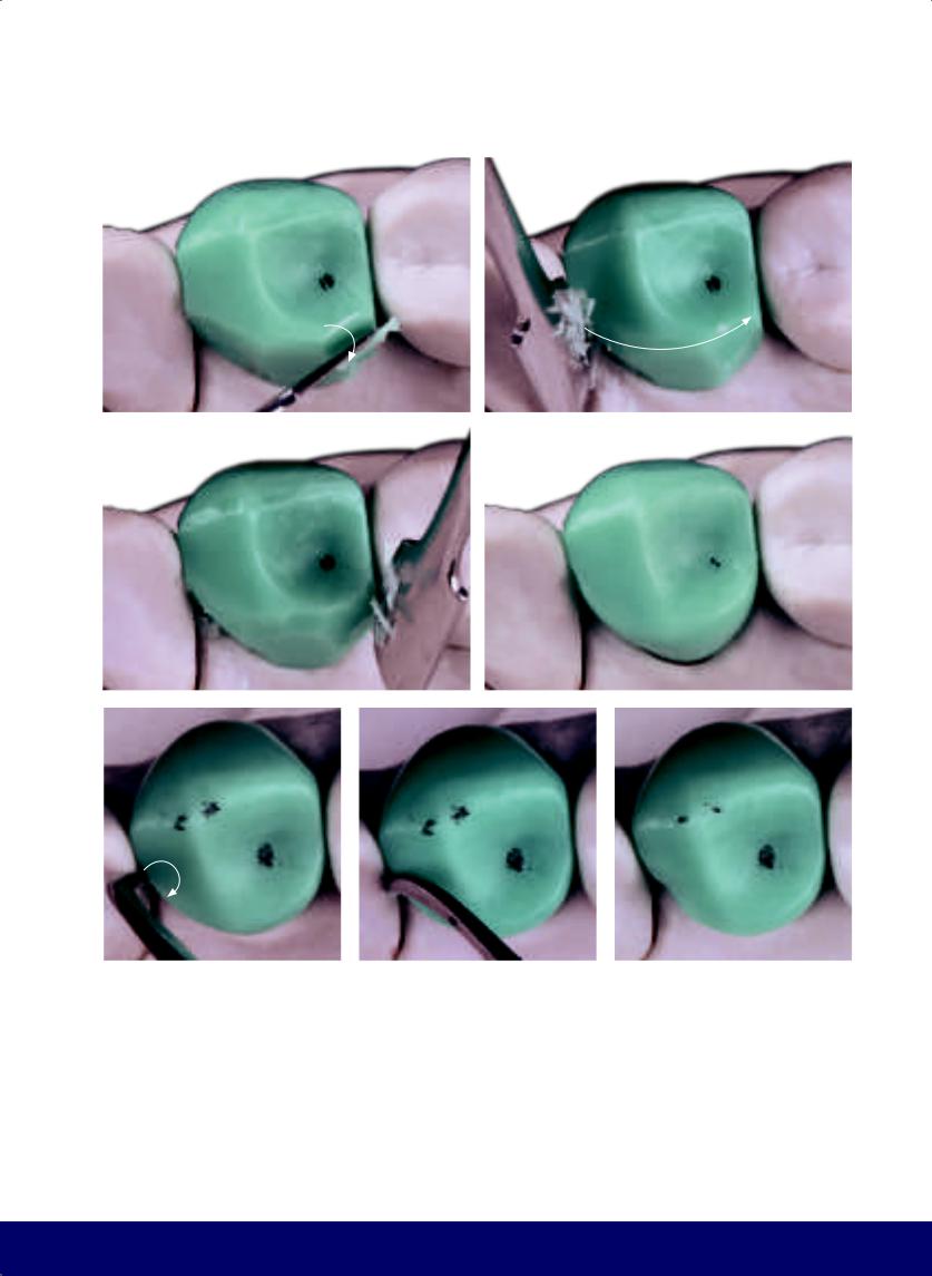
MANDIBULAR FIRST PREMOLAR
g |
|
h |
|
|
|
i |
|
j |
|
|
|
k |
|
l |
|
m |
|
|
|
|
|
159
@dentistinfo стоматологический телеграм канал
CHAPTER 7
a |
|
b |
|
|
|
c
d |
|
e |
|
|
|
Fig 7-7 | (a) Buccal face. Note that the mesial segment of the longitudinal ridge of the buccal cusp is smaller than the distal one, and consequently, the tip of the buccal cusp is slightly toward the mesial side. (b and c) The vertical macro texture consists of two developmental grooves. The mesial developmental groove has a beginning, middle, and end that follows from the area of the mesial papilla to the occlusal third. The distal developmental groove, however, only goes to the middle third. The term groove is used
160
metaphorically, as they are better described as broad depressions. (d and e) The horizontal macro texture is composed of the subtle horizontal depressions, the predominant one being in the middle third and following a trajectory opposite the crown/root line. This results in the narrow waist, which creates the shape of the buccal face and improves its silhouette.
@dentistinfo стоматологический телеграм канал

a |
b |
|
|
c |
d |
|
|
e |
f |
|
|
Fig 7-8 | (a) Observe the buccal macro texture from an occlusal perspective. (b) Mark the future mesial fossa and the distal fossa, where the tip of the lingual cusp of the maxillary first premolar touches. (c and d) The mesiodistal groove is the “smile” connecting the mesial and distal fossae that passes through the high relief of the enamel bridge. (e and f) Create the lingual groove from the mesial fossa.
MANDIBULAR FIRST PREMOLAR
161
@dentistinfo стоматологический телеграм канал
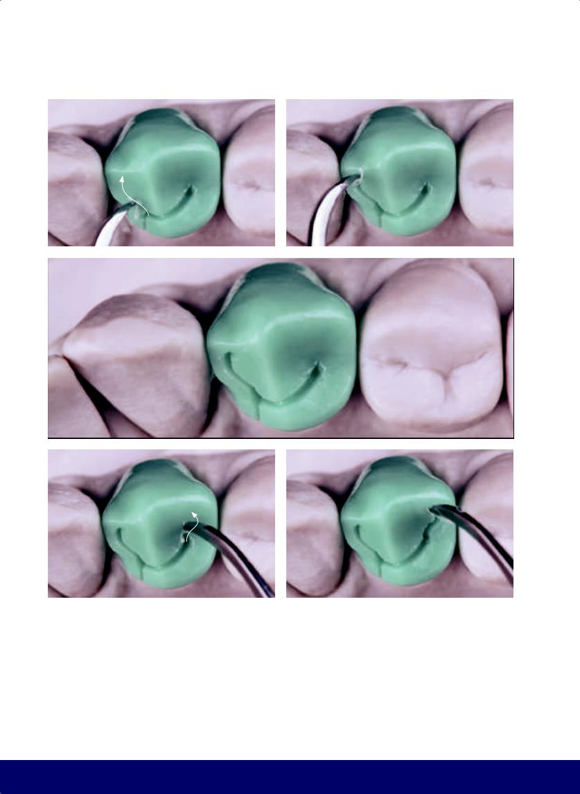
CHAPTER 7
a |
|
b |
|
|
|
c
d |
|
e |
|
|
|
Fig 7-9 | (a to e) The secondary grooves are an S and an inverted S. They begin from the mesial and distal fossae, respectively, and proceed toward the buccal face, forming two inward-curving Zebu horns. (f to i) Create two additional grooves to create a lobe on the distal marginal ridge. (j and k) Round the opening of the lingual groove.
162
@dentistinfo стоматологический телеграм канал
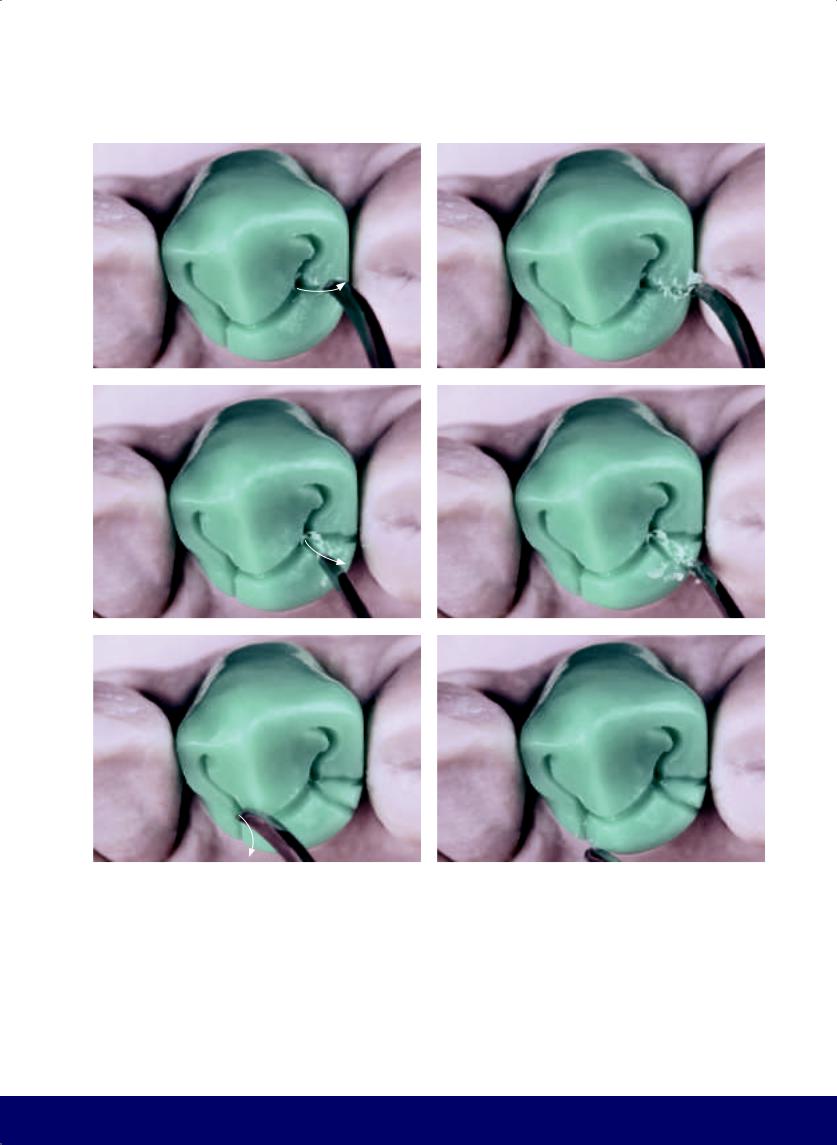
MANDIBULAR FIRST PREMOLAR
f |
|
g |
|
|
|
h |
|
i |
|
|
|
j |
|
k |
|
|
|
163
@dentistinfo стоматологический телеграм канал
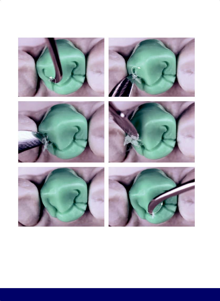
CHAPTER 7
a |
|
b |
|
|
|
c |
|
d |
|
|
|
e |
|
f |
|
|
|
Fig 7-10 | (a to l) Smooth the angles and open the grooves.
Enlarge and round the opening for the secondary grooves.
164
@dentistinfo стоматологический телеграм канал
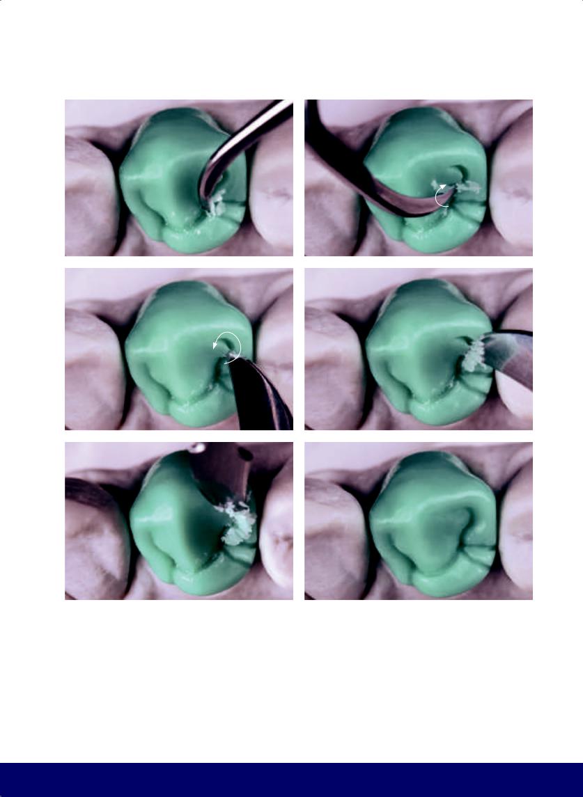
MANDIBULAR FIRST PREMOLAR
g |
|
h |
|
|
|
i |
|
j |
|
|
|
k |
|
l |
|
|
|
165
@dentistinfo стоматологический телеграм канал
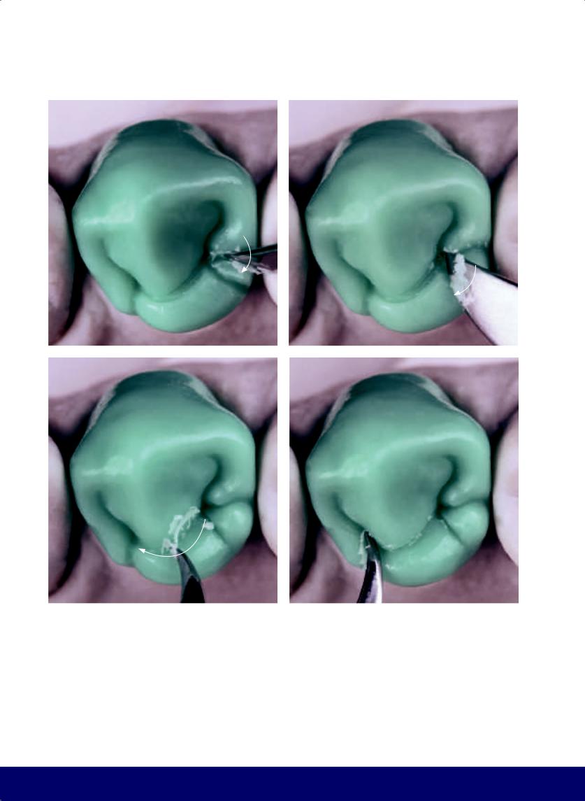
CHAPTER 7
a |
|
b |
|
|
|
c |
|
d |
|
|
|
Fig 7-11 | (a and b) Finish forming the lobe on the distal marginal crest. (c and d) Smooth and round the opening of the mesiodistal groove. (e) Use wax to form a lobe and create the wineglass shape on the distal grinding slope of the buccal cusp. It can be short and wide or thin and long. (f) Form a small depression on the mesial grinding slope of buccal cusp. (g) The definitive occlusal morphology. Note the mesiodistal groove, which separates a sizeable buccal cusp from the more modest lingual cusp. Also note
166
the secondary grooves (ie, S and inverted S), which together form two inward-curving Zebu horns. Note the kidney bean profile in the mesial marginal ridge; the mesial inverted D and the larger distal D, separated by the enamel bridge; the wineglass shape and lobes of the distal marginal ridge. In the occlusal view, the dominance of the mesial developmental groove is noted.
@dentistinfo стоматологический телеграм канал
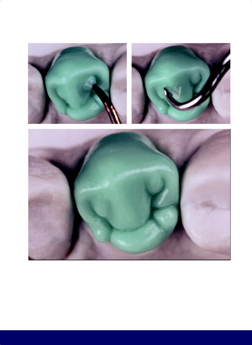
MANDIBULAR FIRST PREMOLAR
e |
|
f |
|
|
|
g
167
@dentistinfo стоматологический телеграм канал
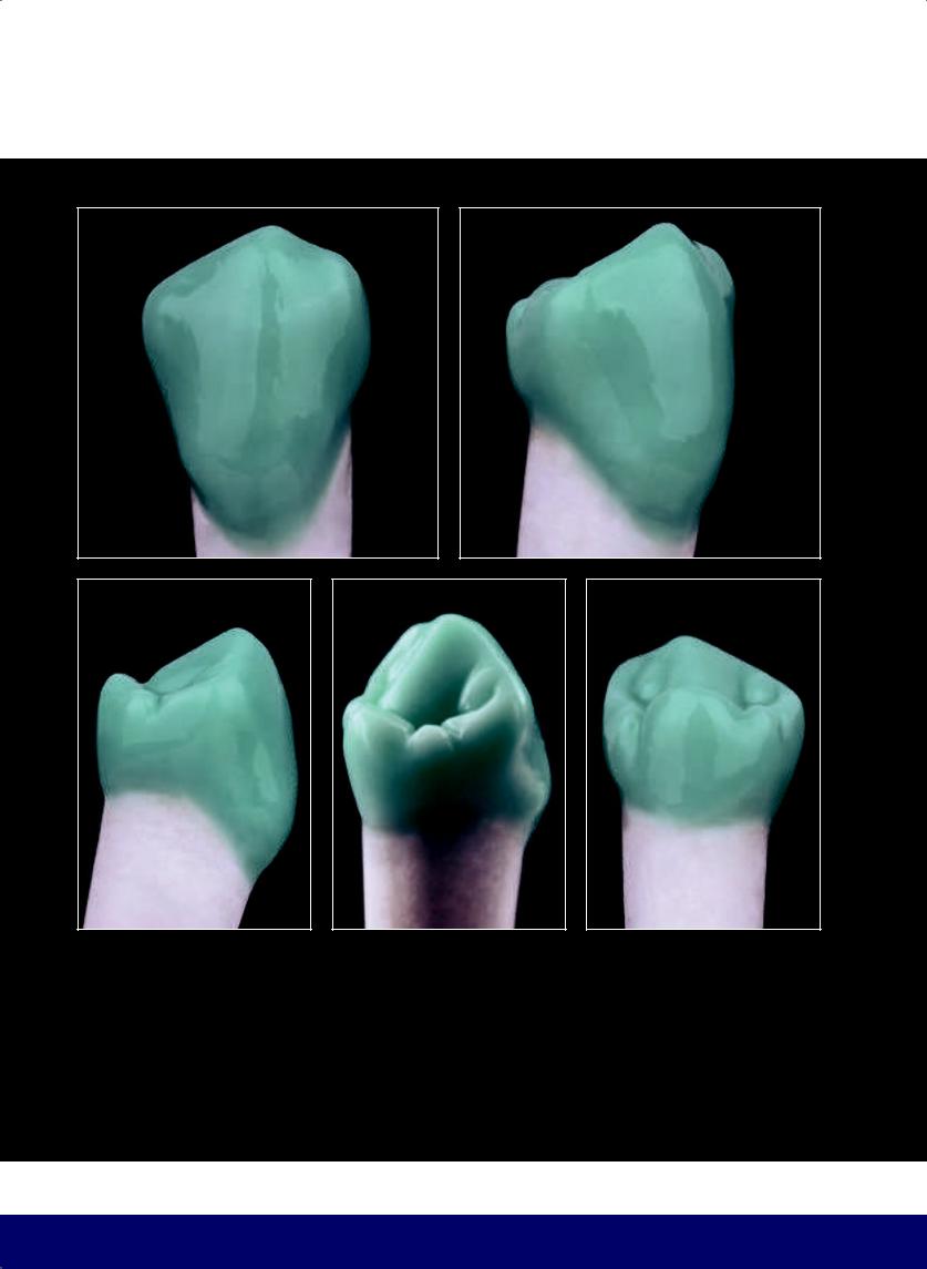
CHAPTER 7
a |
b |
c |
d |
e |
Fig 7-12 | (a to j) Completed contours of the mandibular first premolar.
168
@dentistinfo стоматологический телеграм канал
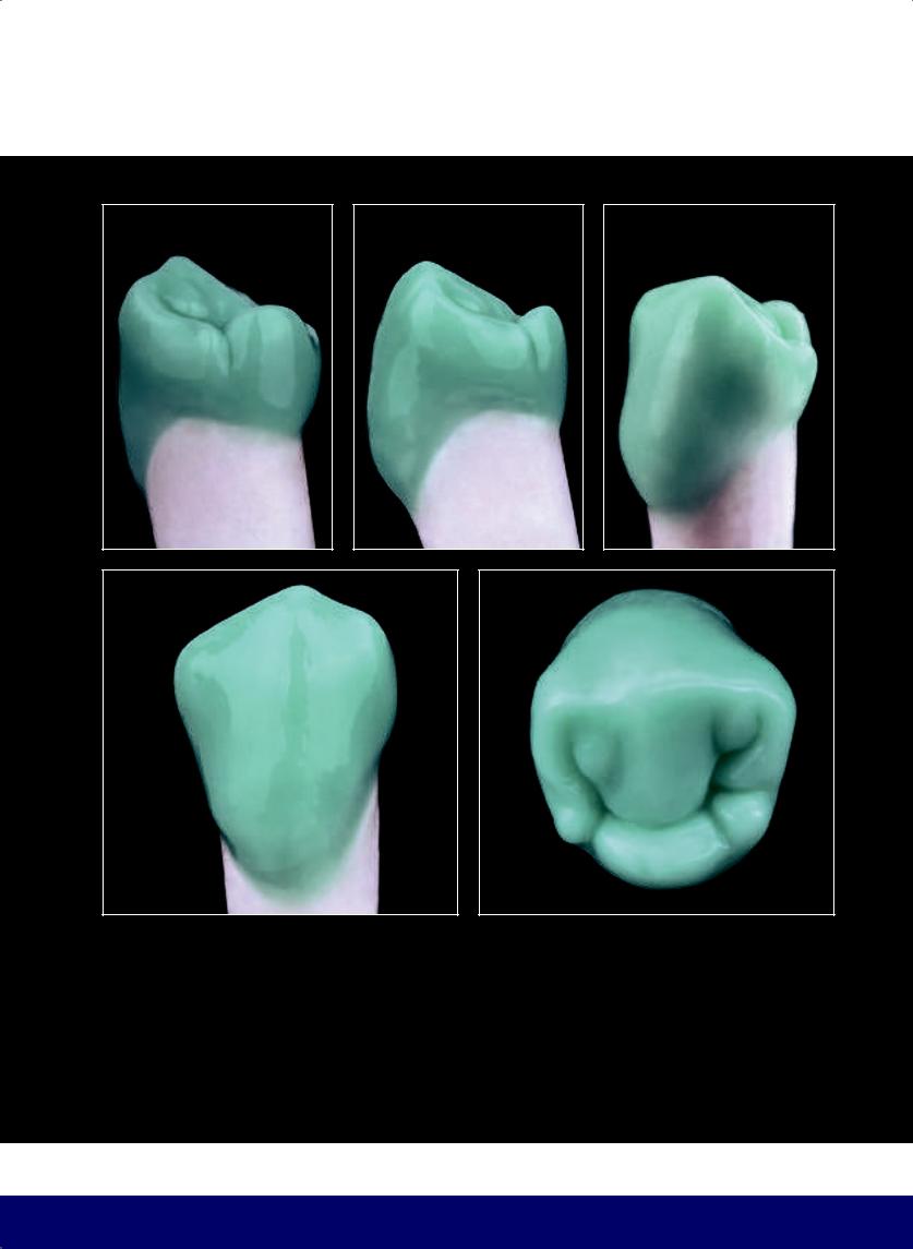
MANDIBULAR FIRST PREMOLAR
f |
g |
h |
i |
j |
169
@dentistinfo стоматологический телеграм канал
Premolar
Mandibular Second
08
170
@dentistinfo стоматологический телеграм канал
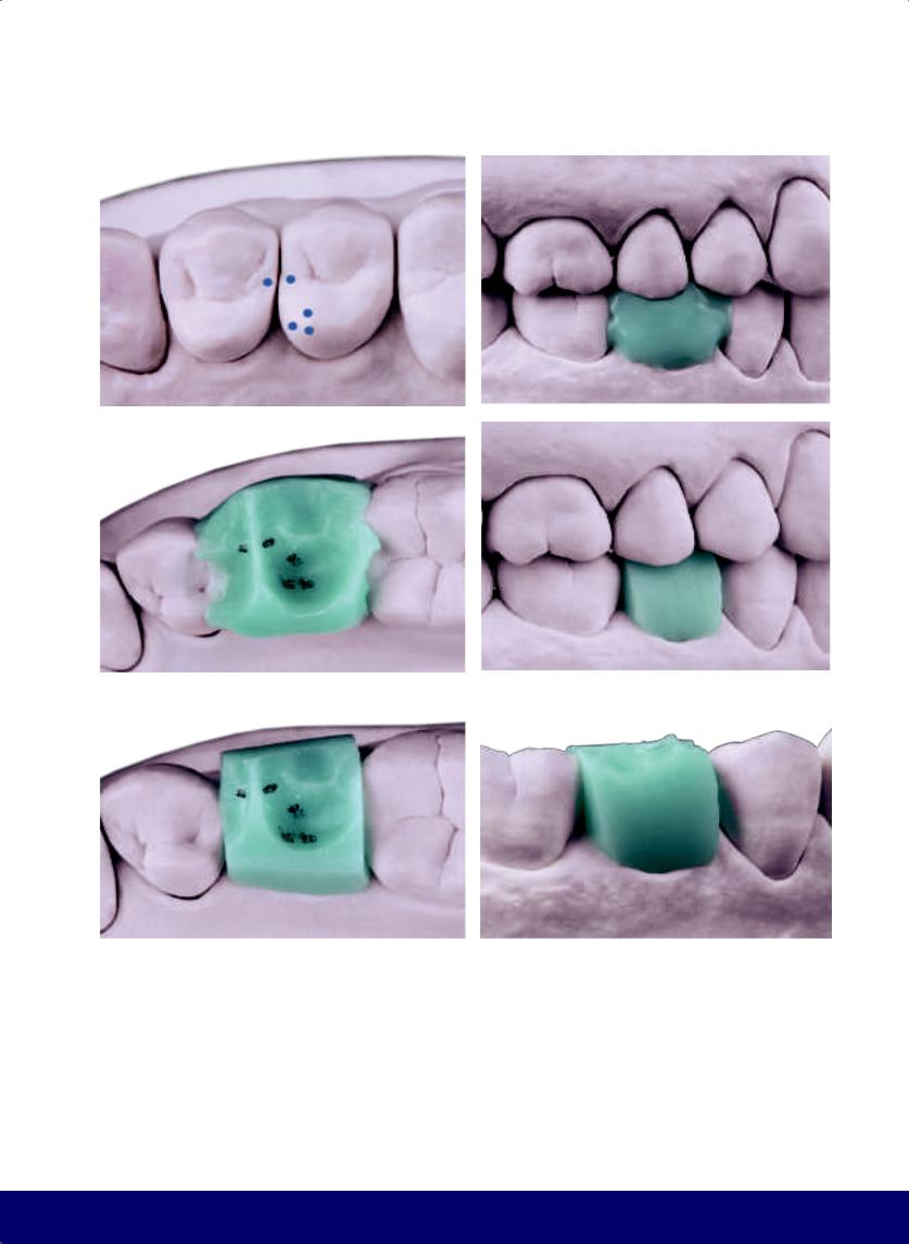
MANDIBULAR SECOND PREMOLAR
a |
|
b |
|
|
|
c |
|
d |
|
|
|
e |
|
f |
|
|
|
Fig 8-1 | (a) Centric contact points in the maxillary dentition. (b and c) With the cast in occlusion, the softened wax records the occlusal morphology of the opposing maxillary teeth. Note the contact points that were marked in pencil on the antagonists were reproduced in the wax. (d to f) Buccal and occlusal views of the wax block after removing excess wax.
171
@dentistinfo стоматологический телеграм канал
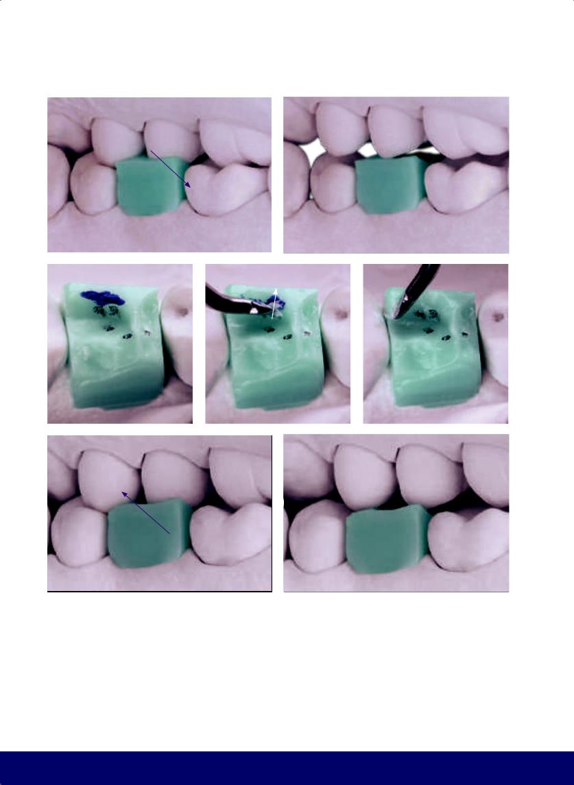
CHAPTER 8
a |
|
b |
|
|
|
c |
|
d |
|
e |
|
|
|
|
|
f |
g |
Fig 8-2 | (a to c) Lingual interference in working movement. (d and e) Carve away lingual interference. (f and g) Buccal interference in balancing movement. (Purple arrows indicate the movement of the mandible, while white arrows indicate the direction of instrumentation.)
172
@dentistinfo стоматологический телеграм канал
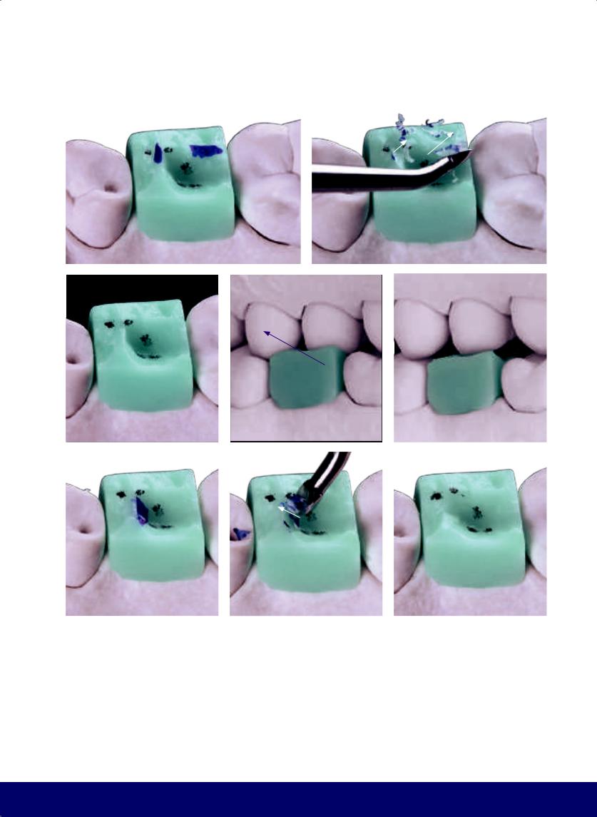
MANDIBULAR SECOND PREMOLAR
a |
|
b |
|
|
|
c |
d |
e |
f |
|
g |
|
h |
|
|
|
|
|
Fig 8-3 | (a to c) Carve away buccal interference. (d and e) Interference in protrusive movement. (f to h) Carve away interference on protrusive movement.
173
@dentistinfo стоматологический телеграм канал
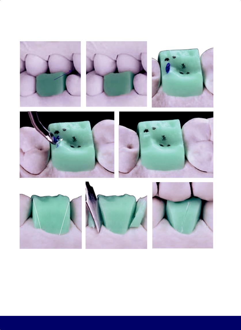
CHAPTER 8
a |
|
b |
|
c |
|
|
|
|
|
d |
e |
f |
|
g |
|
h |
|
|
|
|
|
Fig 8-4 | (a and b) Interference in retrusive movement. (c to e) Carve away interference on retrusive movement. (f and g) Open up the buccal embrasures. (h) Divide the buccal face into a smaller mesial part and a larger distal part.
174
@dentistinfo стоматологический телеграм канал
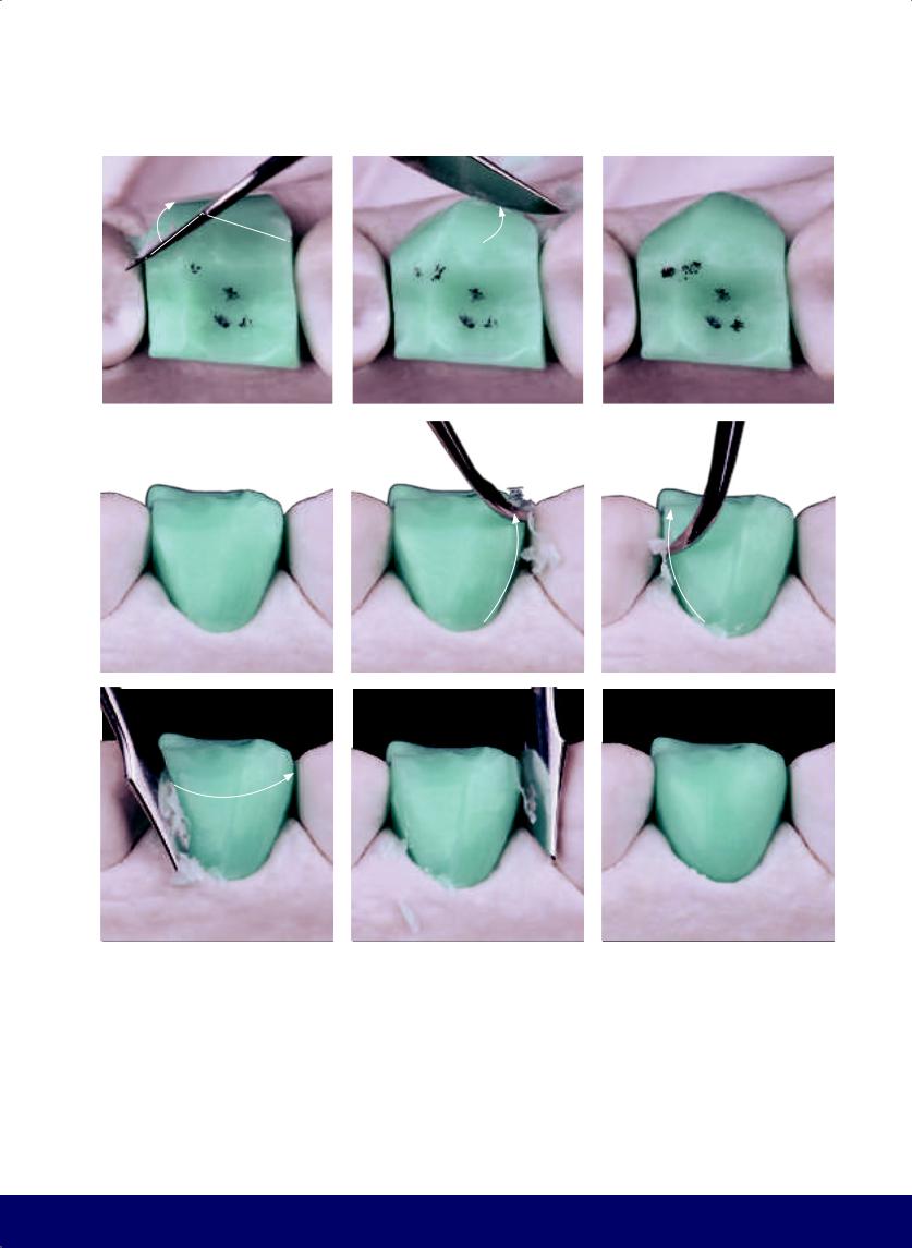
MANDIBULAR SECOND PREMOLAR
a |
|
b |
|
c |
|
|
|
|
|
d |
|
e |
|
f |
|
|
|
|
|
g |
h |
i |
Fig 8-5 | (a to d) Develop the mesial and distal sides of the buccal face. Note the curvature. (e to i) Soften the angles by rounding and finalizing the contour of the buccal face.
175
@dentistinfo стоматологический телеграм канал
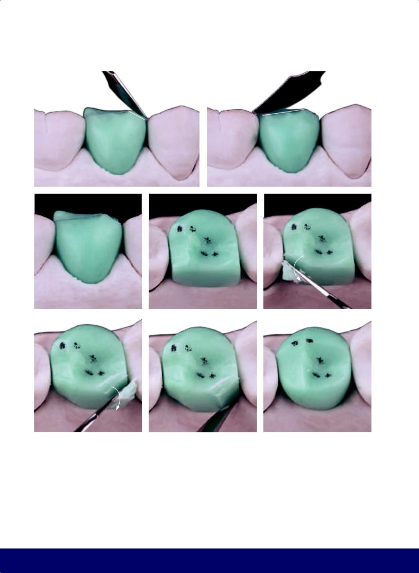
CHAPTER 8
a |
|
b |
|
|
|
c |
d |
e |
f |
|
g |
|
h |
|
|
|
|
|
Fig 8-6 | (a to c) Form the buccal cusp tip by working the mesial and distal segments of the longitudinal ridge of the buccal cusp. (d to h) Open the lingual embrasures and round the lingual surface.
176
@dentistinfo стоматологический телеграм канал
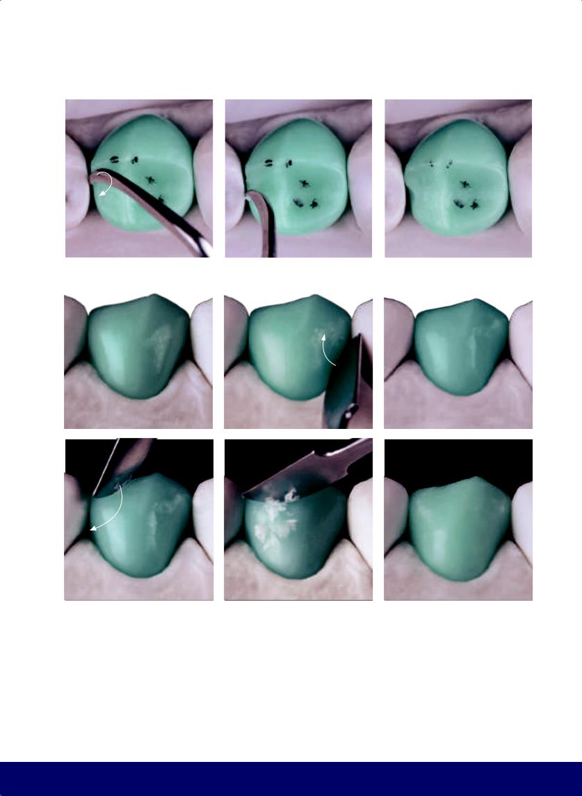
MANDIBULAR SECOND PREMOLAR
a |
|
b |
|
c |
|
|
|
|
|
d |
|
e |
|
f |
|
|
|
|
|
g |
h |
i |
Fig 8-7 | (a and b) Create the kidney bean profile on the mesial marginal ridge. (c) This view shows the division of the occlusal surface into two parts: the smaller inverted D on the mesial and larger D on the distal. (d) As in the first premolar, the mesial segment of the longitudinal ridge of the buccal cusp is smaller than the distal segment, and consequently, the tip of the buccal cusp slightly turns toward the mesial side. Note that the buccal face is divided into two parts, with the mesial side being smaller and the distal
side larger. (e to i) The vertical macro texture is composed of two developmental grooves but with characteristics that are opposite to the first premolar. The mesial developmental groove only goes to the middle third, not reaching the occlusal third. In addition, the distal developmental groove has a beginning, middle, and end, which follows from the distal papilla to the occlusal third. As mentioned previously, these “grooves” are really wide depressions.
177
@dentistinfo стоматологический телеграм канал
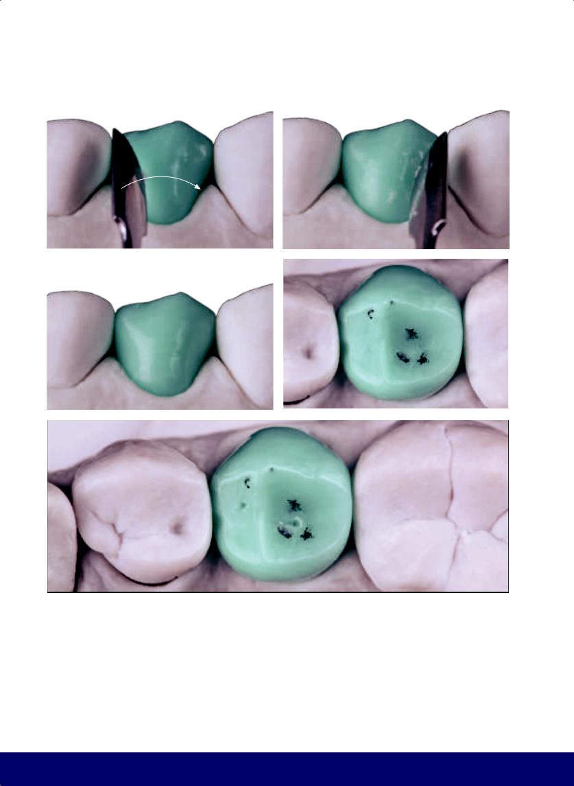
CHAPTER 8
a |
|
b |
|
|
|
c |
|
d |
|
|
|
e
Fig 8-8 | (a and b) The horizontal macro texture is most visible in the shape of the buccal face and its narrowed waist. (c) Note the flat area on the buccal face. (d and e) Mark the distal fossa, where it touches the tip of the lingual cusp of the maxillary second premolar, and the mesial fossa.
178
@dentistinfo стоматологический телеграм канал
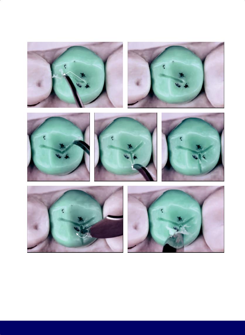
a |
b |
|
|
c |
d |
f |
g |
Fig 8-9 | (a to e) Open the mesiodistal groove and the working groove. Note that three lines start from the distal fossa: the mesial and distal segments of the mesiodistal groove as well as the working groove. Together they form the letter Y. (f and g) Widen the lingual groove without breaking the longitudinal ridge of the lingual cusp, thus maintaining the entire cusp.
MANDIBULAR SECOND PREMOLAR
e
179
@dentistinfo стоматологический телеграм канал
CHAPTER 8
a |
|
b |
|
|
|
c |
|
d |
|
|
|
e |
|
f |
|
|
|
Fig 8-10 | (a to c) Open the distal end of the mesiodistal groove. (d to i) Round and deepen the mesial fossa of the mesiodistal groove toward the buccal and lingual, creating a significant depression. (j to m) Round the buccal and lingual edges of the mesiodistal groove.
180
@dentistinfo стоматологический телеграм канал
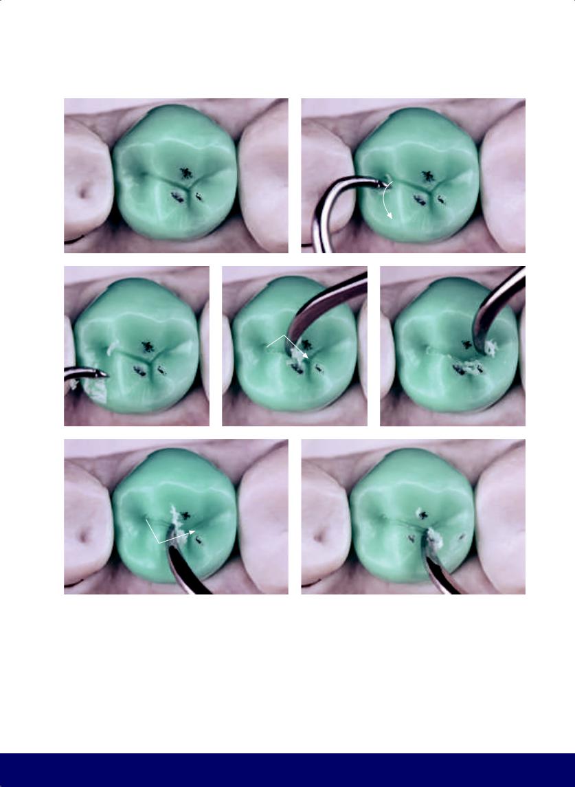
MANDIBULAR SECOND PREMOLAR
g |
|
h |
|
|
|
i |
|
j |
|
k |
|
|
|
|
|
l |
|
m |
|
|
|
181
@dentistinfo стоматологический телеграм канал
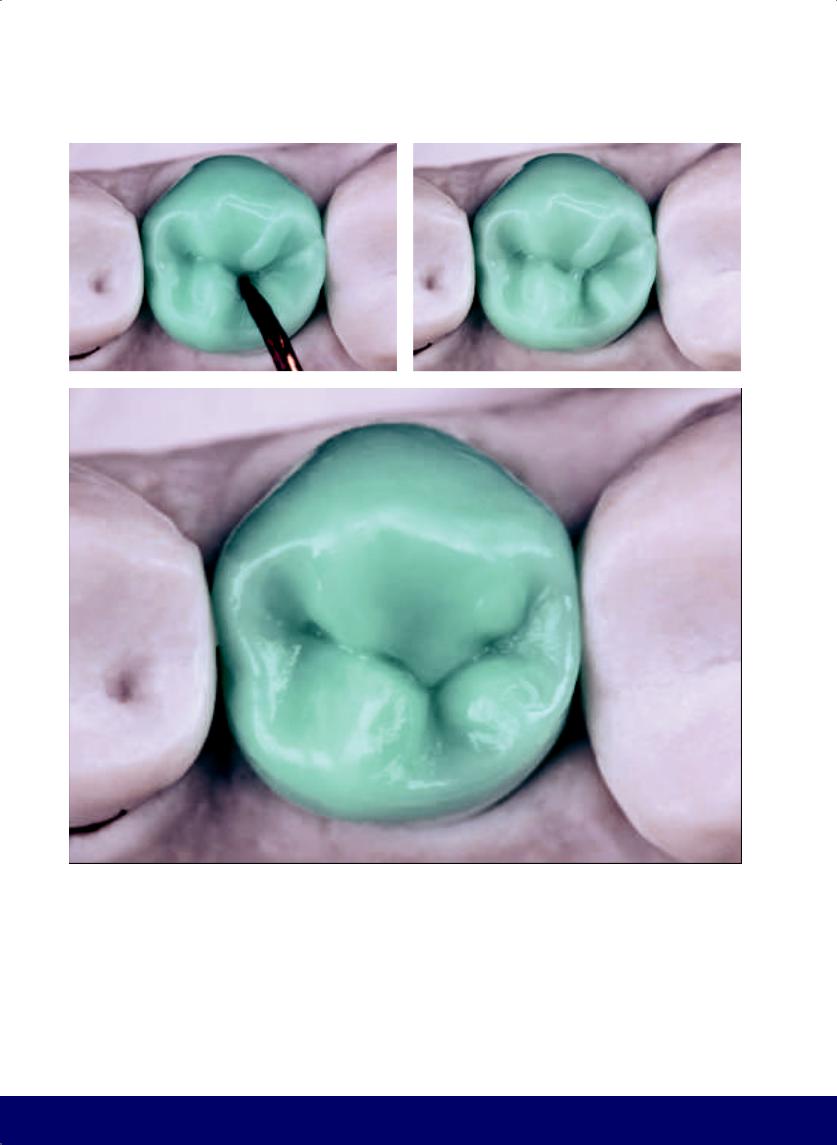
CHAPTER 8
a |
|
b |
|
|
|
c
Fig 8-11 | (a and b) Add wax to create the wineglass shape on the grinding slope distal to the buccal cusp. This lobe can be short and wide or thin and long. (c) Occlusal view of finalized morphology and external contour. Note the Y of the mesiodistal groove positioned at the center of the occlusal surface. It is still slightly lingual, revealing buccal and lingual cusps of similar volumes. Also
182
note the secondary grooves that start tentatively and open at the end in the form of a letter S. Note the kidney bean profile in the mesial marginal ridge, the two Ds (mesial and distal), and the mesiodistal groove forming a smile, which together with the working lingual groove form the letter Y.
@dentistinfo стоматологический телеграм канал
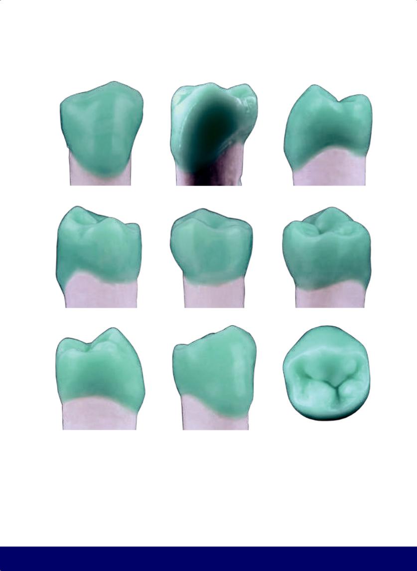
MANDIBULAR SECOND PREMOLAR
a |
|
b |
|
c |
|
|
|
|
|
d |
|
e |
|
f |
|
|
|
|
|
g |
|
h |
|
i |
|
|
|
|
|
Fig 8-12 | (a to i) External contour and occlusal morphology of the mandibular second premolar.
183
@dentistinfo стоматологический телеграм канал

First Molar
Mandibular
09
184
@dentistinfo стоматологический телеграм канал
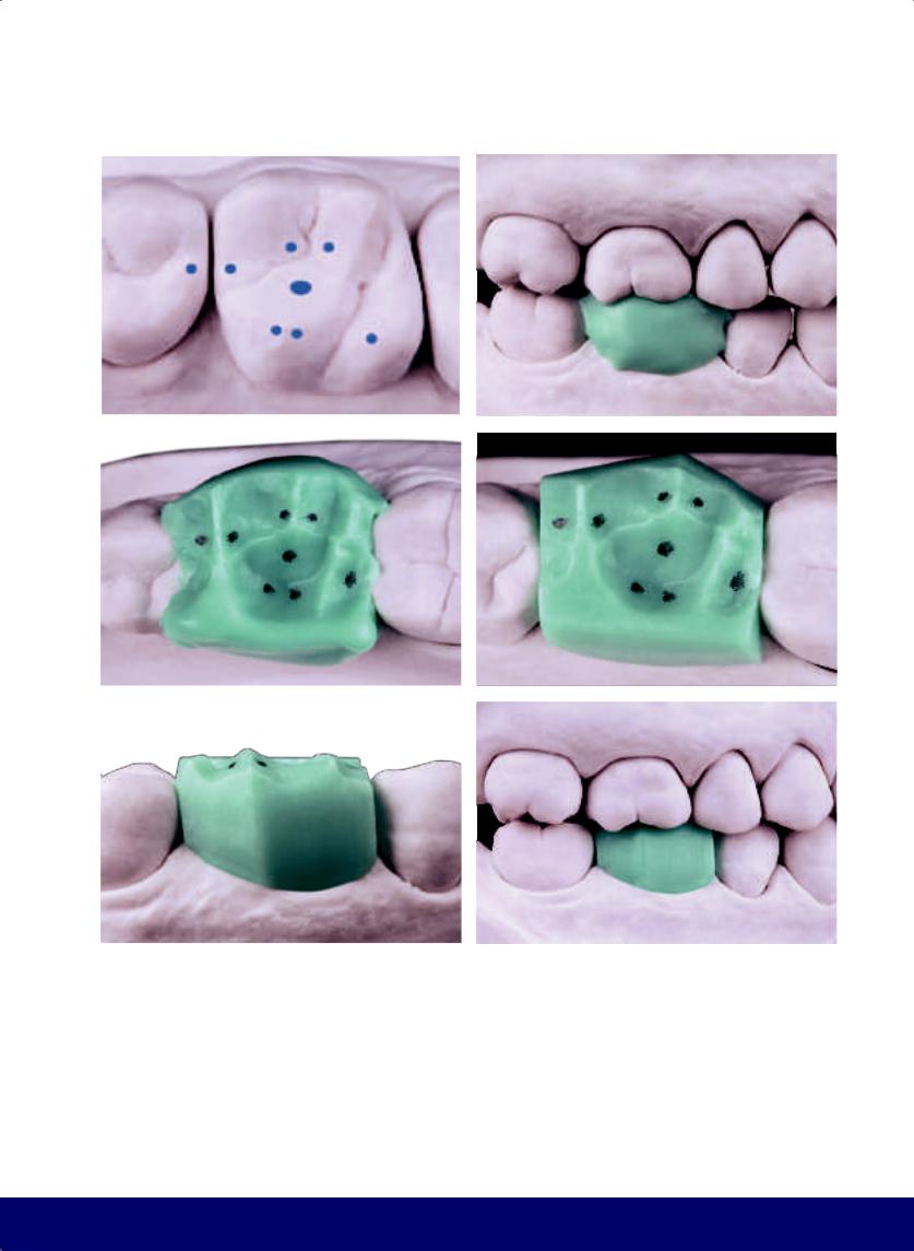
a |
b |
|
|
c |
d |
|
|
e |
f |
|
|
Fig 9-1 | (a) Contact points in the maxillary dentition to be reproduced in the softened wax. (b and c) Buccal and occlusal views. Note that the contact points marked in pencil on the antagonist were reproduced in the wax. (d and e) Buccal and occlusal perspective of the wax block after being trimmed. (f) The buccal face is divided into two parts, with a virtual line situated from the buccal groove of the maxillary first molar to the mesial centric contact point of the longitudinal ridge of the mesiobuccal cusp of the mandibular first molar.
MANDIBULAR FIRST MOLAR
185
@dentistinfo стоматологический телеграм канал
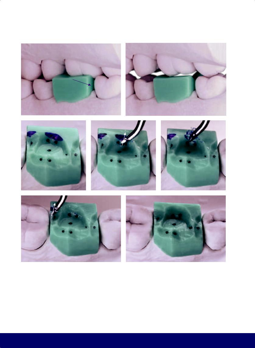
CHAPTER 9
a |
|
b |
|
|
|
c |
|
d |
|
e |
|
|
|
|
|
f |
|
g |
|
|
|
Fig 9-2 | (a to c) Lingual interference in working movement. Observe where the mesioand distobuccal cusps of the maxillary first molar touch the wax. (d to g) Carve away lingual interference. (Purple arrows indicate the movement of the mandible, while white arrows indicate the direction of instrumentation.)
186
@dentistinfo стоматологический телеграм канал
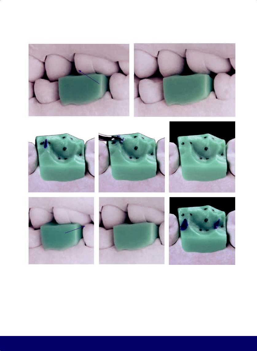
a |
b |
|
|
c |
|
d |
|
|
|
f |
|
g |
|
|
|
Fig 9-3 | (a to c) Buccal interference in balancing movement. (d and e) Carve away buccal interference. (f to h) Interference in protrusive movement.
MANDIBULAR FIRST MOLAR
e
h
187
@dentistinfo стоматологический телеграм канал
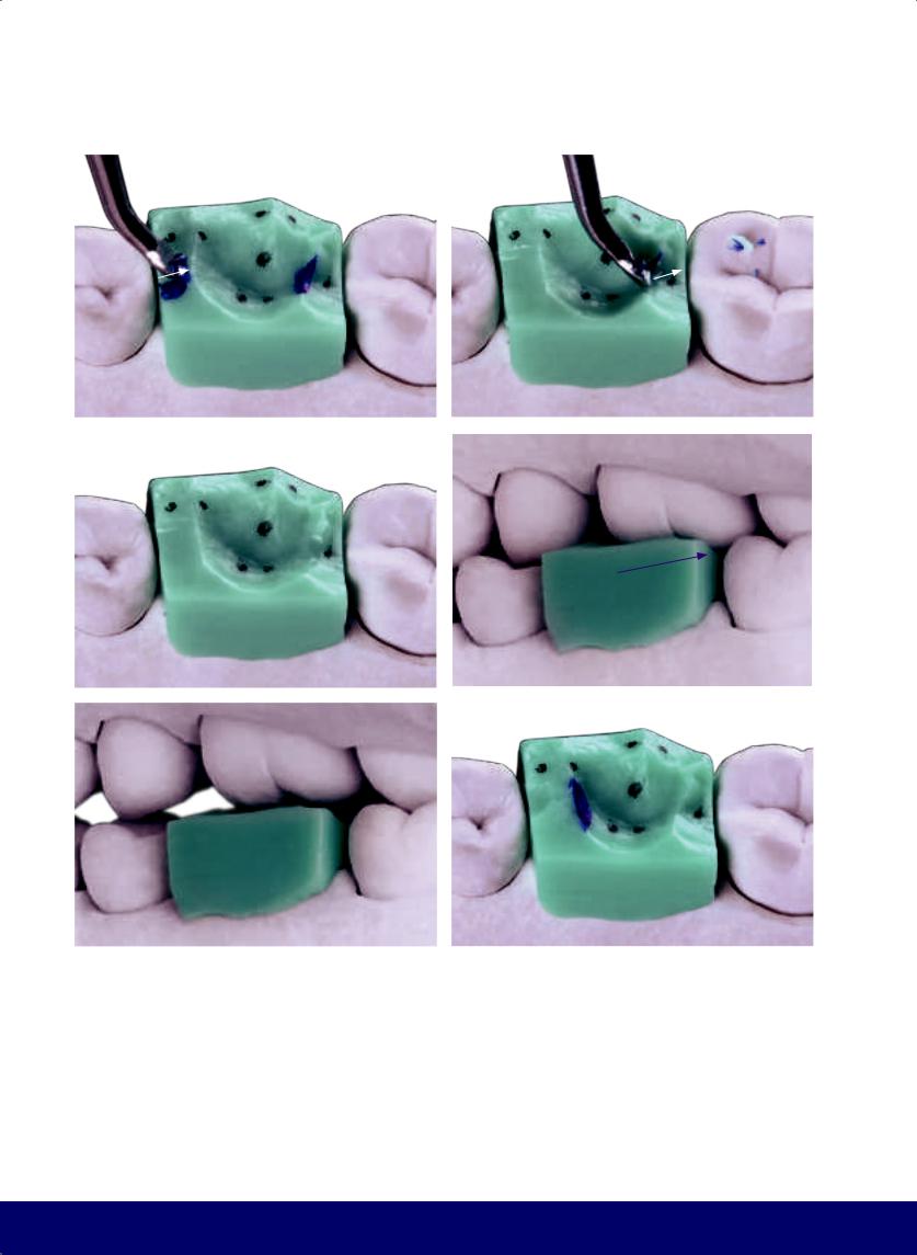
CHAPTER 9
a |
|
b |
|
|
|
c |
|
d |
|
|
|
e |
|
f |
|
|
|
Fig 9-4 | (a to c) Carve away interference on protrusive movement. (d to f) Interference in retrusive movement.
188
@dentistinfo стоматологический телеграм канал
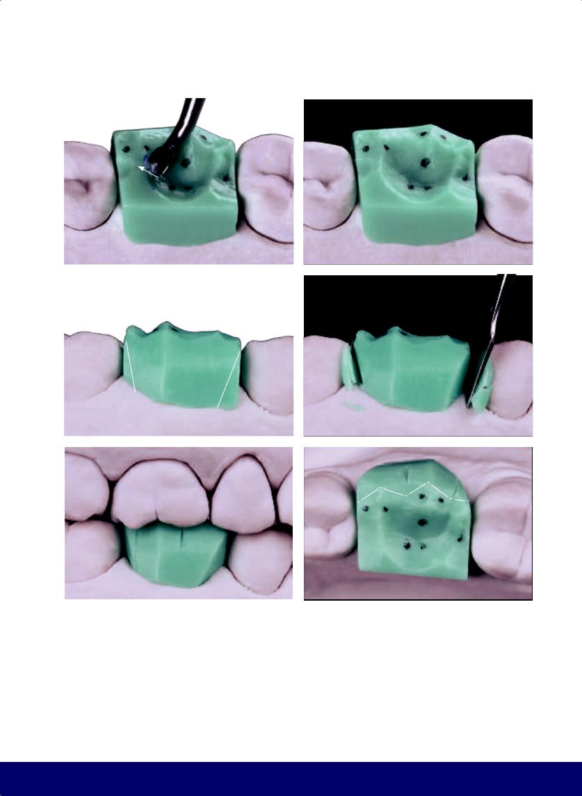
a |
b |
|
|
c |
d |
|
|
e |
f |
|
|
Fig 9-5 | (a and b) Carve away interference on retrusive movement. (c and d) Open the buccal embrasures. (e and f) The references for locating of the mesioand distobuccal grooves of the mandibular first molar are, respectively, the tips of the mesioand distobuccal cusps of the maxillary first molar.
MANDIBULAR FIRST MOLAR
189
@dentistinfo стоматологический телеграм канал
CHAPTER 9
a |
|
b |
|
|
|
c |
|
d |
|
e |
|
|
|
|
|
f |
|
g |
|
h |
|
|
|
|
|
Fig 9-6 | (a to h) Form and define the buccal aspect.
190
@dentistinfo стоматологический телеграм канал
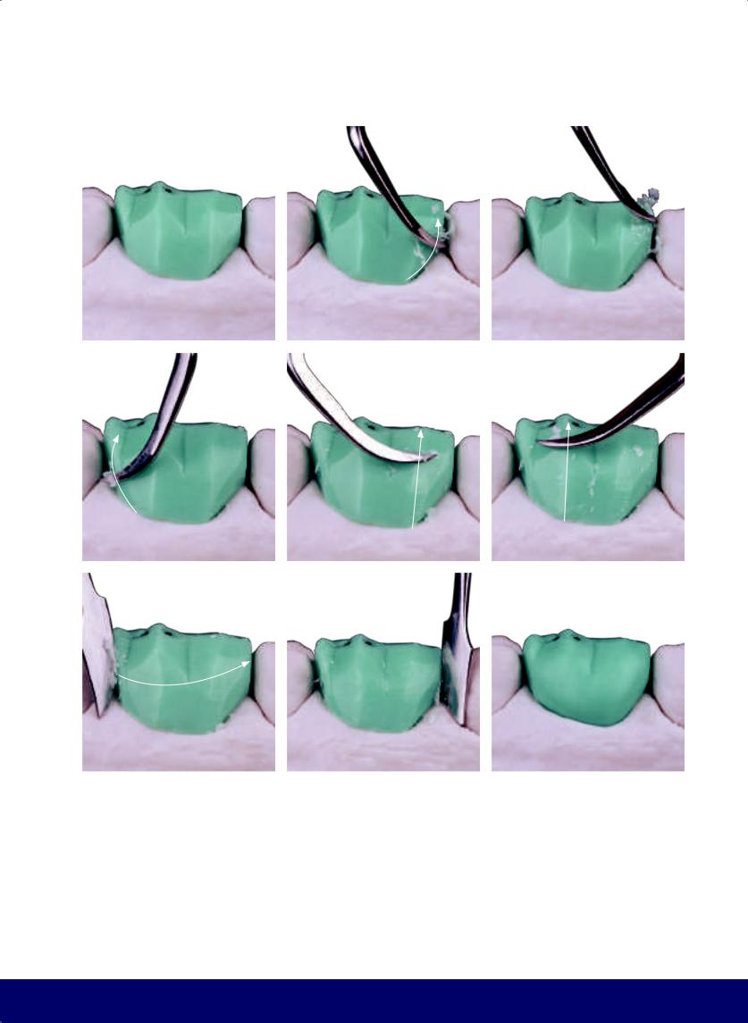
MANDIBULAR FIRST MOLAR
a |
|
b |
|
c |
|
|
|
|
|
d |
|
e |
|
f |
|
|
|
|
|
g |
|
h |
|
i |
|
|
|
|
|
Fig 9-7 | (a to i) Round the angles and complete the buccal face.
191
@dentistinfo стоматологический телеграм канал
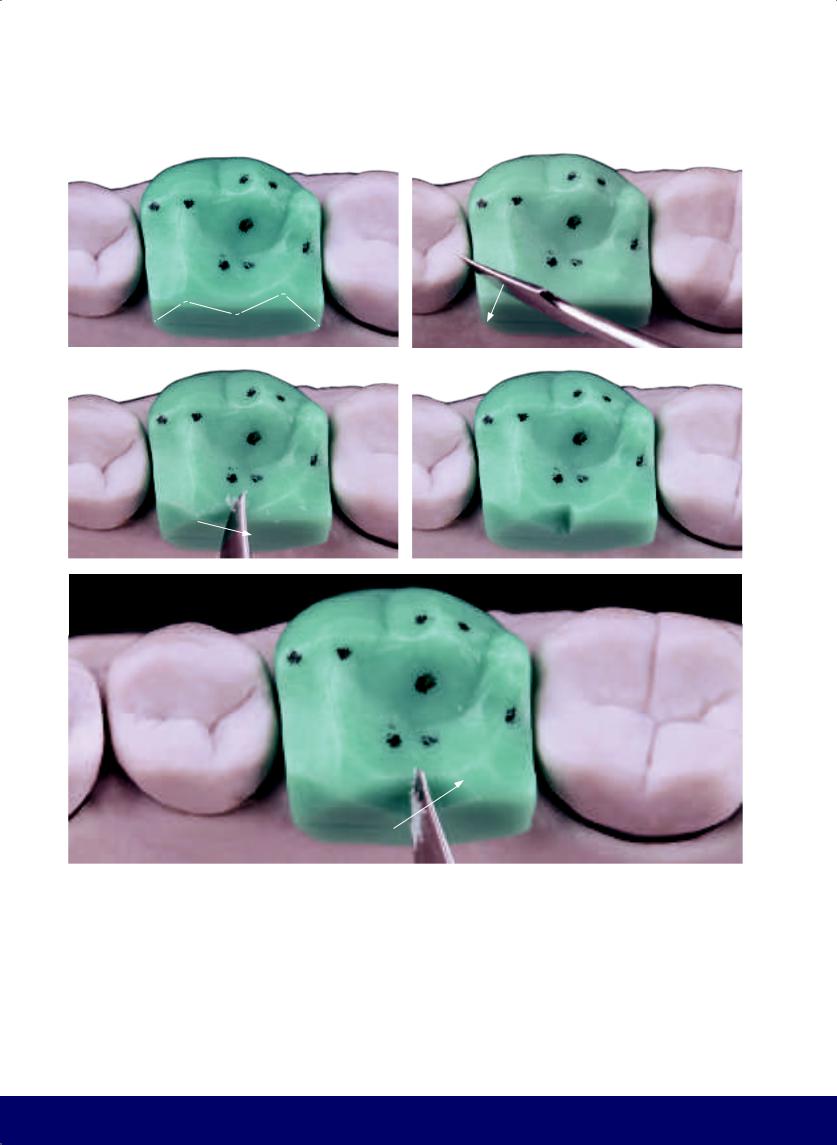
CHAPTER 9
a |
|
b |
|
|
|
c |
|
d |
|
|
|
e
Fig 9-8 | (a to f) Carve the lingual ridges to form the cusps. (g to j) Open the lingual embrasures.
192
@dentistinfo стоматологический телеграм канал
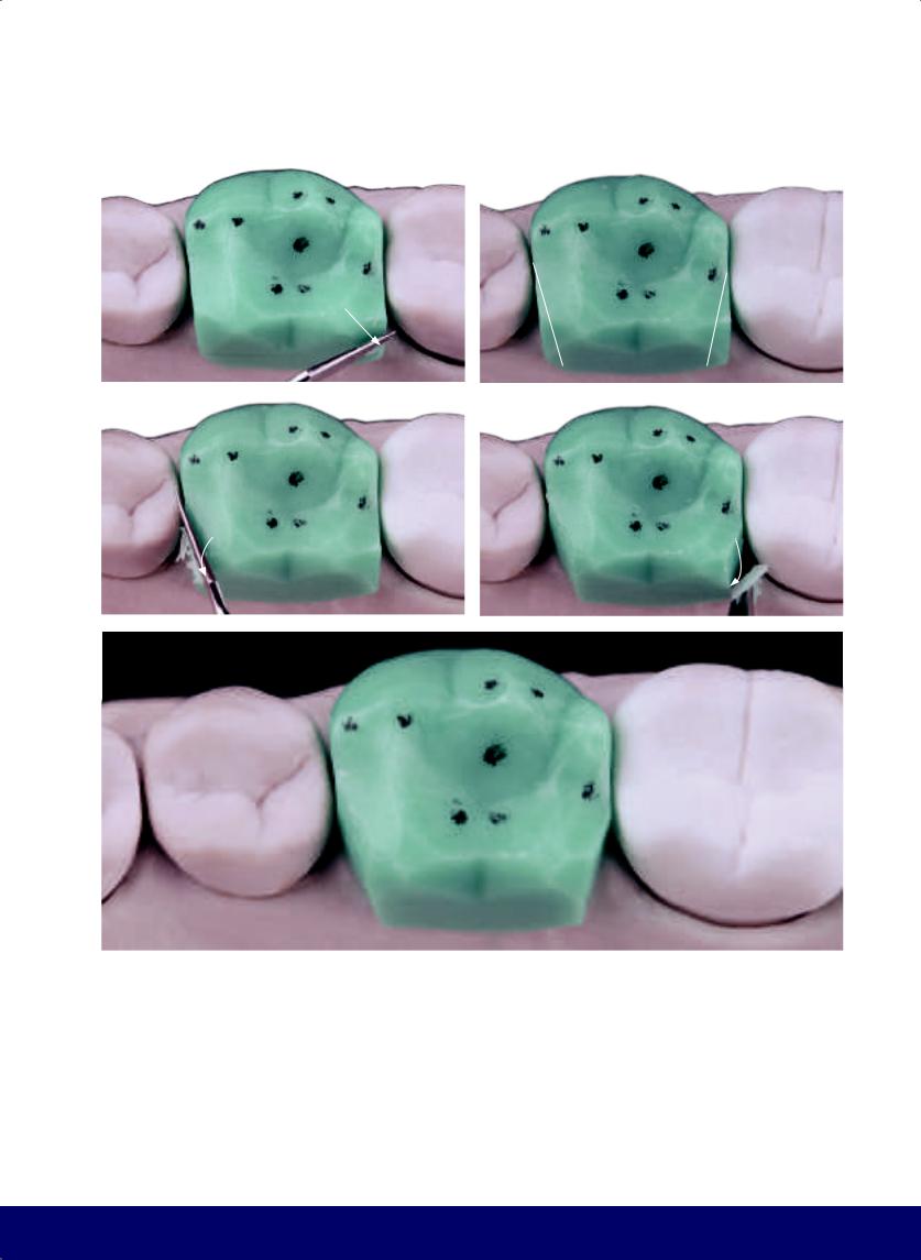
MANDIBULAR FIRST MOLAR
f |
|
g |
|
|
|
h |
|
i |
|
|
|
j
193
@dentistinfo стоматологический телеграм канал
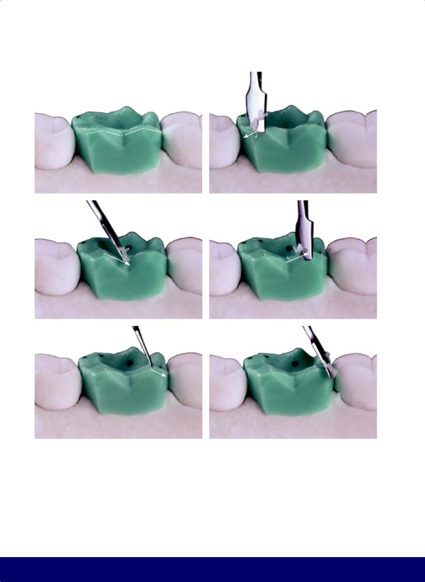
CHAPTER 9
a |
|
b |
|
|
|
c |
|
d |
|
|
|
e |
|
f |
|
|
|
Fig 9-9 | (a to f) Finalize the positioning of the cusp tips.
194
@dentistinfo стоматологический телеграм канал
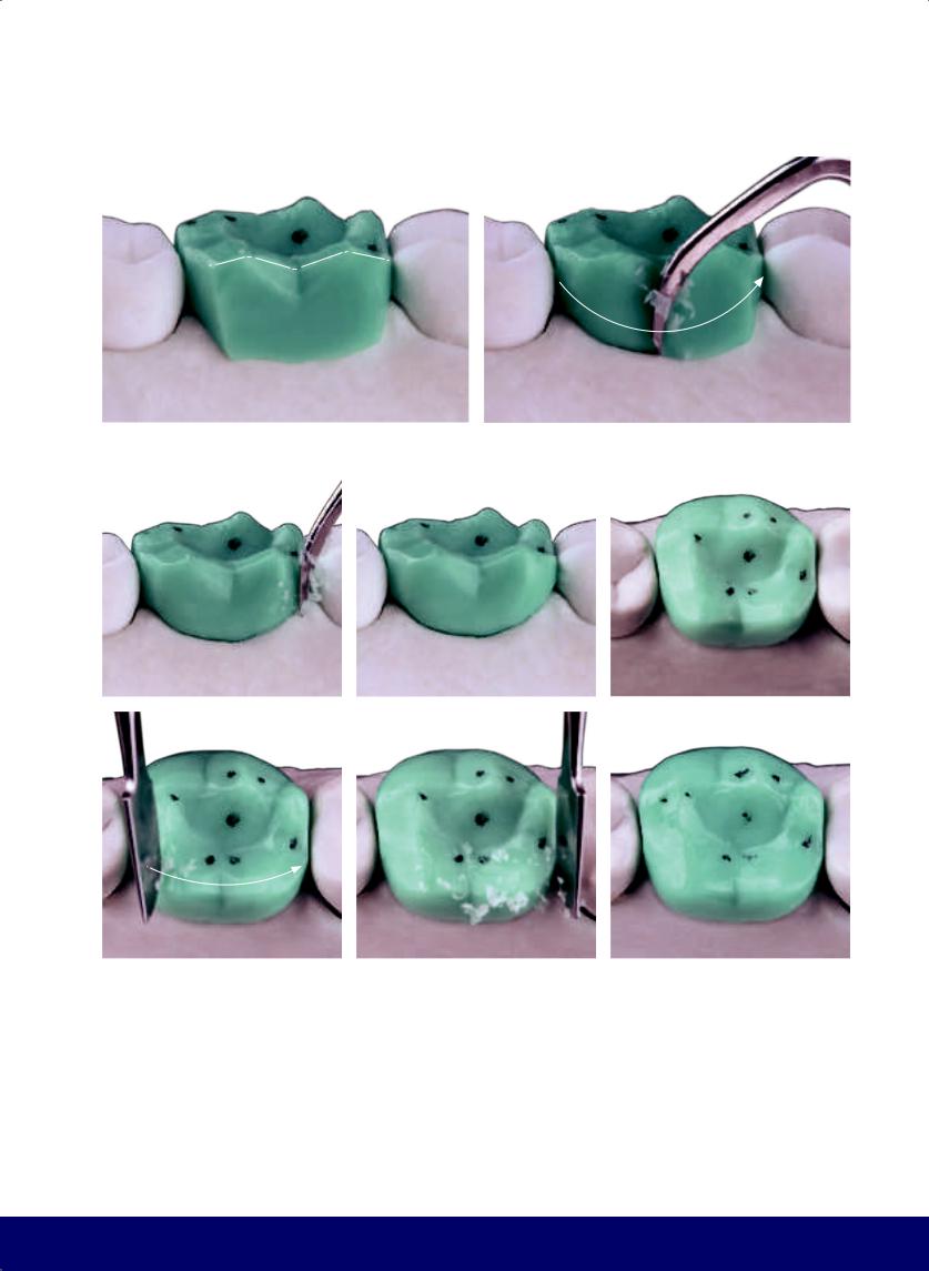
MANDIBULAR FIRST MOLAR
a |
|
b |
|
|
|
c |
|
d |
|
e |
|
|
|
|
|
f |
|
g |
|
h |
|
|
|
|
|
Fig 9-10 | (a to h) Soften the whole lingual aspect as well as the cusp tips.
195
@dentistinfo стоматологический телеграм канал
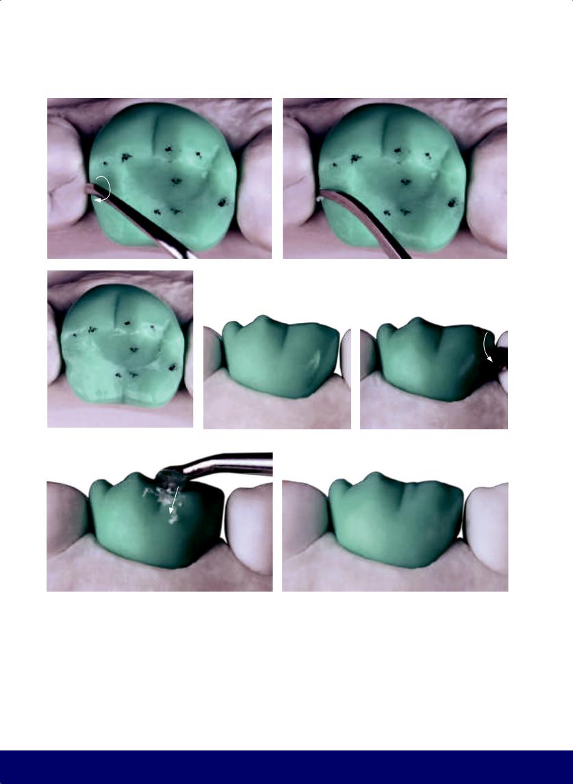
CHAPTER 9
a |
|
b |
|
|
|
c |
|
d |
|
e |
|
|
|
|
|
f |
|
g |
|
|
|
Fig 9-11 | (a to c) Place the kidney bean profile on the mesial marginal ridge above the contact area. (d to g) The macro vertical texture includes the mesial and distal pseudo developmental grooves on the mesiobuccal cusp.
196
@dentistinfo стоматологический телеграм канал
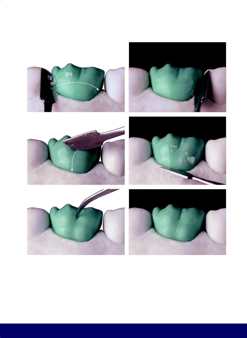
a |
b |
|
|
c |
d |
|
|
e |
f |
|
|
Fig 9-12 | (a and b) Macro horizontal texture shapes the buccal face with the resulting narrow waist. (c and d) The buccal face is divided by a depression running from the buccal groove to the furcation of the mesioand distobuccal roots. (e and f) Form the buccal fossa, the double hook that results from the juxtaposition of the horizontal macro texture and the mesiobuccal groove.
MANDIBULAR FIRST MOLAR
197
@dentistinfo стоматологический телеграм канал
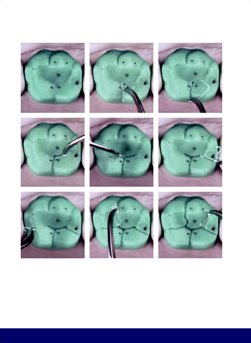
CHAPTER 9
a |
|
b |
|
c |
|
|
|
|
|
d |
|
e |
|
f |
|
|
|
|
|
g |
|
h |
|
i |
|
|
|
|
|
Fig 9-13 | (a) Occlusal view of the final outer contour shows the macro vertical and horizontal textures. Note the three external points that will open into the central grooves. (b and c) Form the lingual groove (ie, working groove) that connects the central fossa to the vertex of the angle formed between the lingual cusps. (d and e) Form groove extensions from the bottom of the mesial fossa to create the mesiobuccal groove. Note the Mercedes star. The distal segment is slightly longer than the mesial segment. (f) Open the protrusive groove. Note that it is directed toward the
198
distolingual embrasure. (g) Conclude the mesiodistal sulcus and finish the letter M. (h) Connect the mesial vertex of the letter M with the mesial external reference point to create the mesiobuccal sulcus. Note that the first segment of this groove turns to the center before turning back to the buccal aspect. (i) Connect the distal vertex of the letter M with the distal external reference point to create the distobuccal groove. Note that the first segment of this groove turns to the center before turning back to the buccal aspect.
@dentistinfo стоматологический телеграм канал
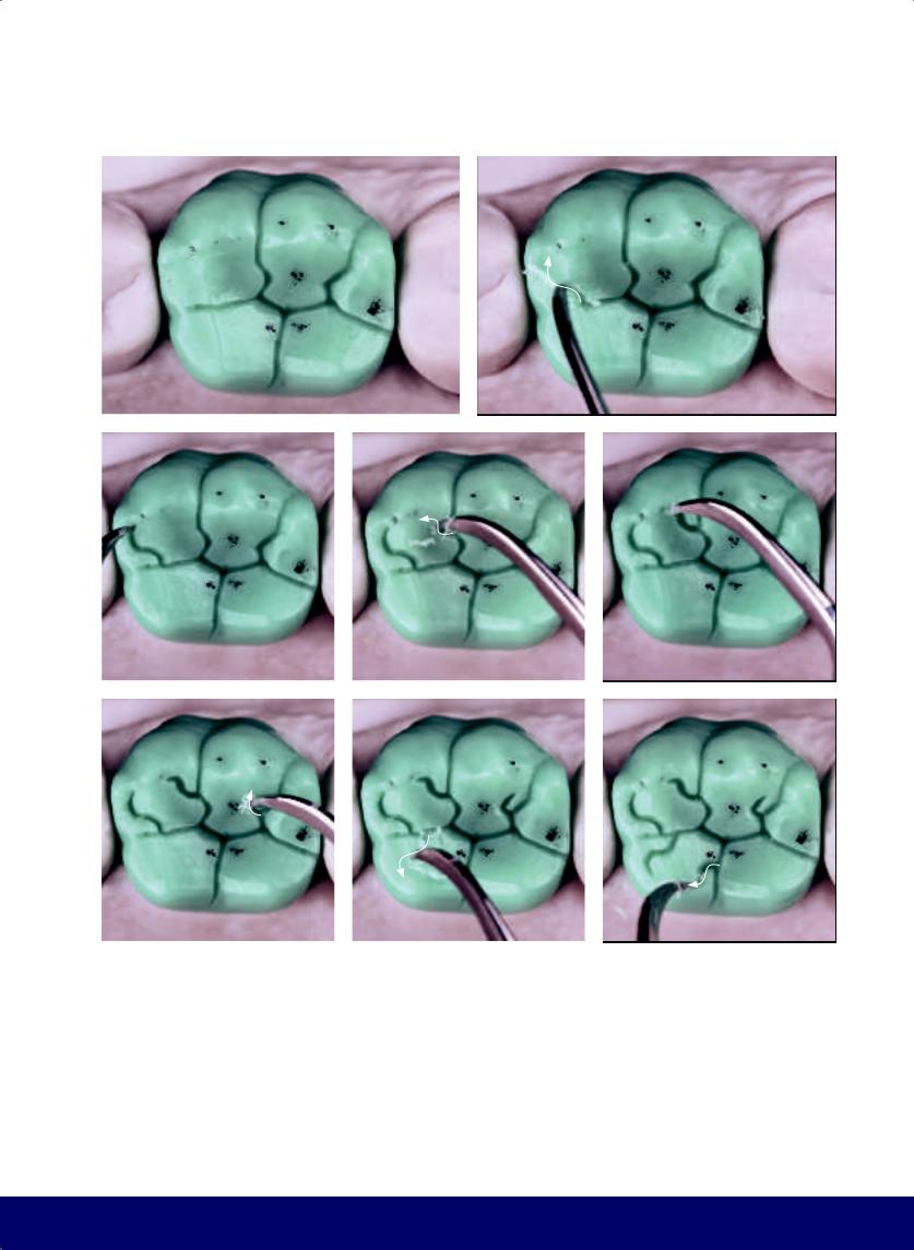
a |
b |
|
|
c |
|
d |
|
|
|
f |
|
g |
|
|
|
Fig 9-14 | (a) Completed primary grooves. (b to e) Create the inward-curving horns on the mesiobuccal cusp. The letter S next to the mesiodistal sulcus as well as the mesial arm of the Mercedes star form the retrusive groove. (f) Create a secondary groove from the distobuccal groove to form a J hook. (g and h) Create the inward-curving horns on the mesiolingual cusp.
MANDIBULAR FIRST MOLAR
e
h
199
@dentistinfo стоматологический телеграм канал
CHAPTER 9
a |
|
b |
|
|
|
c |
|
d |
|
|
|
e |
|
f |
|
|
|
Fig 9-15 | (a to d) Create the inward-curving horns on the distolingual cusp. (e to l) Soften the angles and open up the ends of the grooves, still preserving the transverse and longitudinal ridges.
200
@dentistinfo стоматологический телеграм канал
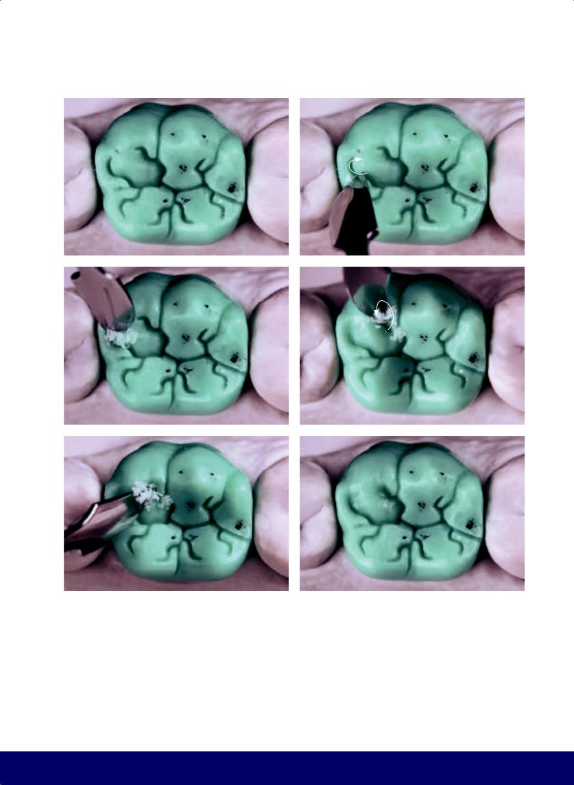
MANDIBULAR FIRST MOLAR
g |
|
h |
|
|
|
i |
|
j |
|
|
|
k |
|
l |
|
|
|
201
@dentistinfo стоматологический телеграм канал
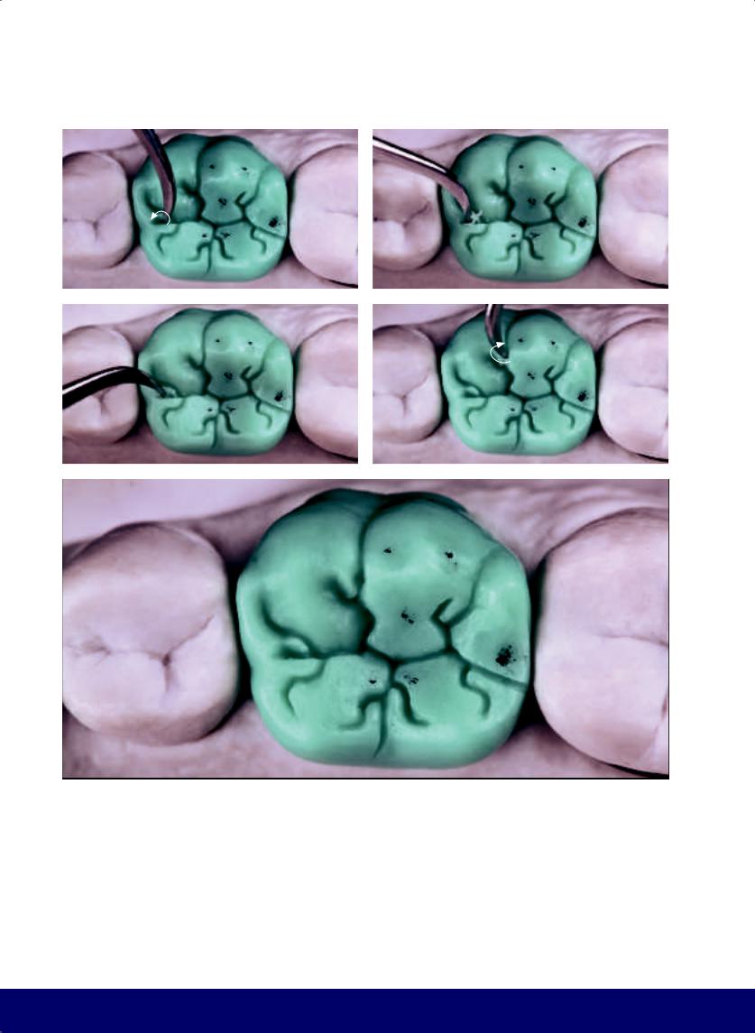
CHAPTER 9
a |
|
b |
|
|
|
c |
|
d |
|
|
|
e
Fig 9-16 | (a to e) Gently slope the furrows and corners of the primary grooves and create an “elbow.”
202
@dentistinfo стоматологический телеграм канал
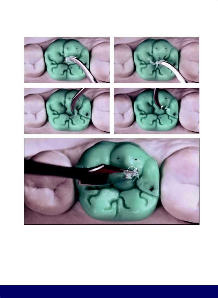
MANDIBULAR FIRST MOLAR
a |
|
b |
|
|
|
c |
|
d |
|
|
|
e
Fig 9-17 | (a to e) Round the corners of the mesial cusp and open the end of the J hook.
203
@dentistinfo стоматологический телеграм канал
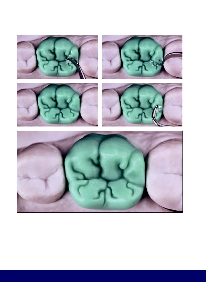
CHAPTER 9
a |
|
b |
|
|
|
c |
|
d |
|
|
|
e
Fig 9-18 | (a to c) Round the corners of the distobuccal cusp. (d and e) Create a minimal depression on the distobuccal cusp.
204
@dentistinfo стоматологический телеграм канал
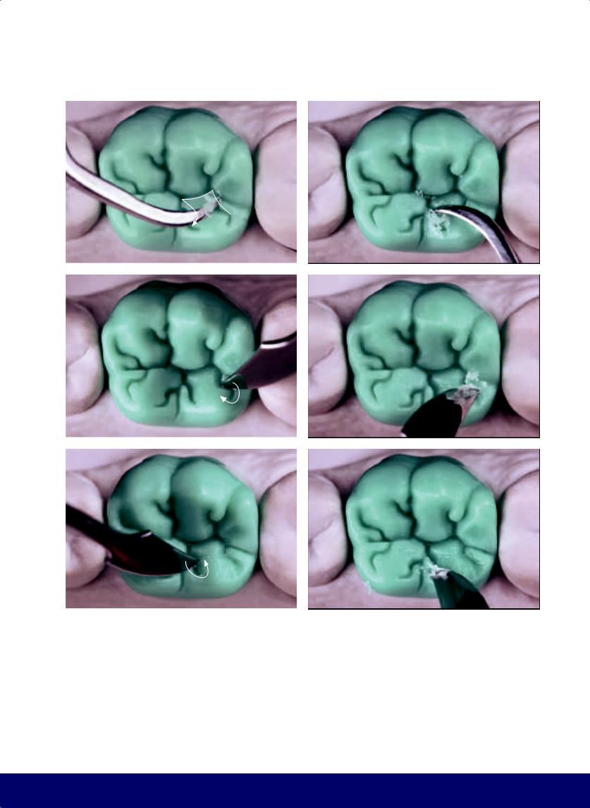
a |
b |
|
|
c |
d |
|
|
e |
f |
|
|
Fig 9-19 | (a to f) Round the corners and open the ends of the grooves on the distolingual cusp, preserving all transverse and longitudinal ridges.
MANDIBULAR FIRST MOLAR
205
@dentistinfo стоматологический телеграм канал
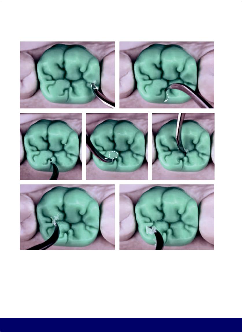
CHAPTER 9
a |
|
b |
|
|
|
c |
|
d |
|
e |
|
|
|
|
|
f |
|
g |
|
|
|
Fig 9-20 | (a to c) Gently slope the furrows and corners of the primary grooves and create an elbow. (d to k) Round the corners and open the ends of the grooves, preserving all the transverse and longitudinal ridges of the mesiolingual cusp. (l and m) The inclined plane of the ridge and the corner from the primary groove creates an elbow. (n) Occlusal morphology and finished external contour. The mesiodistal groove, or letter M positioned at the center of the occlusal surface, delineates the vestibular and lingual cusps with similar dimensions and ends in the distolingual groove.
206
Note the absence of mesiobuccal and mesiolingual grooves. Also the secondary grooves start tentatively and open at the end in the form of a J (on the mesial cusp) and an S (on the distal cusp). Together, these secondary grooves constitute the inward-curving horns. Note the kidney bean profile on the marginal mesial ridge. The profile of the longitudinal ridge of the lingual cusps resembles a key. The vertical and horizontal macro texture is visible from this occlusal perspective.
@dentistinfo стоматологический телеграм канал
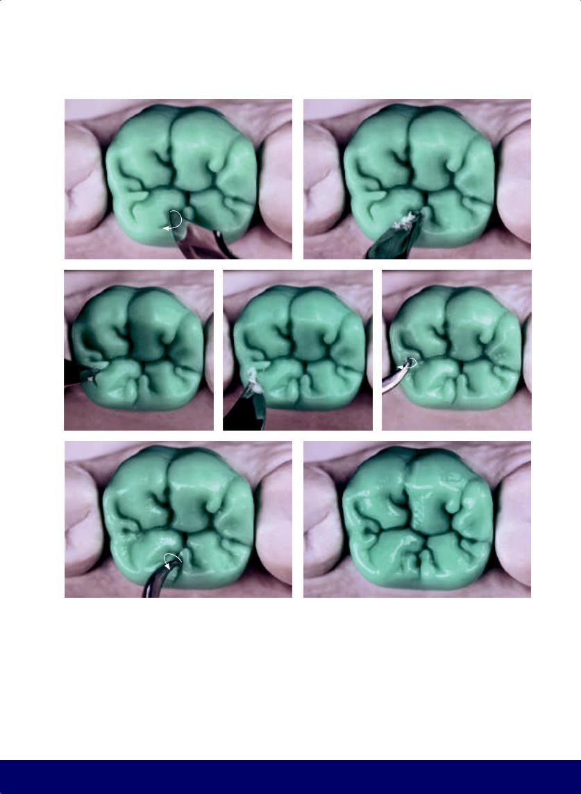
MANDIBULAR FIRST MOLAR
h |
|
i |
|
|
|
j |
|
k |
|
l |
|
|
|
|
|
m |
|
n |
|
|
|
207
@dentistinfo стоматологический телеграм канал
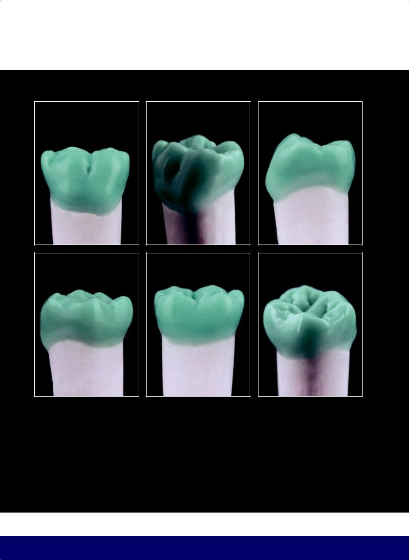
CHAPTER 9
a |
b |
c |
|
|
|
d |
e |
f |
Fig 9-21 | (a to j) Completed mandibular first molar.
208
@dentistinfo стоматологический телеграм канал
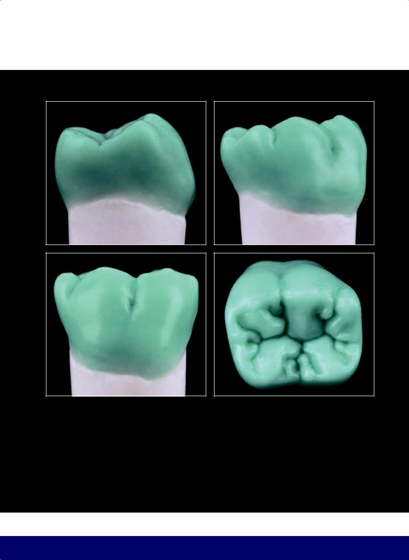
MANDIBULAR FIRST MOLAR
g |
h |
|
|
i |
j |
209
@dentistinfo стоматологический телеграм канал
Second Molar
Mandibular
10
210
@dentistinfo стоматологический телеграм канал
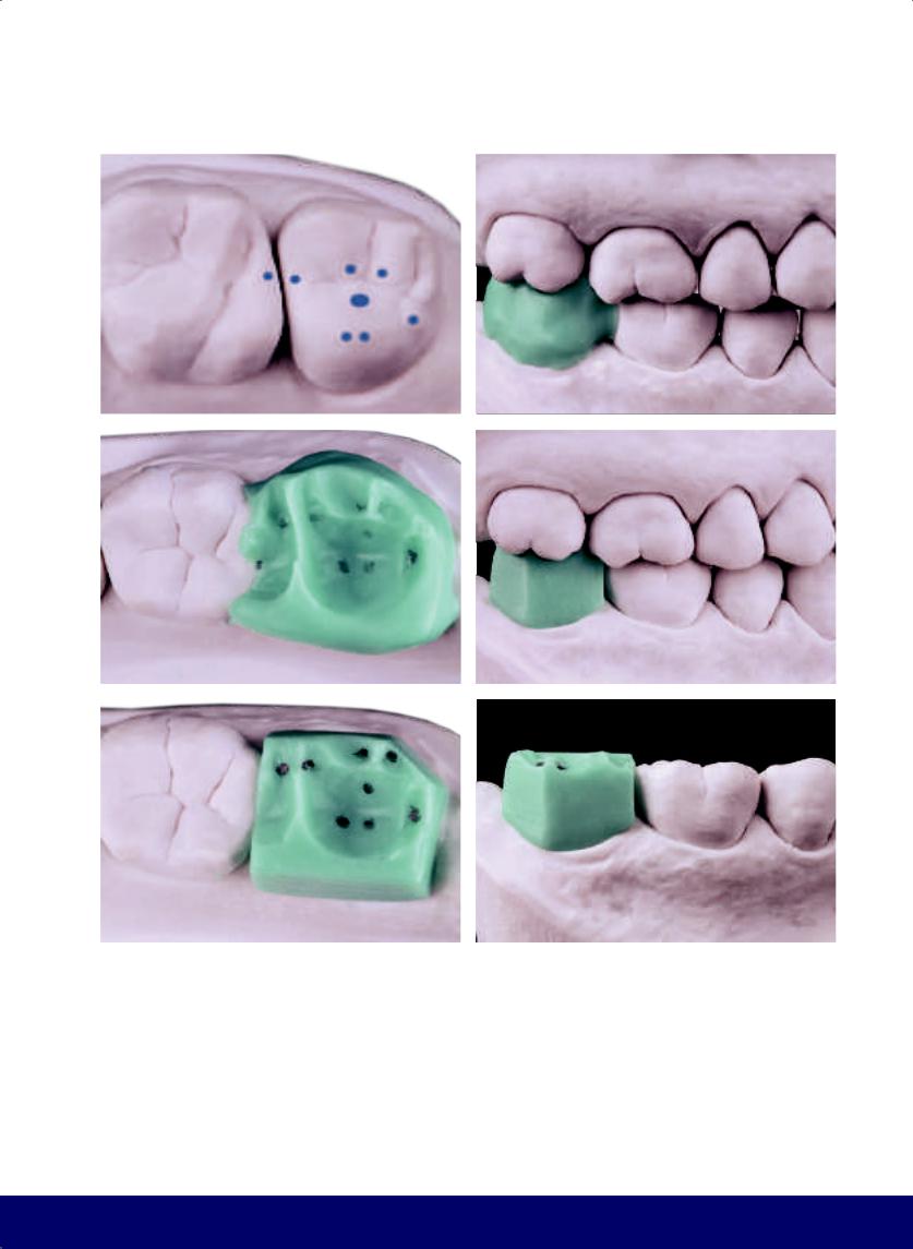
a |
b |
|
|
c |
d |
|
|
e |
f |
|
|
Fig 10-1 | (a) Maxillary centric contact points. (b and c) Buccal and occlusal views of cast in occlusion. Note that the contact points that were marked in pencil on the antagonist were reproduced in the wax. (d and e) Buccal and occlusal perspectives of the wax block after being trimmed. (f) Divide the buccal face into two parts, using as reference a virtual line situated in the direction of the buccal groove of the maxillary first molar to the centric mesial contact point of the longitudinal ridge of the mesiobuccal cusp of the mandibular second molar.
MANDIBULAR SECOND MOLAR
211
@dentistinfo стоматологический телеграм канал
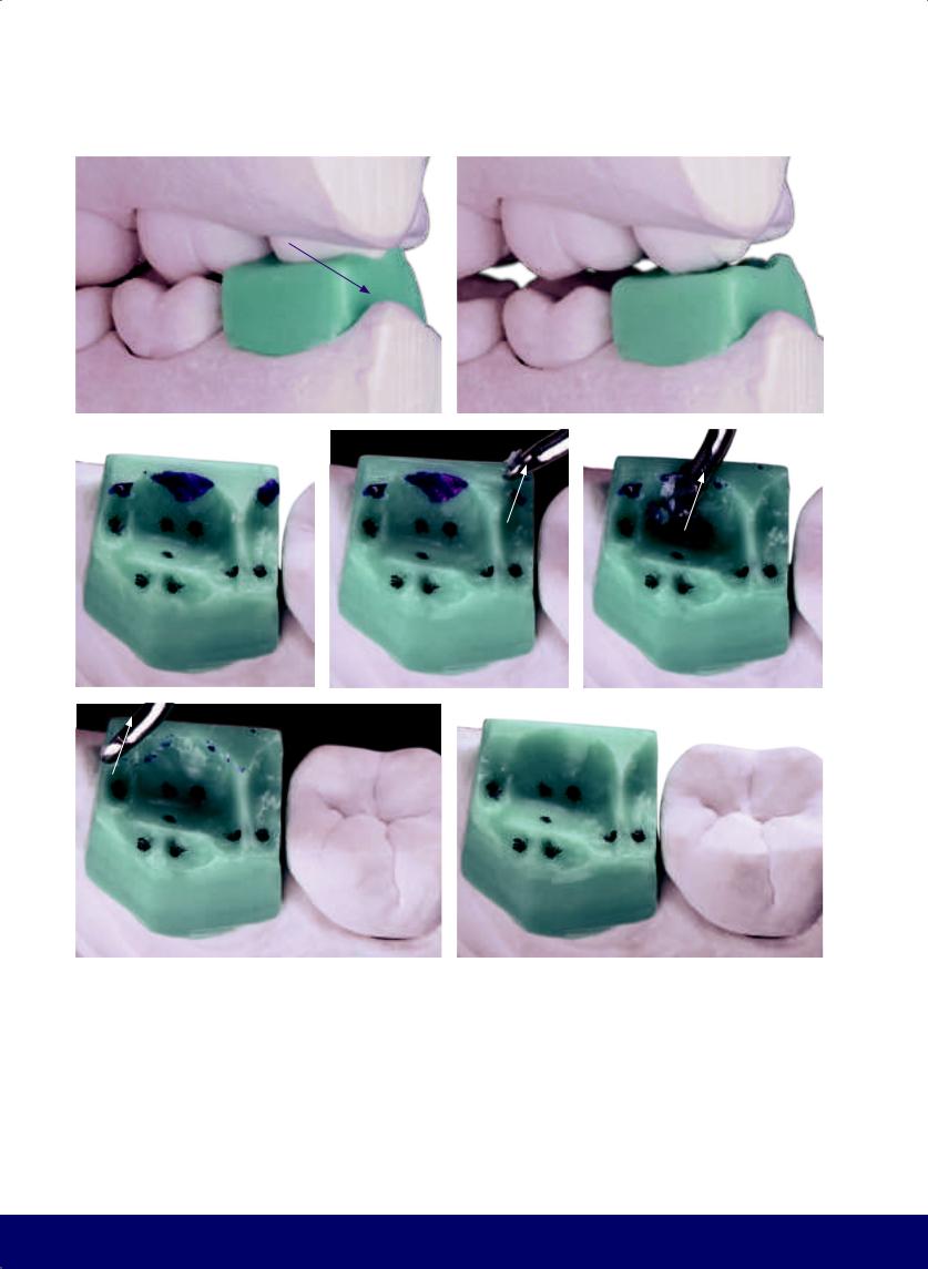
CHAPTER 10
a |
|
b |
|
|
|
c |
d |
e |
|
|
|
f |
g |
|
|
Fig 10-2 | (a to c) Lingual interference in working movement. Observe where the mesioand distobuccal cusps of the maxillary first molar touch the wax. (d to g) Carve away the lingual interference. (Purple arrows indicate the movement of the mandible, while white arrows indicate the direction of instrumentation.)
212
@dentistinfo стоматологический телеграм канал
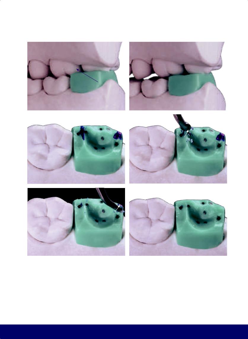
MANDIBULAR SECOND MOLAR
a |
|
b |
|
|
|
c |
|
d |
|
|
|
e |
f |
|
|
Fig 10-3 | (a to c) Buccal interference in balancing movement. (d to f) Carve away buccal interference on balancing movement.
213
@dentistinfo стоматологический телеграм канал
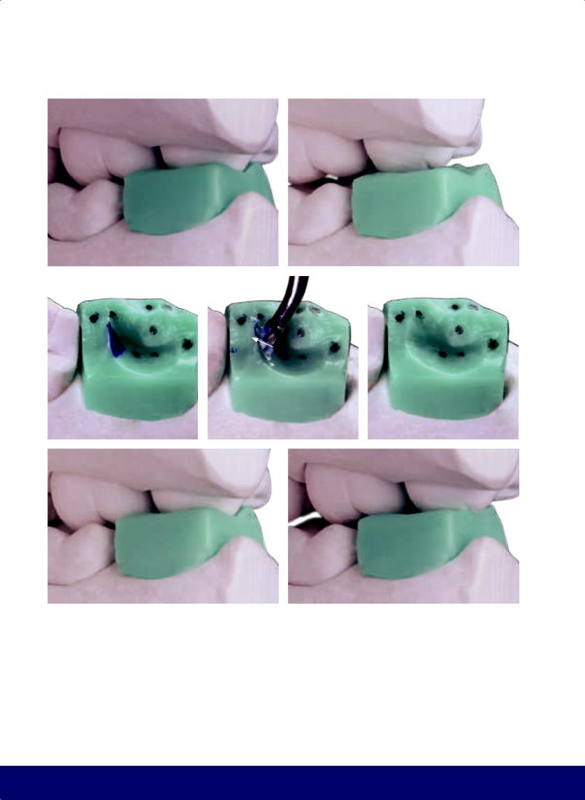
CHAPTER 10
a |
|
b |
|
|
|
c |
|
d |
|
e |
|
|
|
|
|
f |
|
g |
|
|
|
Fig 10-4 | (a to c) Interference in retrusive movement. (d and e) Carve away interference on retrusive movement. (f and g) Interference in protrusive movement.
214
@dentistinfo стоматологический телеграм канал
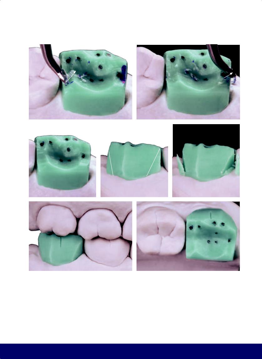
a |
b |
|
|
c |
|
d |
|
|
|
f |
g |
|
|
Fig 10-5 | (a to c) Carve away interference on protrusive movement. (d and e) Open the buccal embrasures. (f and g) The location reference for the buccal groove of the mandibular second molar is the tip of the mesiobuccal cusp of the maxillary second molar.
MANDIBULAR SECOND MOLAR
e
215
@dentistinfo стоматологический телеграм канал
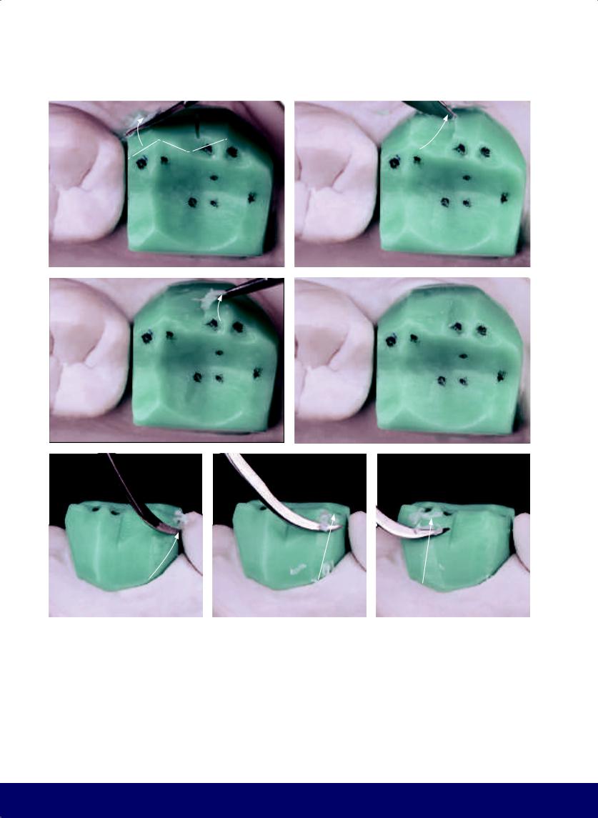
CHAPTER 10
a |
b |
|
|
c |
d |
|
|
e |
f |
g |
Fig 10-6 | (a to d) Define buccal aspect. (e to g) Round the angles and finish the shape of the buccal face.
216
@dentistinfo стоматологический телеграм канал
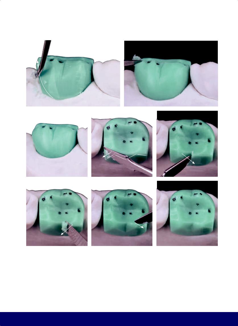
a |
b |
|
|
c |
|
d |
|
|
|
f |
|
g |
|
|
|
Fig 10-7 | (a to c) Eliminate the gull shape from the buccal of the maxillary second molar. (d to h) Carve the lingual wedges to form cusps.
MANDIBULAR SECOND MOLAR
e
h
217
@dentistinfo стоматологический телеграм канал
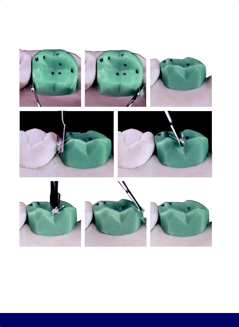
CHAPTER 10
a |
|
b |
|
c |
|
|
|
|
|
d |
e |
f |
|
g |
|
h |
|
|
|
|
|
Fig 10-8 | (a and b) Open the lingual embrasures. (c to h) Finalize placement and angulation of the cusp tips.
218
@dentistinfo стоматологический телеграм канал
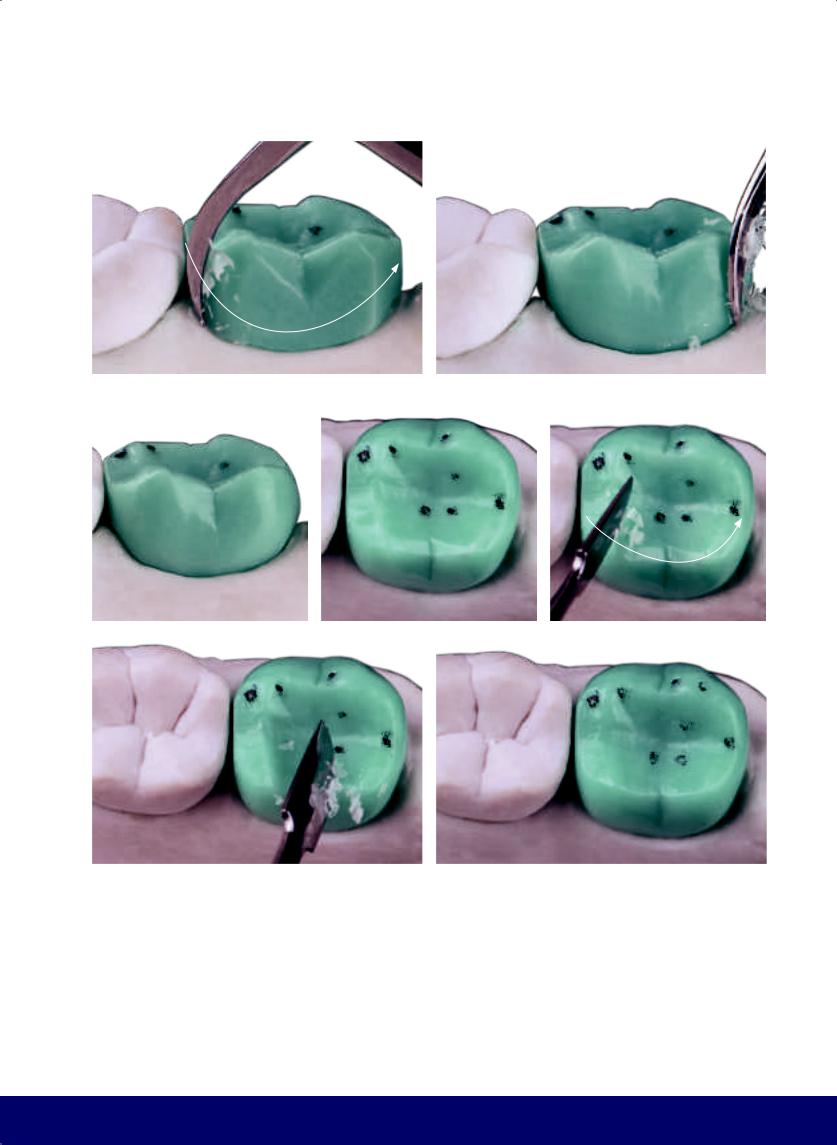
MANDIBULAR SECOND MOLAR
a |
|
b |
|
|
|
c |
|
d |
|
e |
|
|
|
|
|
f |
|
g |
|
|
|
Fig 10-9 | (a to g) Round the entire lingual aspect and the cusp tips.
219
@dentistinfo стоматологический телеграм канал
CHAPTER 10
a
b |
|
c |
|
|
|
d |
|
e |
|
|
|
Fig 10-10 | (a to c) The kidney bean profile is placed on the mesial marginal crest above the contact area. (d and e) The overall vertical macro texture includes mesial and distal pseudo developmental grooves on the mesiobuccal cusp. (f and g) The horizontal macro texture has the effect of cinching and narrowing the buccal face. (h to j) Divide the buccal face with a depression that runs from the buccal groove to the furcation of the mesioand
220
distobuccal roots. Observe the buccal fossa, the double hook that results from the juxtaposition of the horizontal macro texture and the buccal groove.
@dentistinfo стоматологический телеграм канал
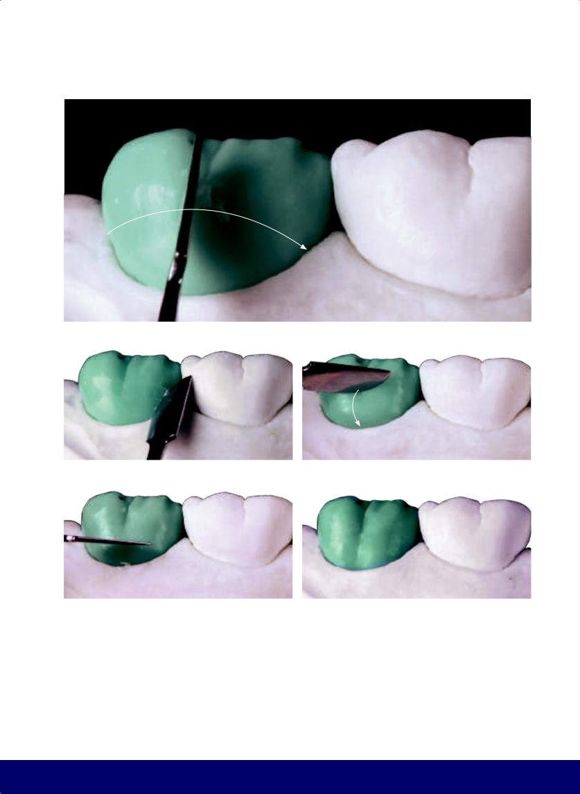
MANDIBULAR SECOND MOLAR
f
g |
|
h |
|
|
|
i |
|
j |
|
|
|
221
@dentistinfo стоматологический телеграм канал
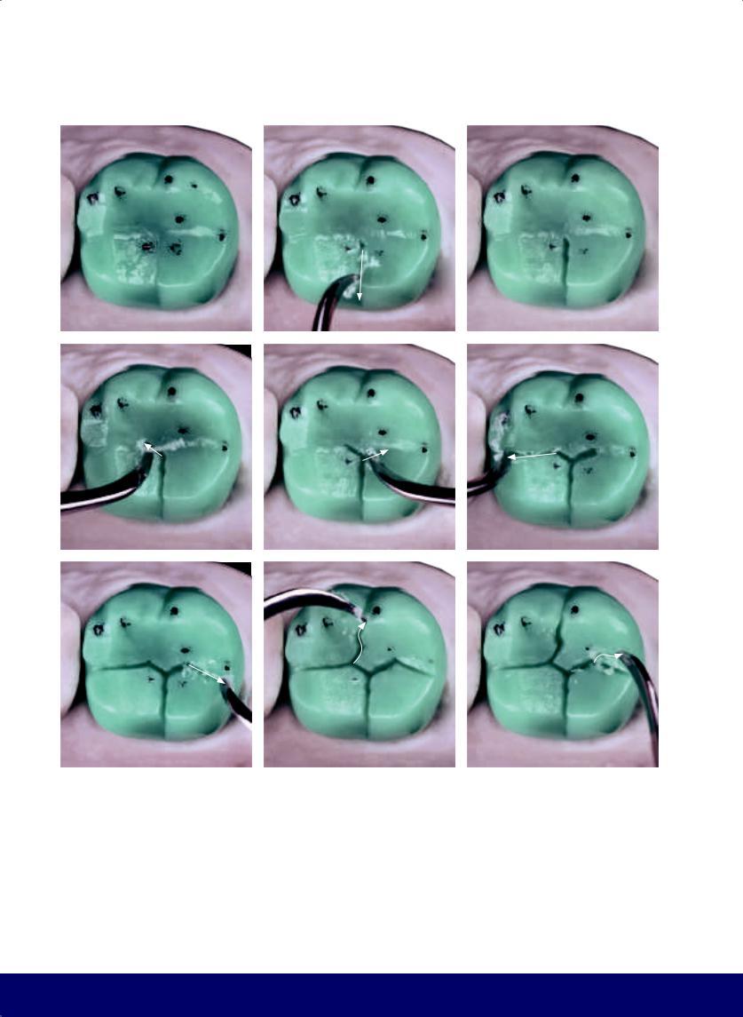
CHAPTER 10
a |
|
b |
|
c |
|
|
|
|
|
d |
e |
|
f |
|
|
|
|
g |
h |
|
i |
|
|
|
|
Fig 10-11 | (a) Occlusal view of the final external contour. Note the two external points for placing grooves as well as the vertical and horizontal macro texturing. (b and c) Open the lingual groove connecting the central fossa to the vertex of the angle formed between the lingual cusps (ie, the working groove). (d to f) Create two groove extensions from the central fossa to form the Mercedes star. The distal segment is slightly longer than the mesial segment. (g) Conclude the mesiodistal groove (the letter M) by
222
opening the protrusive groove, which is directed toward the lingual aspect. (h) Connect the mesial vertex of the M with the external mesial reference point, creating the buccal groove. Note that the first segment of this groove is directed distobuccally before turning toward the buccal aspect. (i) From the distal apex of the M, a secondary groove is created. The primary grooves of the second molar are now complete.
@dentistinfo стоматологический телеграм канал
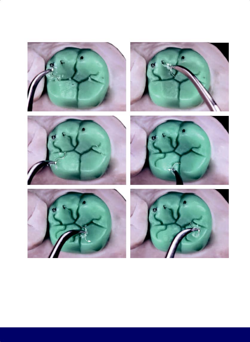
a |
b |
|
|
c |
d |
|
|
e |
f |
|
|
Fig 10-12 | (a and b) Place the inward-curving horns on the mesiobuccal cusp. The S near the mesiodistal groove and the mesial arm of the Mercedes star form the retrusive groove. (c and d) Place the inward-curving horns on the mesiolingual cusp. (e and f) Place the inward-curving horns on the distolingual cusp.
MANDIBULAR SECOND MOLAR
223
@dentistinfo стоматологический телеграм канал
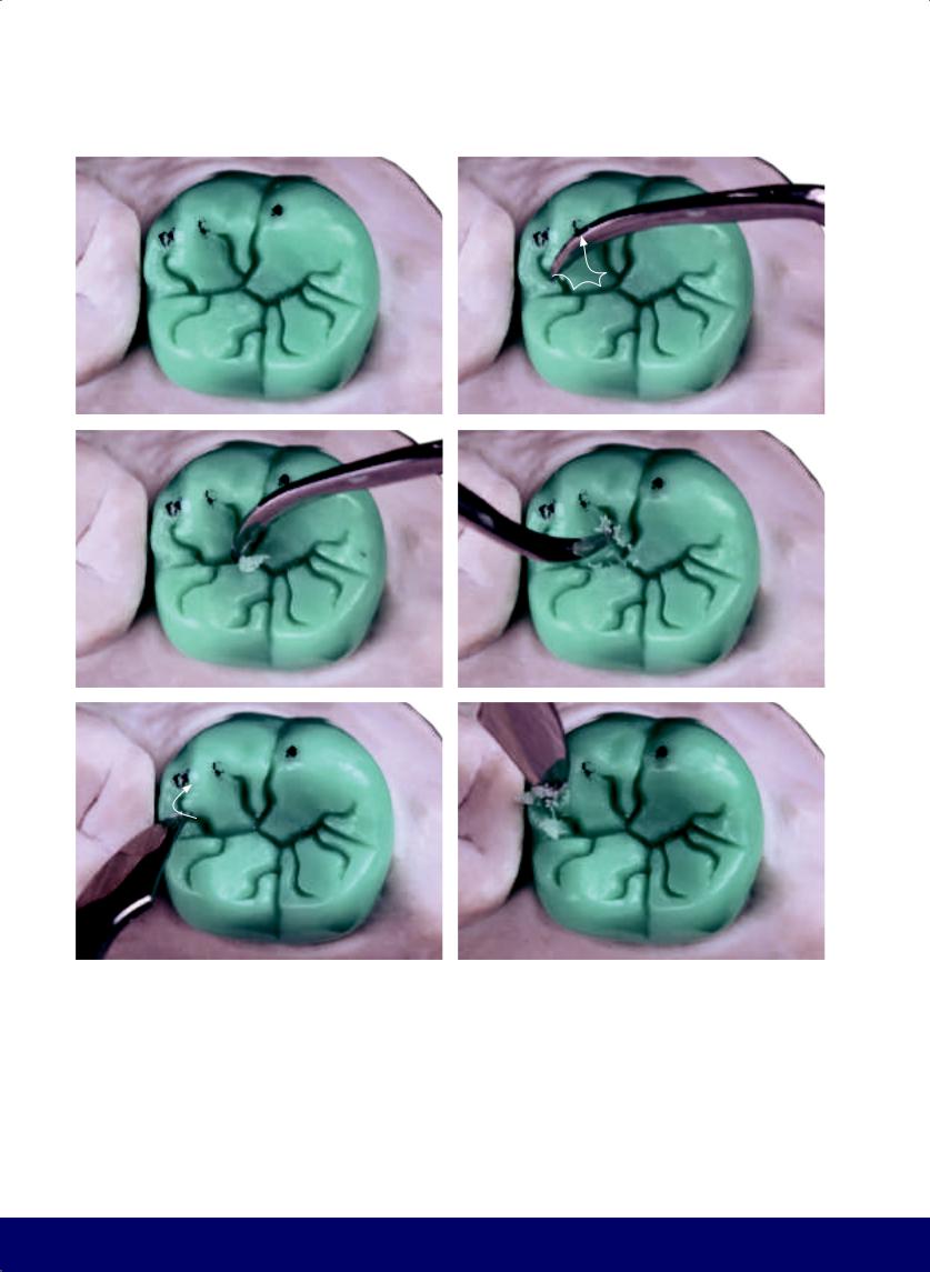
CHAPTER 10
a |
|
b |
|
|
|
c |
|
d |
|
|
|
e |
|
f |
|
|
|
Fig 10-13 | (a to w) Round the corners and angles of the occlusal morphology.
224
@dentistinfo стоматологический телеграм канал

MANDIBULAR SECOND MOLAR
g |
|
h |
|
|
|
i |
|
j |
|
|
|
k |
|
l |
|
|
|
225
@dentistinfo стоматологический телеграм канал
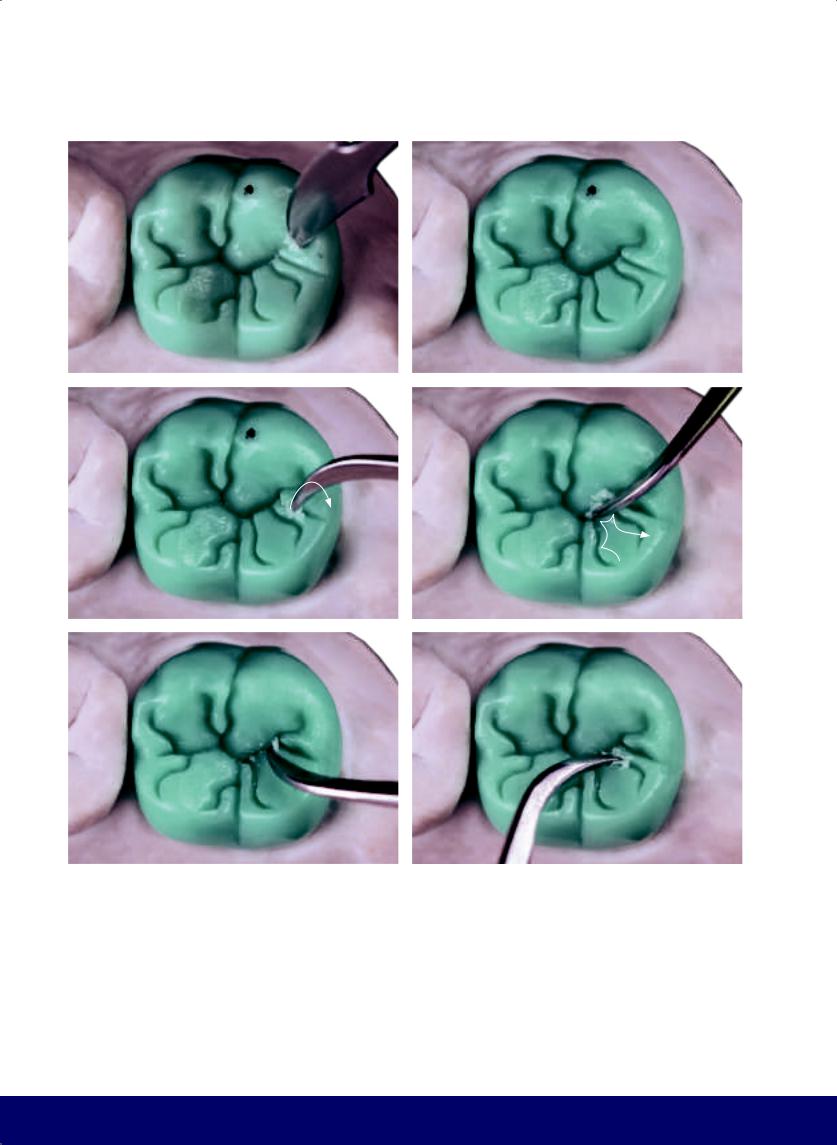
CHAPTER 10
m |
|
n |
|
|
|
o |
|
p |
|
|
|
q |
|
r |
|
|
|
226
@dentistinfo стоматологический телеграм канал
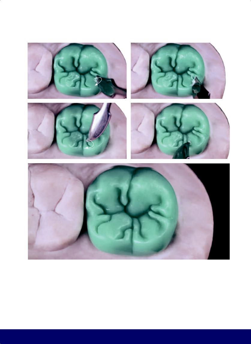
MANDIBULAR SECOND MOLAR
s |
|
t |
|
|
|
u |
|
v |
|
|
|
w
227
@dentistinfo стоматологический телеграм канал
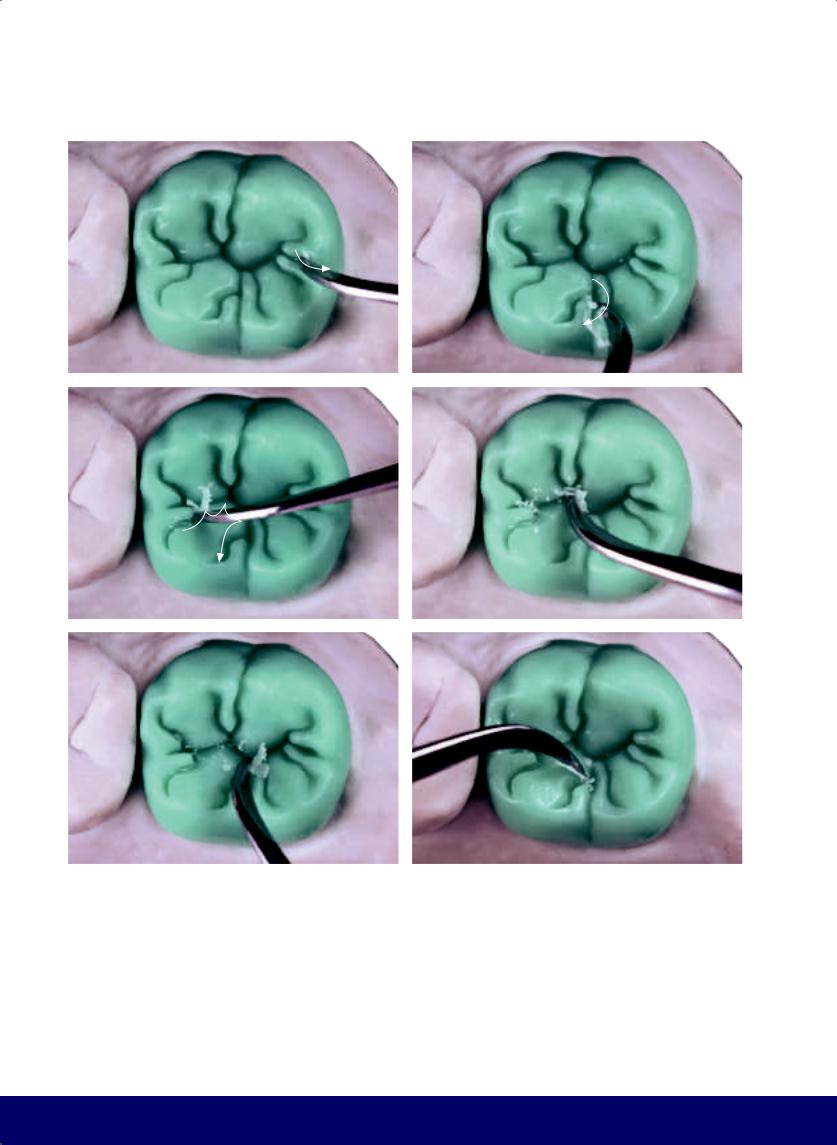
CHAPTER 10
a |
|
b |
|
|
|
c |
|
d |
|
|
|
e |
|
f |
|
|
|
Fig 10-14 | (a to n) Open the ends of the secondary grooves while preserving the transverse and longitudinal ridges and creating the “elbow” in all accessory lobes.
228
@dentistinfo стоматологический телеграм канал
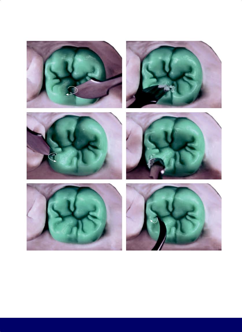
MANDIBULAR SECOND MOLAR
g |
|
h |
|
|
|
i |
|
j |
|
|
|
k |
|
l |
|
|
|
229
@dentistinfo стоматологический телеграм канал
CHAPTER 10
m |
|
n |
|
|
|
o
Fig 10-14 | (o) Occlusal morphology and final external contour. Note that the mesiodistal sulcus, which forms an M, is positioned at the center of the occlusal surface, ensuring buccal and lingual cusp of similar volume. The buccal and lingual grooves do not meet, reproducing the pattern of grooving of the first molar. Also note that the secondary grooves start tentatively and open at the end, in the form of an S and an inverted S. Together, these secondary grooves evoke inward-curving horns. The kidney bean profile
230
is present on the mesial marginal ridge. From the buccal view, the profile of a key is evident on the longitudinal ridge of the lingual cusps. Also note that the lingual groove is more open than the buccal groove.
@dentistinfo стоматологический телеграм канал
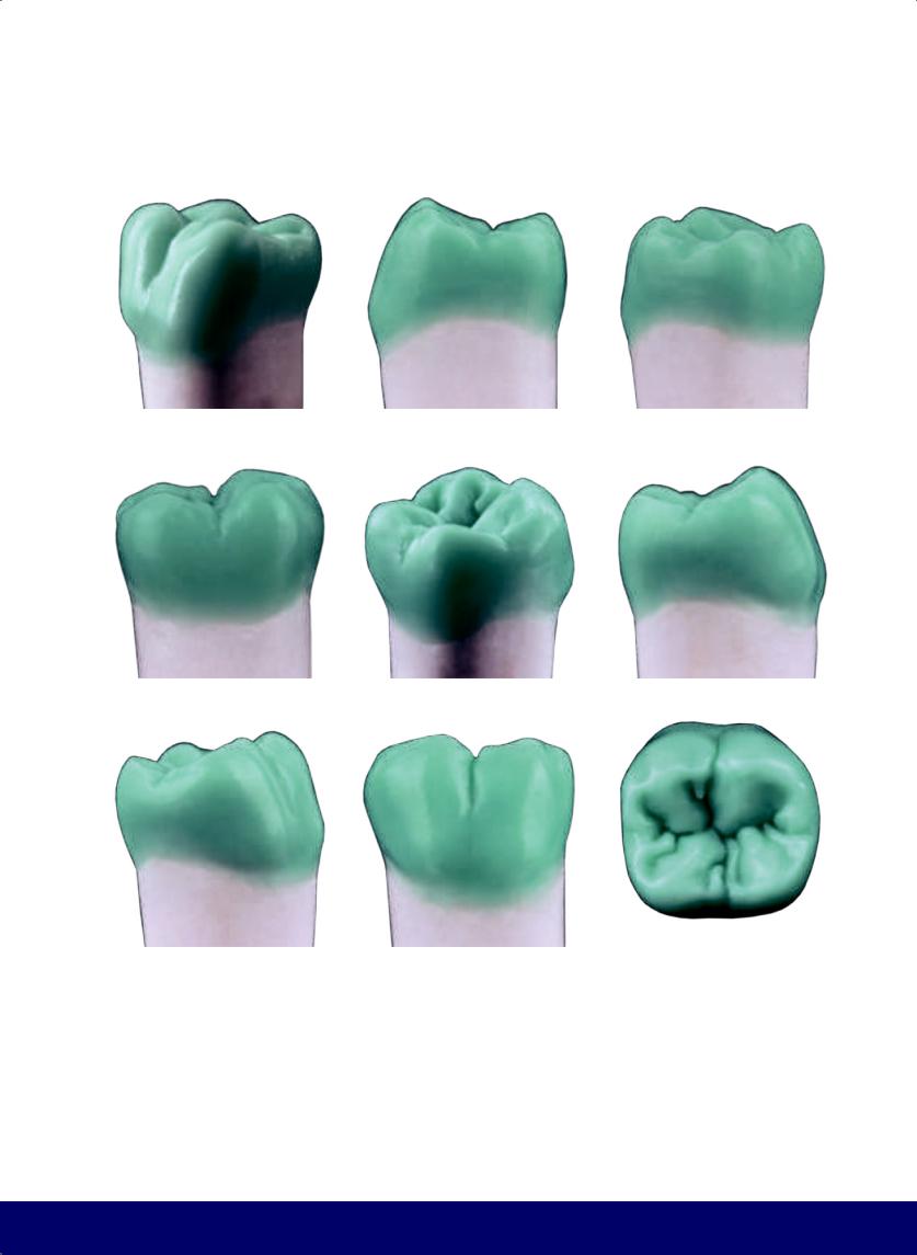
MANDIBULAR SECOND MOLAR
a |
|
b |
|
c |
|
|
|
|
|
d |
|
e |
|
f |
|
|
|
|
|
g |
|
h |
|
i |
|
|
|
|
|
Fig 10-15 | (a to i) Completed morphology and exterior contour of the mandibular second molar.
231
@dentistinfo стоматологический телеграм канал

Quadrant
Mandibular Posterior
11
232
@dentistinfo стоматологический телеграм канал
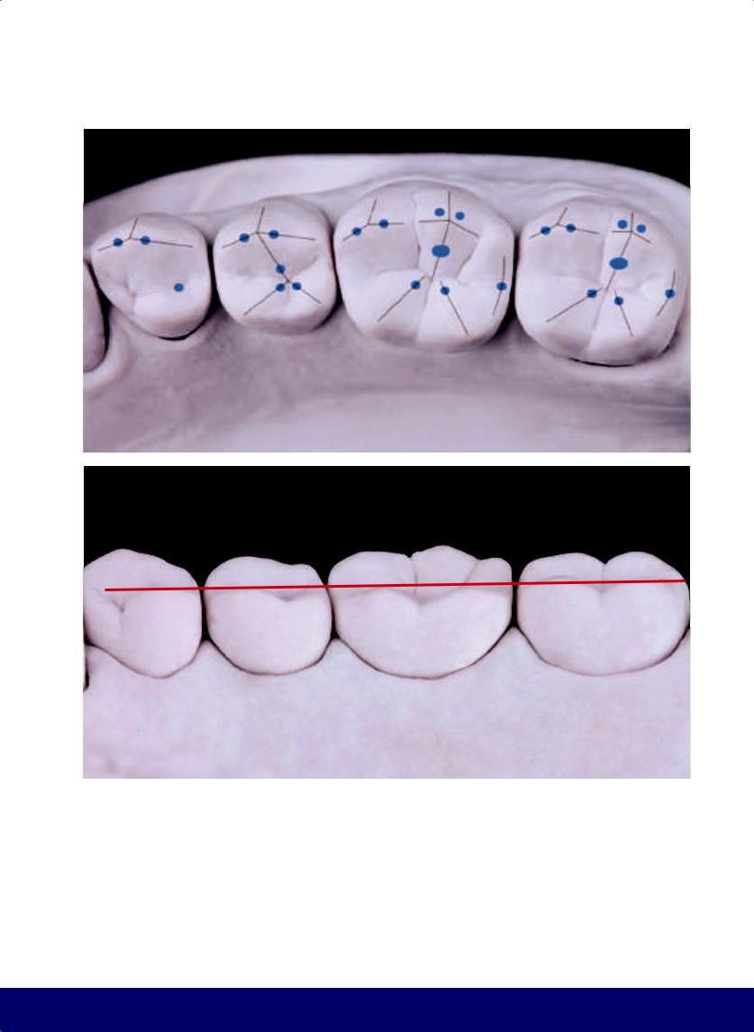
MANDIBULAR POSTERIOR QUADRANT
a
b
Fig 11-1 | (a) Reference for the smooth ridges, grinding slopes, and the centric contact points (Angle Class I) that must be recorded in the wax-up. (b) Height reference for cusps. Note the line touches the tips of the buccal cusps of the first and second premolars and the distobuccal cusp of the first molar.
233
@dentistinfo стоматологический телеграм канал
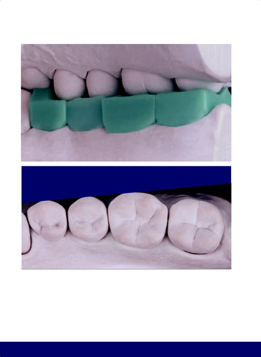
CHAPTER 11
a
b
Fig 11-2 | (a) Wax blocks are carved individually following the standard dimensions. (b) Reference for the buccal volume. Note that the straight line touches the buccal aspect of the first and second premolars and first molar, from the cervical to occlusal.
234
@dentistinfo стоматологический телеграм канал
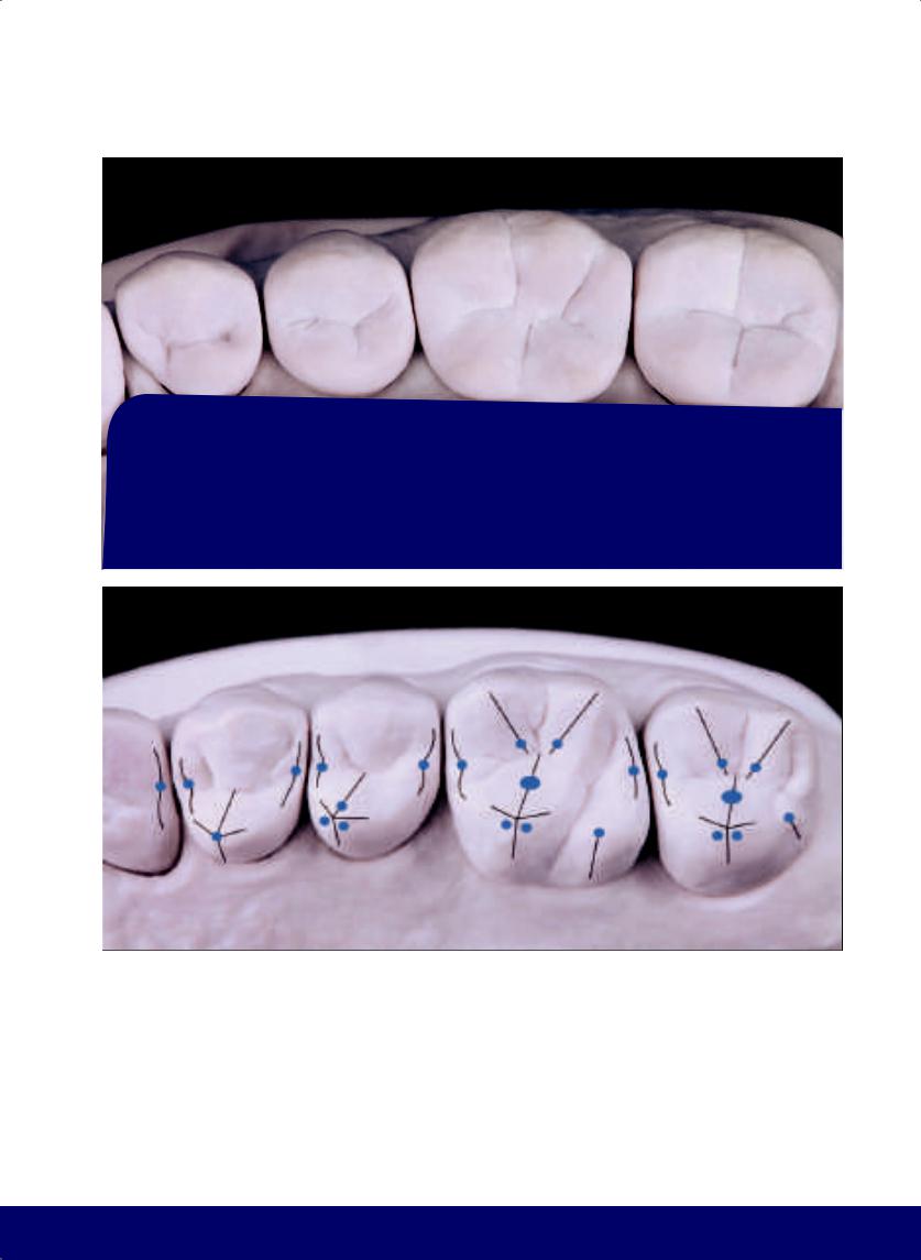
MANDIBULAR POSTERIOR QUADRANT
a
b
Fig 11-3 | (a) Reference for the lingual volume. Note that the straight line touches the lingual aspect of the first premolar and first and second molars, from the cervical to the occlusal. The lingual aspect of the second premolar is 1 mm from the line, which opens the dental arch. (b) Centric contact points are already referenced to be marked on the antagonist.
235
@dentistinfo стоматологический телеграм канал
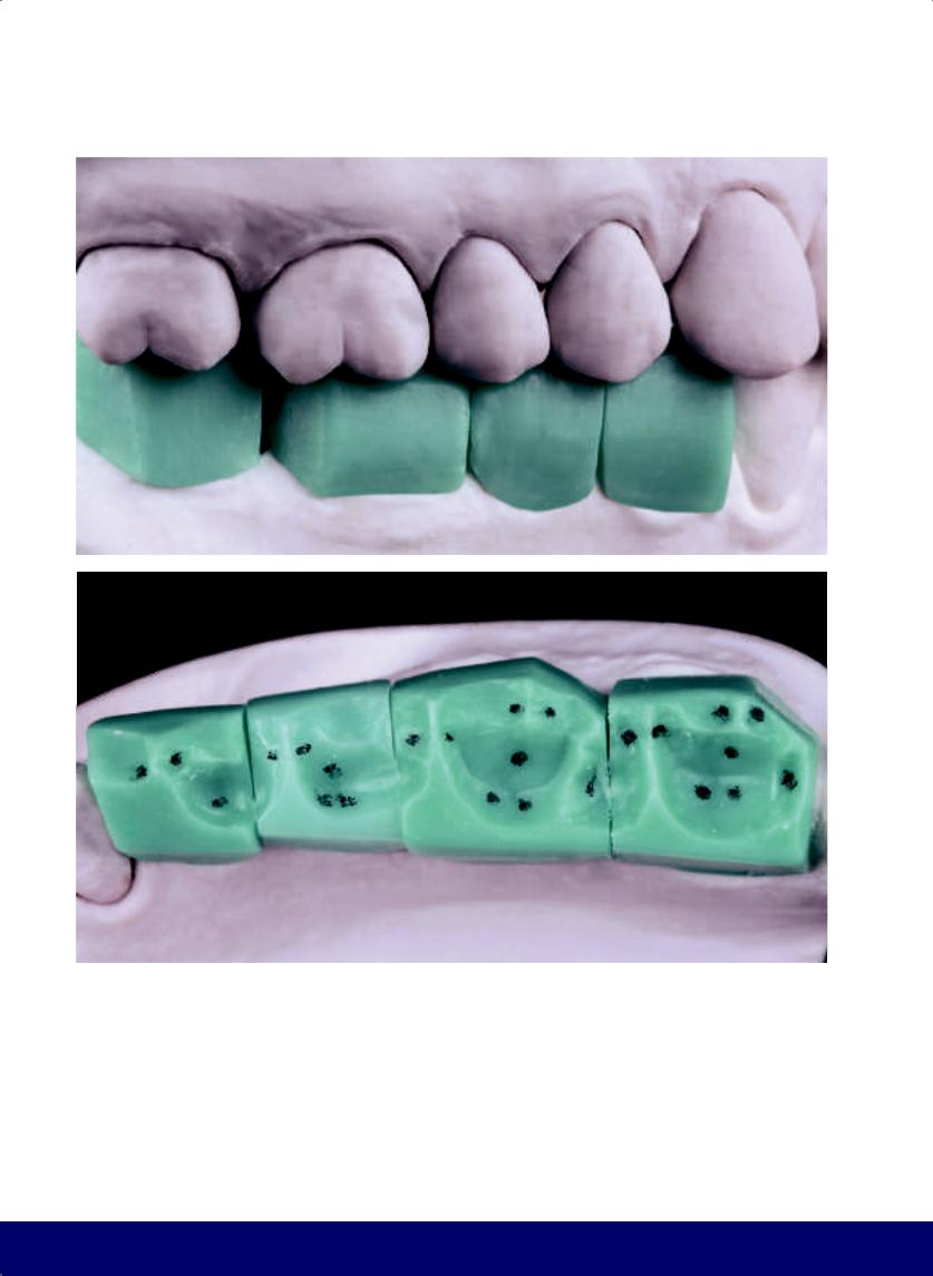
CHAPTER 11
a
b
Fig 11-4 | (a and b) Buccal and lingual excess is removed. Note that the centric contact points have been reproduced in pencil in the wax.
236
@dentistinfo стоматологический телеграм канал
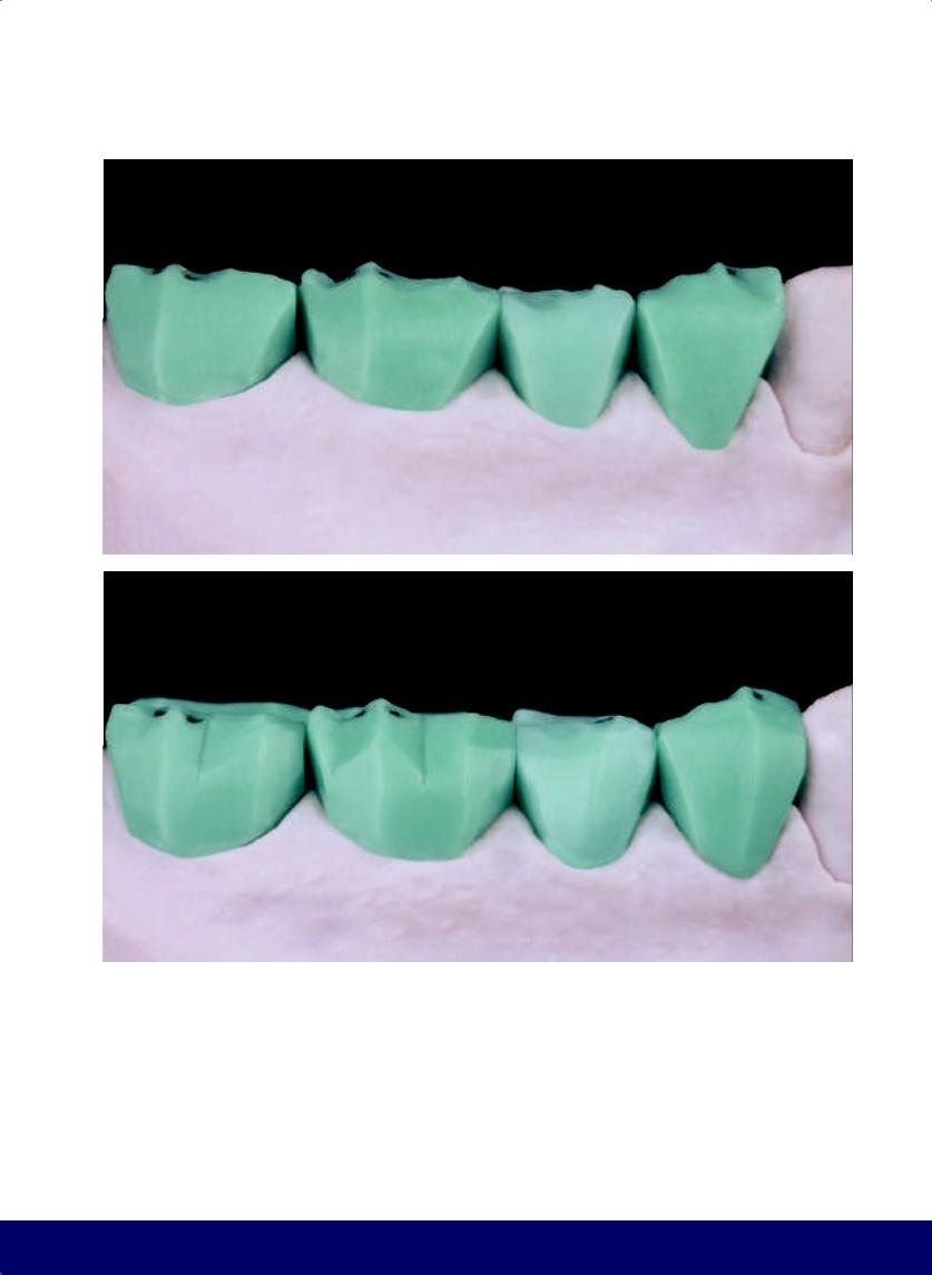
MANDIBULAR POSTERIOR QUADRANT
a
b
Fig 11-5 | (a) Open the embrasures and define the cusp tips. (b) Divide the buccal aspect into different planes. In the first premolar, the mesial plane is smaller than the distal plane. In the second premolar, the two planes are equivalent. In the molars, smaller planes better define the tips of the buccal cusps.
237
@dentistinfo стоматологический телеграм канал
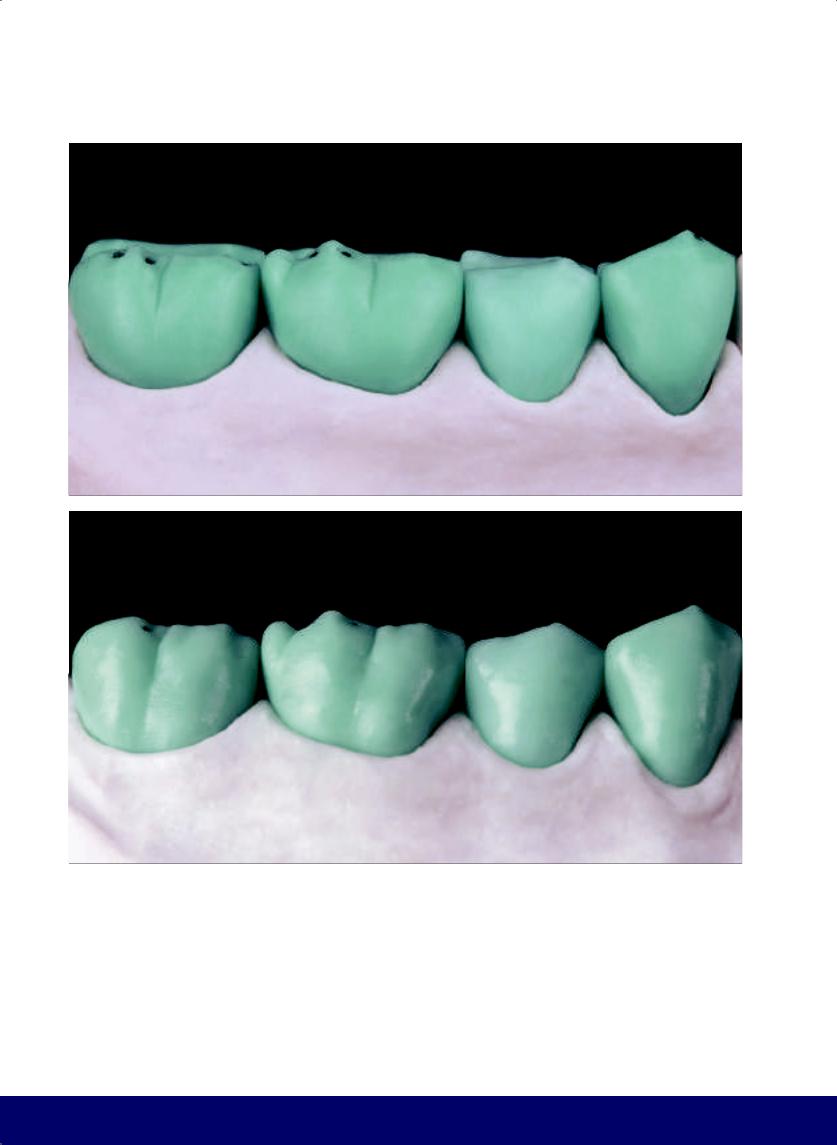
CHAPTER 11
a
b
Fig 11-6 | (a) Round the buccal aspects. Note the barrel shape.
(b) Complete the buccal face with vertical developmental grooves, a more narrow-waisted silhouette, and horizontal macro depressions in every tooth.
238
@dentistinfo стоматологический телеграм канал
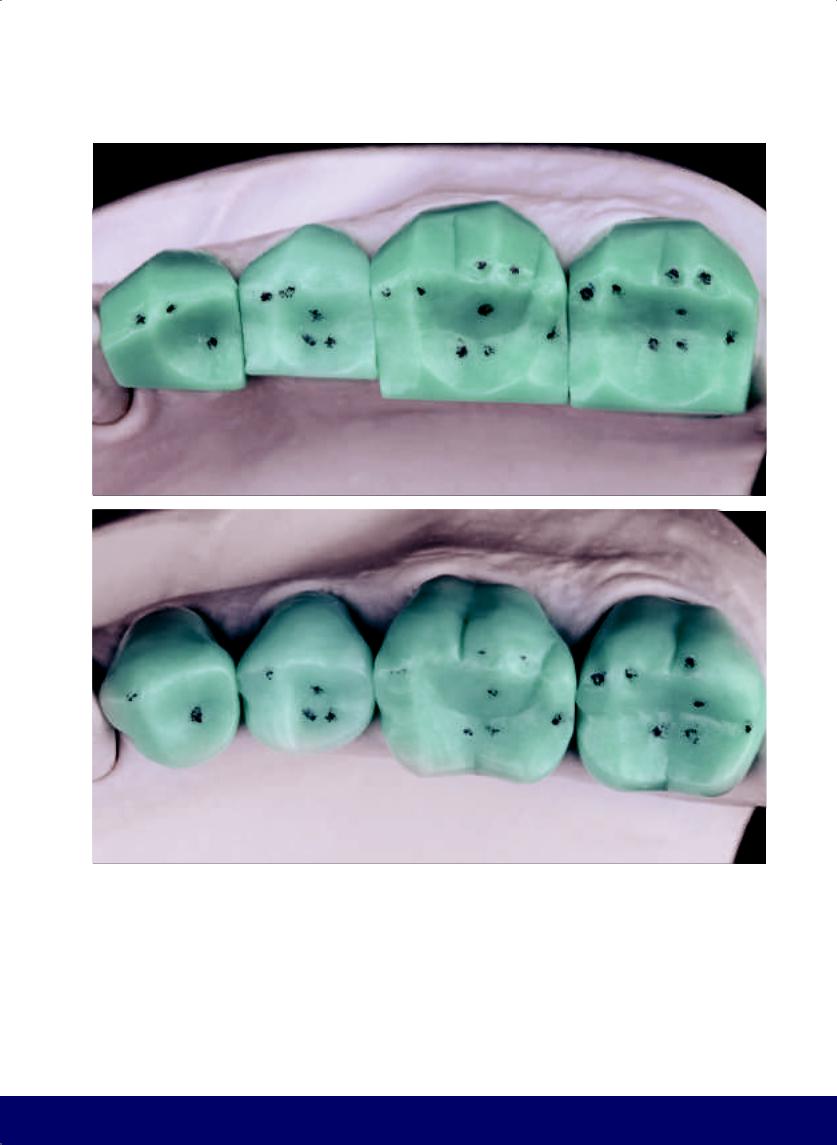
MANDIBULAR POSTERIOR QUADRANT
a
b
Fig 11-7 | (a) Note the different buccal planes. (b) The external and occlusal contours of the finished teeth have been completed with a kidney bean profile in each marginal mesial ridge.
239
@dentistinfo стоматологический телеграм канал
CHAPTER 11
a |
|
b |
|
|
|
c |
|
d |
|
|
|
e |
|
f |
|
|
|
Fig 11-8 | (a to j) Tooth by tooth sequence of positioning the main and secondary grooves and final morphology of the occlusal surface.
240
@dentistinfo стоматологический телеграм канал
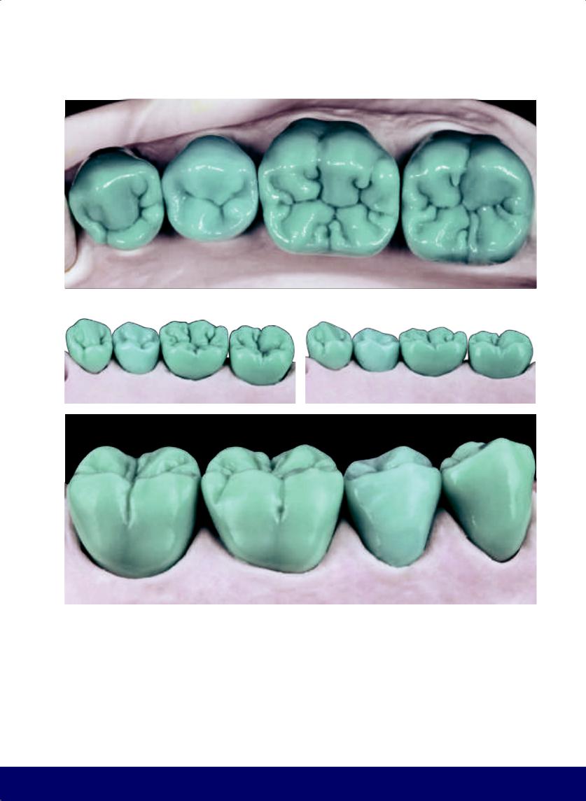
MANDIBULAR POSTERIOR QUADRANT
g
h |
|
i |
|
|
|
j
241
@dentistinfo стоматологический телеграм канал
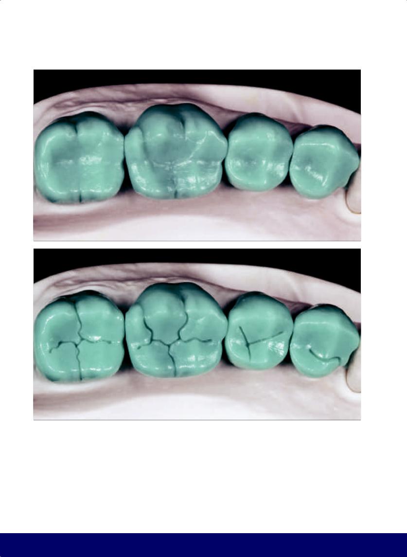
CHAPTER 11
a
b
Fig 11-9 | (a) Definitive external contours. (b) Positioning of the fossae. (c) Delineation of the grooves. (d) Completed occlusal surface without secondary grooves. Note the three characteristics that define a tooth: well-defined external contours, well-located ridges, and well-positioned primary grooves.
242
@dentistinfo стоматологический телеграм канал
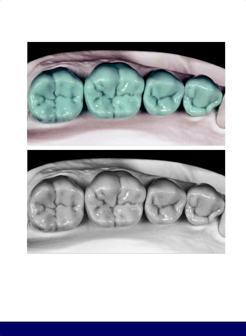
MANDIBULAR POSTERIOR QUADRANT
c
d
243
@dentistinfo стоматологический телеграм канал
SECTION II
Anterior Teeth
@dentistinfo стоматологический телеграм канал

The Esthetic and Functional Parameters of Anterior Teeth
12
245
@dentistinfo стоматологический телеграм канал
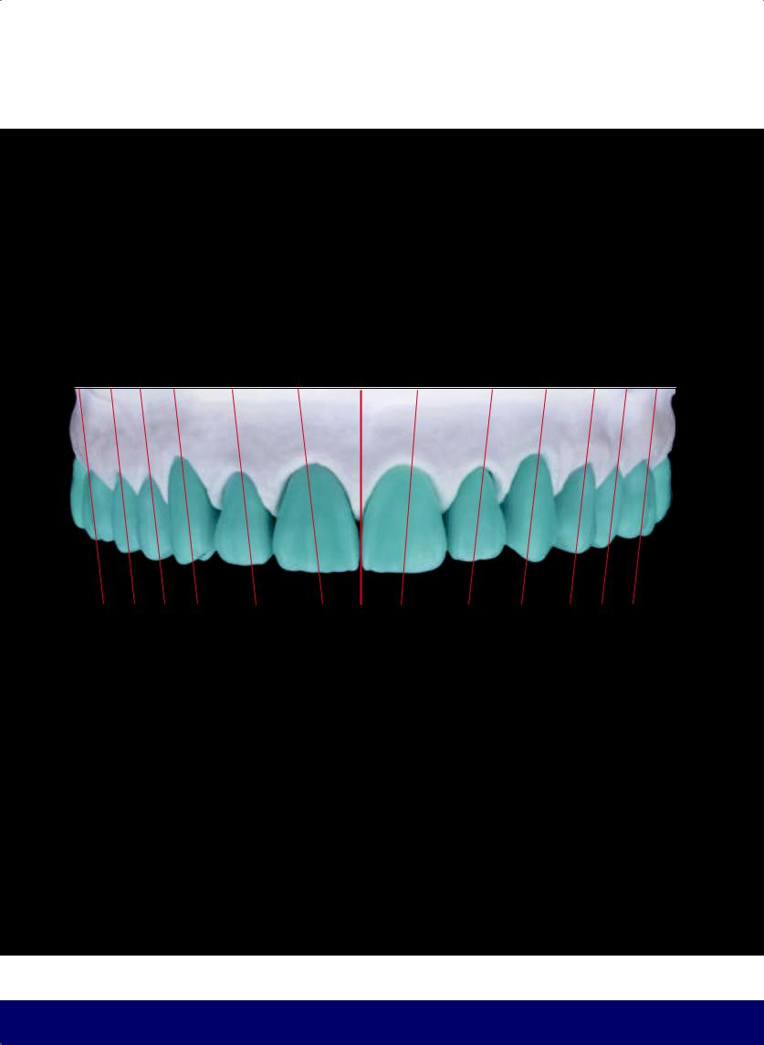
CHAPTER 12
BILATERAL AXIAL ALIGNMENT OF ANTERIOR AND POSTERIOR TEETH
The phenomenon of linear balance from the central fulcrum (ie, the midline).
246
@dentistinfo стоматологический телеграм канал
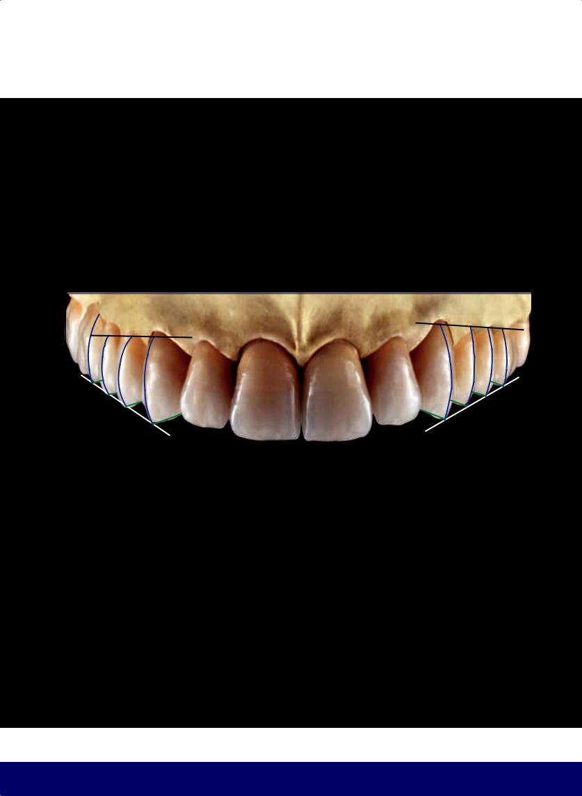
THE ESTHETIC AND FUNCTIONAL PARAMETERS OF ANTERIOR TEETH
PARALLELISM OF THE STRAIGHT LINE
The anteroposterior progression requires the alignment of the buccal contours of the canine, first and second premolars, and first molars, as well as parallelism between the mesial segment of the longitudinal ridges of the canine cusp, the buccal cusp of the first and second premolar, and the mesiobuccal cusp of the first molar.
247
@dentistinfo стоматологический телеграм канал
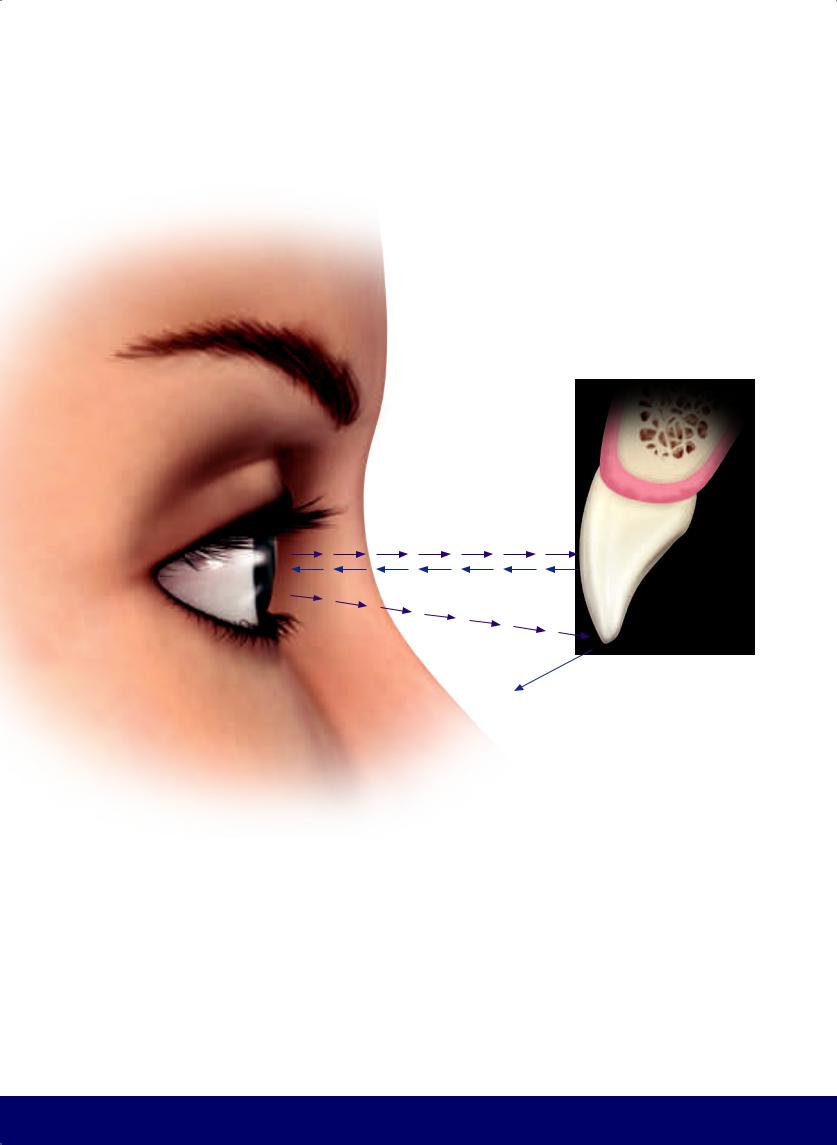
CHAPTER 12
REFLECTED LIGHT
DEFLECTED LIGHT
Modifying the quality of light that is reflected or deflected from objects affects their perception. This can be used to create the phenomenon of illusion.
248
@dentistinfo стоматологический телеграм канал
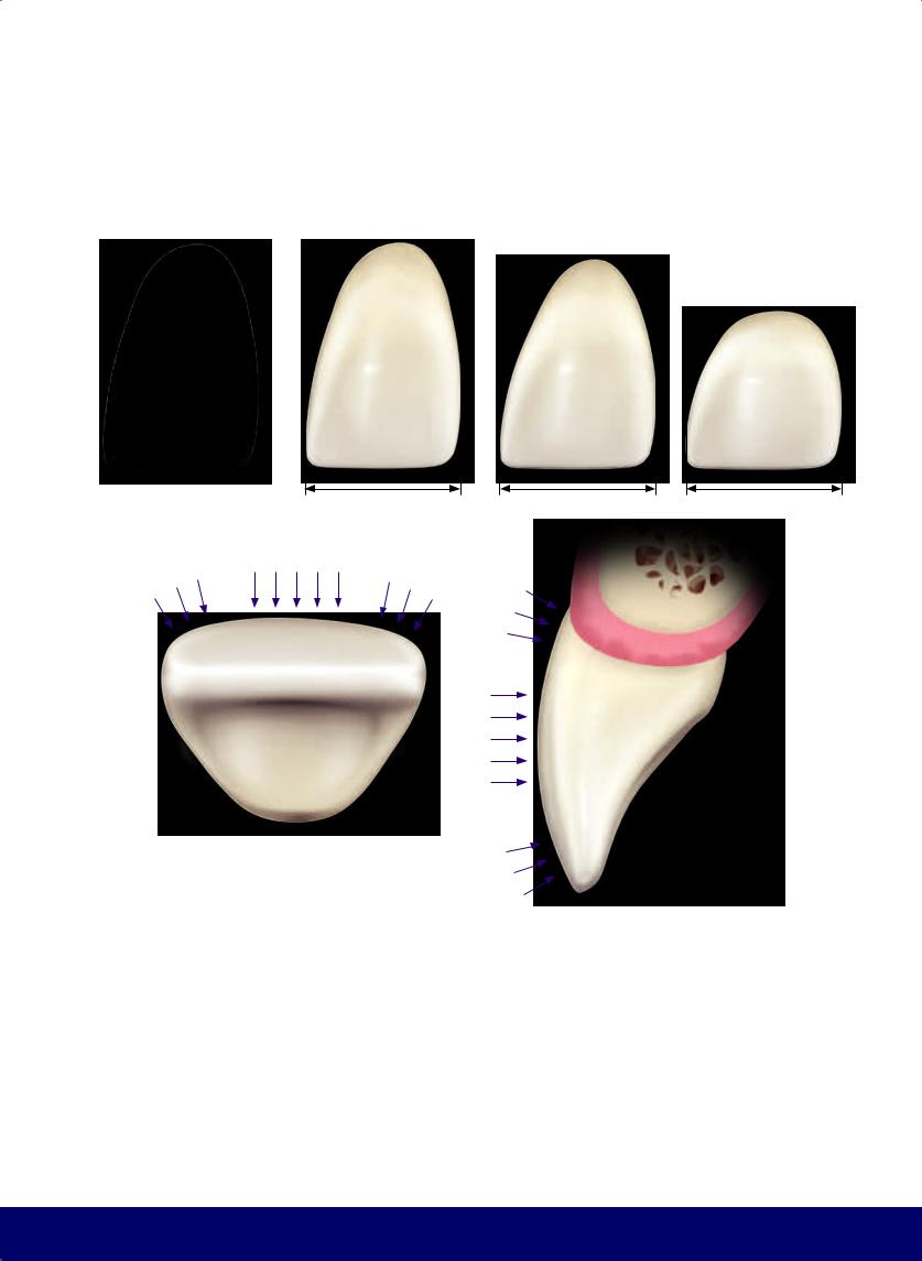
THE ESTHETIC AND FUNCTIONAL PARAMETERS OF ANTERIOR TEETH
Differences in color value affect the perception of size. The brighter the tooth, the bigger it will look, and the opposite is also true. Differences in height affect the perception of width.
The incidence of light on the buccal aspects of the incisors reveals three planes with different inclinations in the cervico-incisal and mesiodistal directions.
The amount of light reflected or deflected in the vertical and horizontal planes is responsible for the perception of larger or smaller surfaces.
249
@dentistinfo стоматологический телеграм канал
CHAPTER 12
THE PERCEPTION OF HEIGHT AND WIDTH IS THE RESULT OF THE RELATIONSHIP BETWEEN THE REFLECTIVE AND DEFLECTIVE AREAS.
250
—ILLUSION OF STRETCHING
The flattening of the gingivo-incisal area of the labial surface increases reflection, decreases deflection, and increases the length of the central third.
@dentistinfo стоматологический телеграм канал
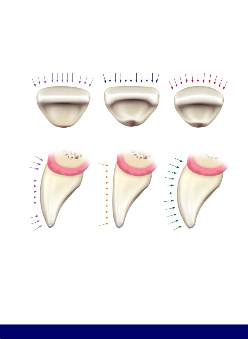
THE ESTHETIC AND FUNCTIONAL PARAMETERS OF ANTERIOR TEETH
|
|
|
|
|
|
|
|
|
|
|
|
|
|
|
|
|
|
|
|
|
|
|
|
|
|
|
|
|
|
|
|
|
|
|
|
|
|
|
|
|
|
|
|
|
|
|
|
|
|
|
|
|
|
|
|
|
|
|
|
|
|
|
|
|
|
|
|
|
|
|
|
|
|
|
|
|
|
|
|
|
|
|
|
|
|
|
|
|
|
|
|
|
|
|
|
|
|
|
|
|
|
|
|
|
|
|
|
|
|
|
|
|
|
|
|
|
|
|
|
|
|
|
|
|
|
|
|
|
|
|
|
|
|
|
|
|
|
|
|
|
|
|
|
|
|
|
|
|
|
|
|
|
|
|
|
|
|
|
|
|
|
|
|
|
|
|
|
|
|
|
|
|
|
|
|
|
|
|
|
|
|
|
|
|
|
|
|
|
|
|
|
|
|
|
|
|
|
|
|
|
|
|
|
|
|
|
|
|
|
|
|
|
|
|
|
|
|
|
|
|
|
|
|
|
|
|
|
|
|
|
|
|
|
|
|
|
|
|
|
|
|
|
|
|
|
|
|
|
|
|
|
|
|
|
|
|
|
|
|
|
|
|
|
|
|
|
|
|
|
|
|
|
|
|
|
|
|
|
|
|
|
|
|
|
— ILLUSION OF SHORTENING |
— ILLUSION OF ENLARGMENT |
— ILLUSION OF NARROWING |
|
|||||||||||
Accentuation of the curve of the buccal surface decreases the length of the central third, increases deflection, and decreases reflection.
Moving the transitional line angles proximally increases the reflective area and reduces the deflective area.
Moving the transitional line angles toward the center reduces the reflective area and increases the deflective area.
251
@dentistinfo стоматологический телеграм канал
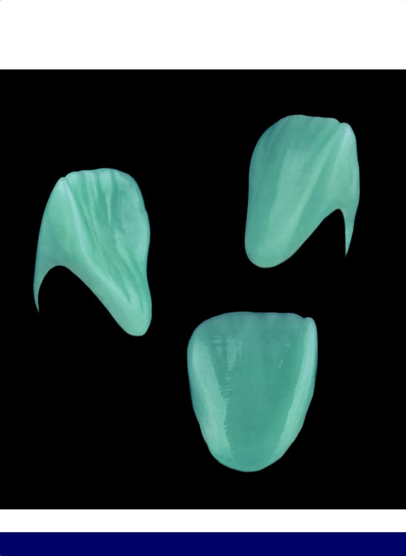
CHAPTER 12
SURFACES
Lingual
Buccal
Cervical
Distal |
Mesial |
Incisal
252
@dentistinfo стоматологический телеграм канал
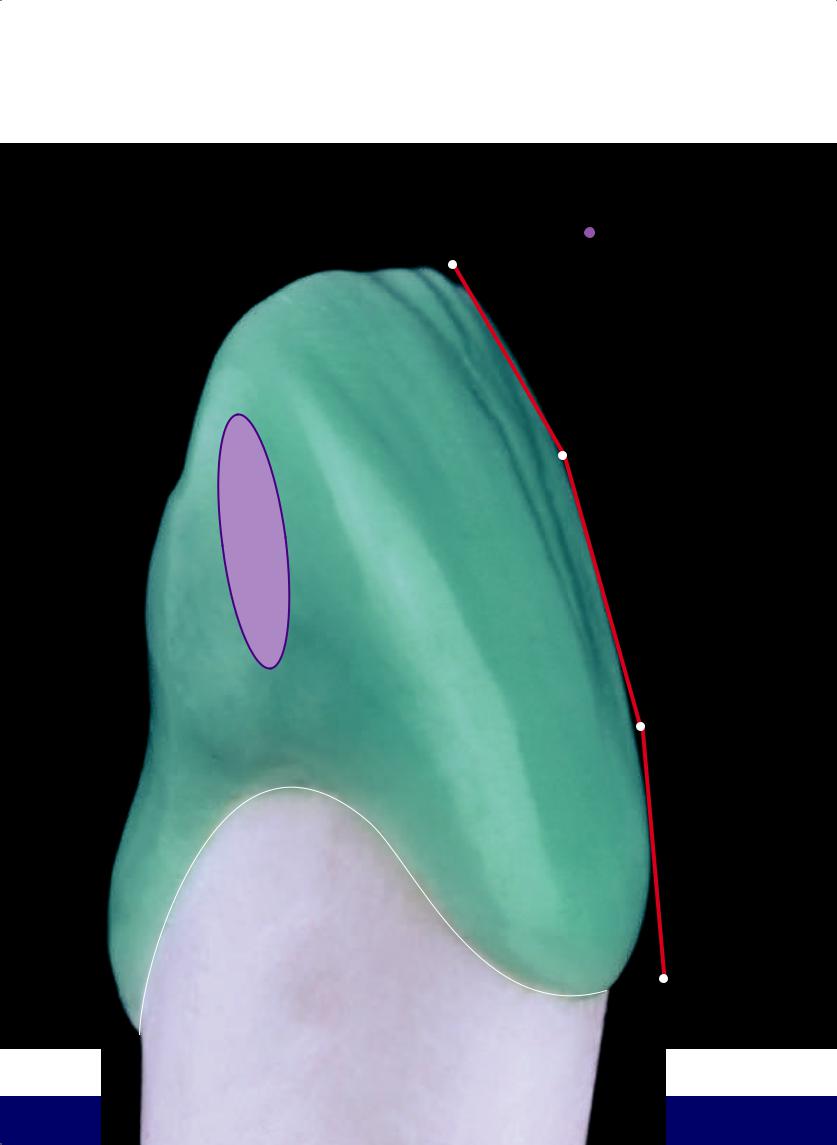
THE ESTHETIC AND FUNCTIONAL PARAMETERS OF ANTERIOR TEETH
I
 AREA OF CONTACT
In anterior teeth, the contact area is a vertical ellipse.
 SLOPE OF THE LABIAL FACE
IN THE VERTICAL PLANE
M
C
253
@dentistinfo стоматологический телеграм канал
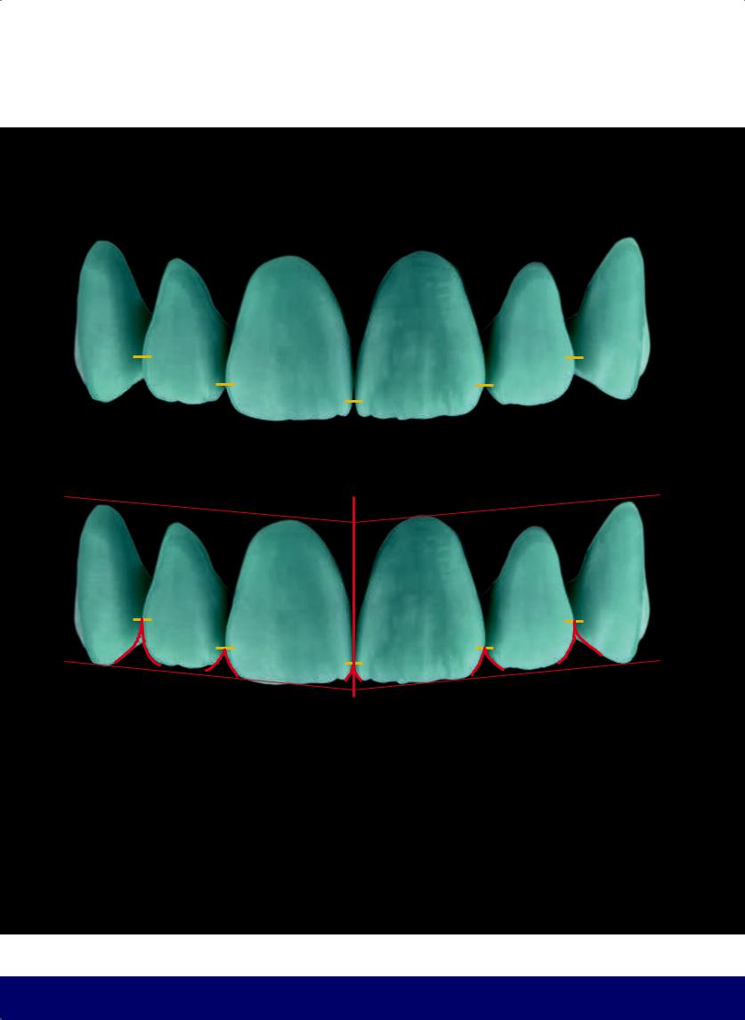
CHAPTER 12
—Positions of contact areas move from the incisal toward the cervical, in progression from the central incisors to the canines.
—From the frontal view, the displacement of areas of contact toward the cervical increases the incisal embrasures.
254
@dentistinfo стоматологический телеграм канал
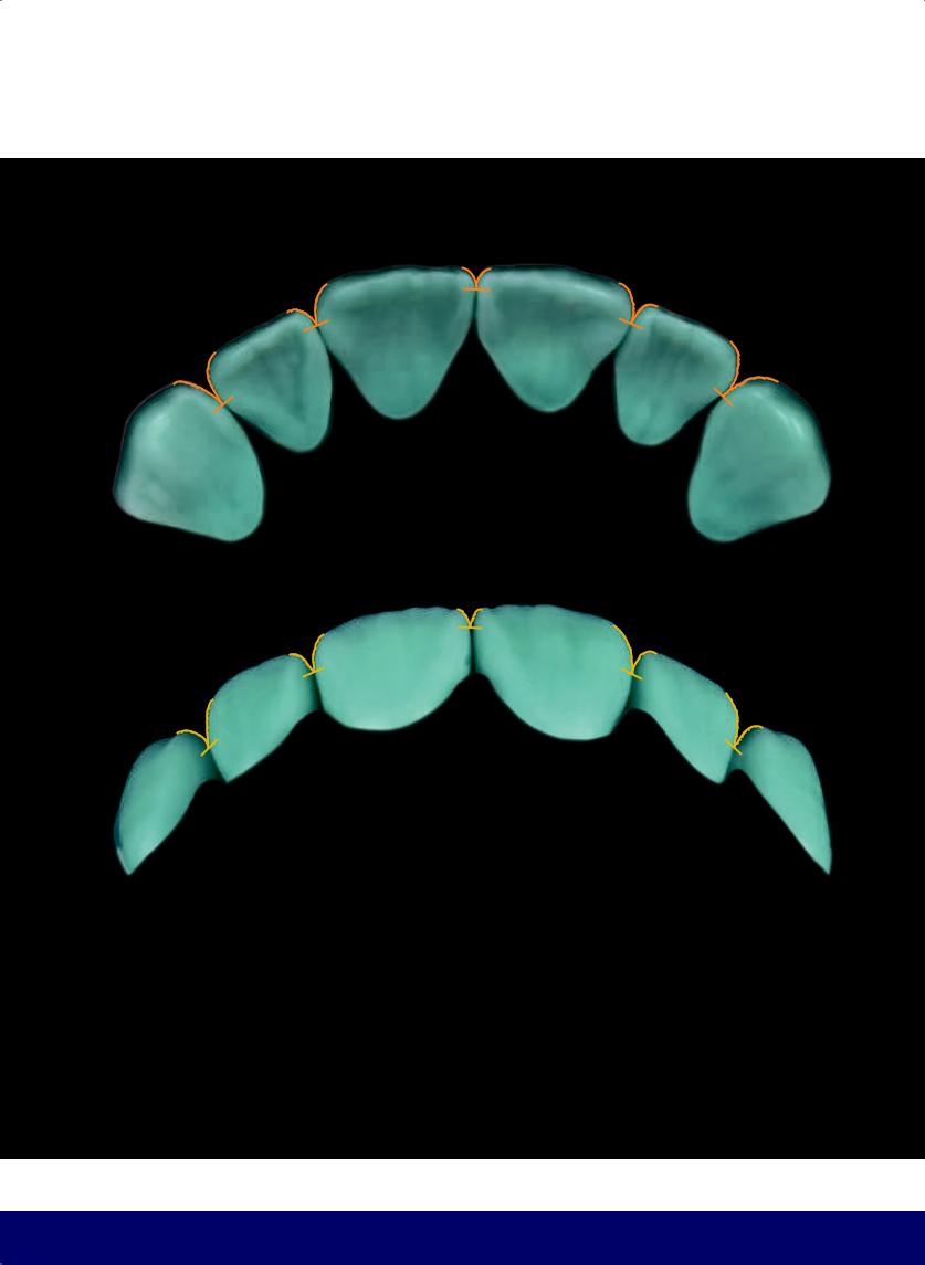
THE ESTHETIC AND FUNCTIONAL PARAMETERS OF ANTERIOR TEETH
—From the occlusal view, displacement of areas of contact toward the lingual increases the labial/buccal embrasures.
—Position of anterior contact areas from a craniocaudal view.
255
@dentistinfo стоматологический телеграм канал
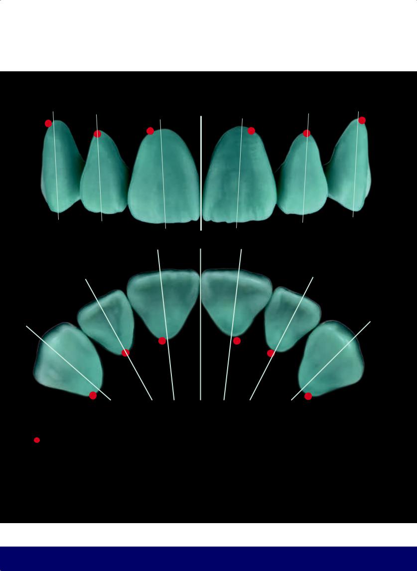
CHAPTER 12
In relation to the vertical axis of the tooth, the location of the gingival zenith (highest point of the crown near the gingiva) is distal in maxillary central incisors and canines and coincident in the lateral incisors (from both the labial and the lingual).
256
@dentistinfo стоматологический телеграм канал
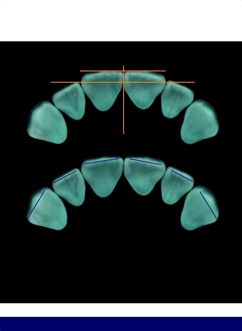
THE ESTHETIC AND FUNCTIONAL PARAMETERS OF ANTERIOR TEETH
—From an occlusal view of the anterior teeth, the central incisors are in front and the lateral incisors are behind.
—Curvature of the incisal edges from canine to canine in an occlusal view.
257
@dentistinfo стоматологический телеграм канал
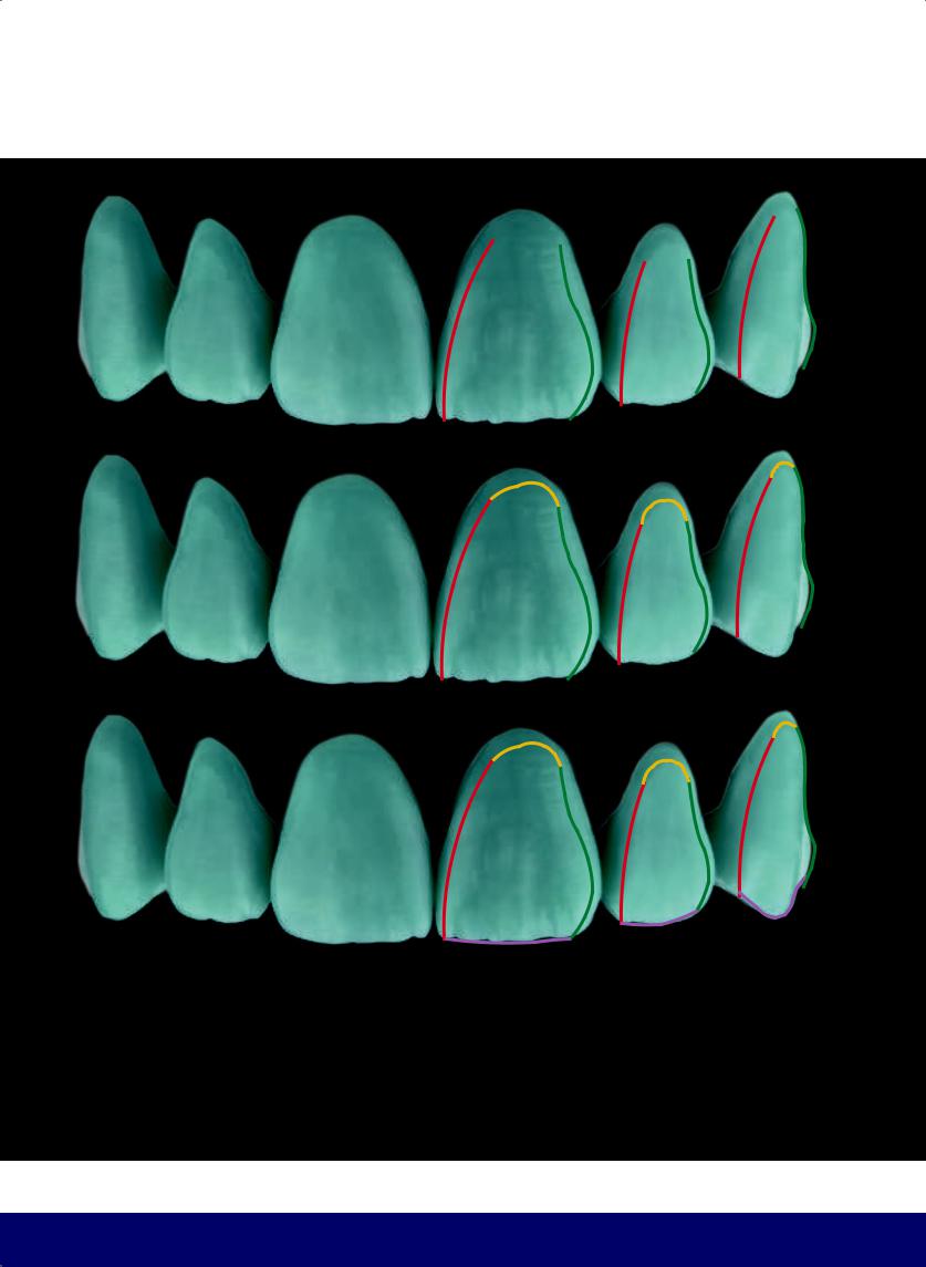
CHAPTER 12
TRANSITIONAL LINE ANGLES |
|
These lines are the mesial and distal borders of the face of the tooth. |
— |
— Mesial border resembles a C. |
— |
— Distal border resembles an S. |
|
Cervical border follows the position of the gingival zenith.
Incisal border and cusp tip.
258
@dentistinfo стоматологический телеграм канал
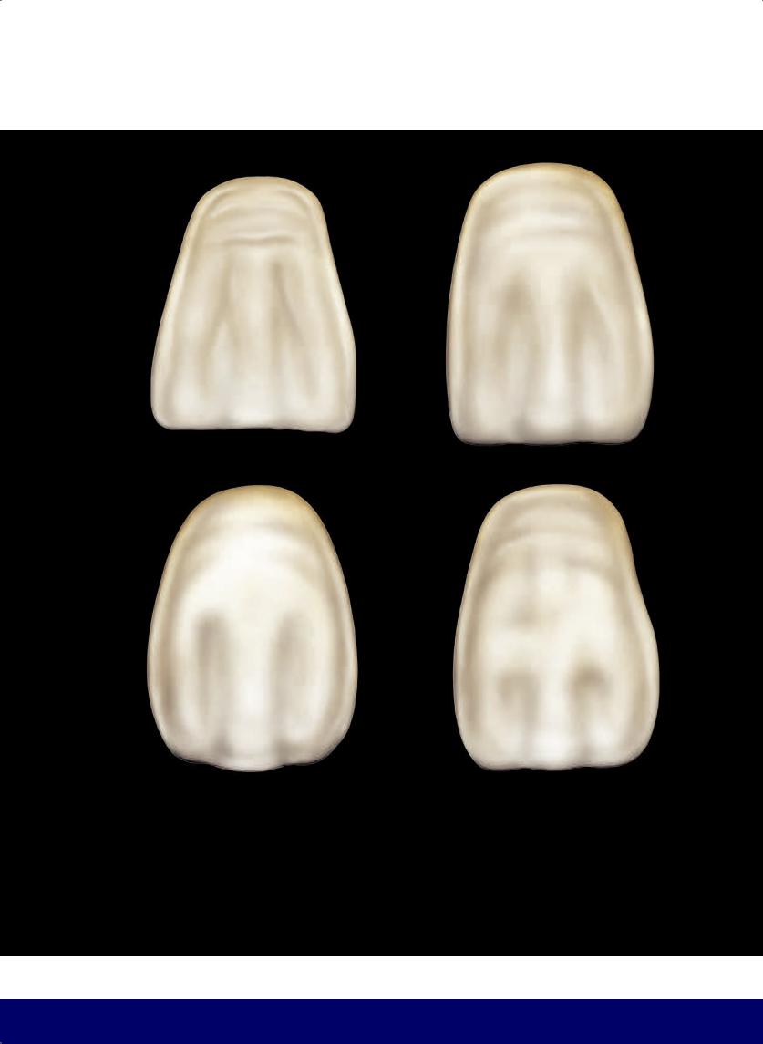
THE ESTHETIC AND FUNCTIONAL PARAMETERS OF ANTERIOR TEETH
TRANSITIONAL LINE ANGLES
These are the borders that delimit the flat area on the face of the tooth. They follow the basic shape of the tooth.
259
@dentistinfo стоматологический телеграм канал
CHAPTER 12
—The face of a tooth is the reflective area on the labial/buccal surface.
—Deflective area. Note that, in a frontal view, the areas of mesial deflection increase from central incisor to canine.
—Vertical macro texture. Mesial and distal developmental grooves often resemble two inverted Ys in the central incisors or a mesial inverted Y and a slight distal depression in the lateral incisors.
260
@dentistinfo стоматологический телеграм канал
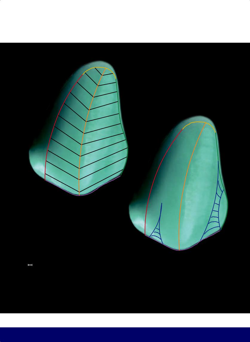
THE ESTHETIC AND FUNCTIONAL PARAMETERS OF ANTERIOR TEETH
—
—
The buccal face of the canine is divided into two parts by a curved labial ridge that extends from the cusp tip to the
gingival zenith. The mesial segment is smaller and the distal segment is larger.
Vertical macro texture of the canine. The mesial developmental groove is a slight depression, while the distal developmental groove is broader.
261
@dentistinfo стоматологический телеграм канал
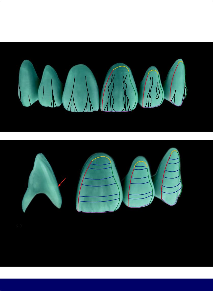
CHAPTER 12
—Schematic for vertical macro texture: These grooves have irregular borders. The mesial and distal developmental grooves resemble two Ys in the central incisors, while on the lateral incisors there is only one mesial Y and a slight distal depression. In addition, there are smaller vertical grooves near the incisal edge.
—Schematic for horizontal macro texture: The depressions (up to five in number) follow a trajectory opposite to the cementoenamel line and are present in all the teeth. They are not necessarily parallel.
262
@dentistinfo стоматологический телеграм канал
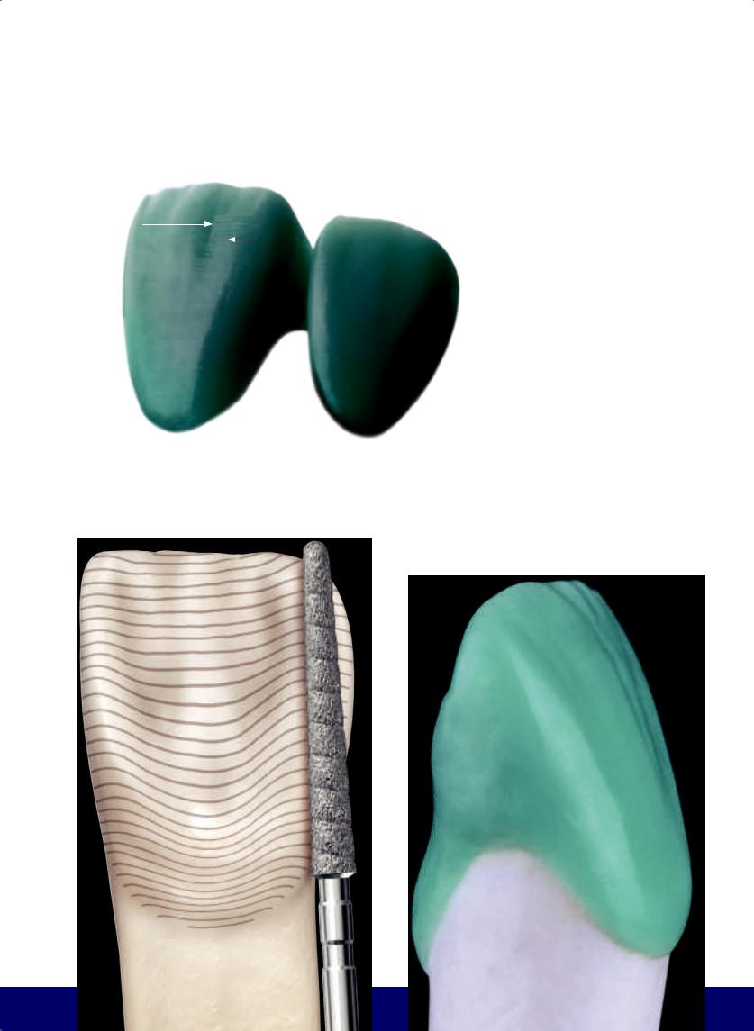
THE ESTHETIC AND FUNCTIONAL PARAMETERS OF ANTERIOR TEETH
CRACKS |
|
The HORIZONTAL MICRO TEXTURE of |
|
STRIAE |
|
|
STRIAE OF RETZIUS is present on all tooth |
|
|
|
|
|
|
faces and follow a trajectory parallel to |
|
|
the crown/root line. The curvature of |
|
|
striae (which is more curved at the |
|
|
cervix) decreases and becomes straight |
|
|
at the incisal edge. |
|
|
The VERTICAL MICRO TEXTURE is repre- |
|
|
sented by cracks and grooves. |
|
|
|
|
|
|
|
|
|
263
@dentistinfo стоматологический телеграм канал
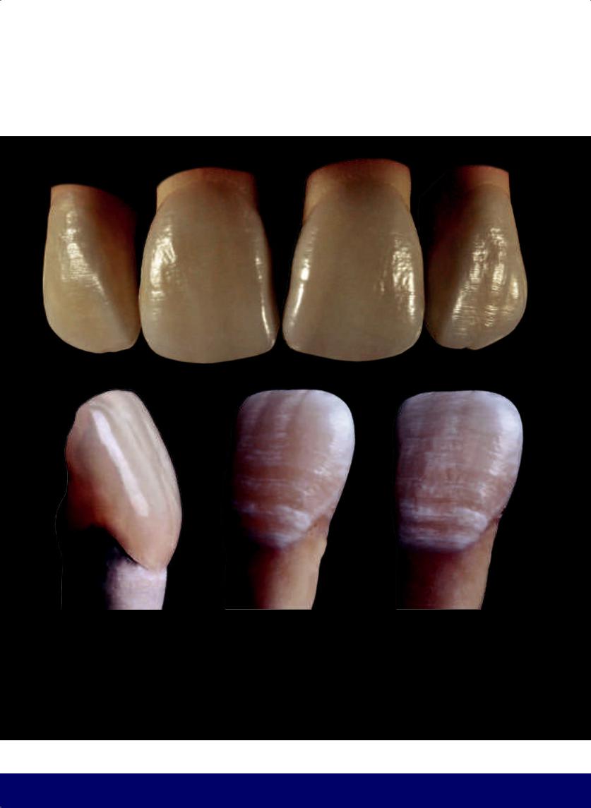
CHAPTER 12
HORIZONTAL AND VERTICAL MICRO TEXTURE
264
@dentistinfo стоматологический телеграм канал
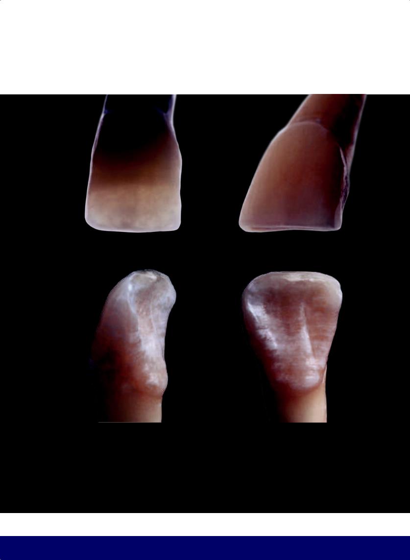
THE ESTHETIC AND FUNCTIONAL PARAMETERS OF ANTERIOR TEETH
265
@dentistinfo стоматологический телеграм канал
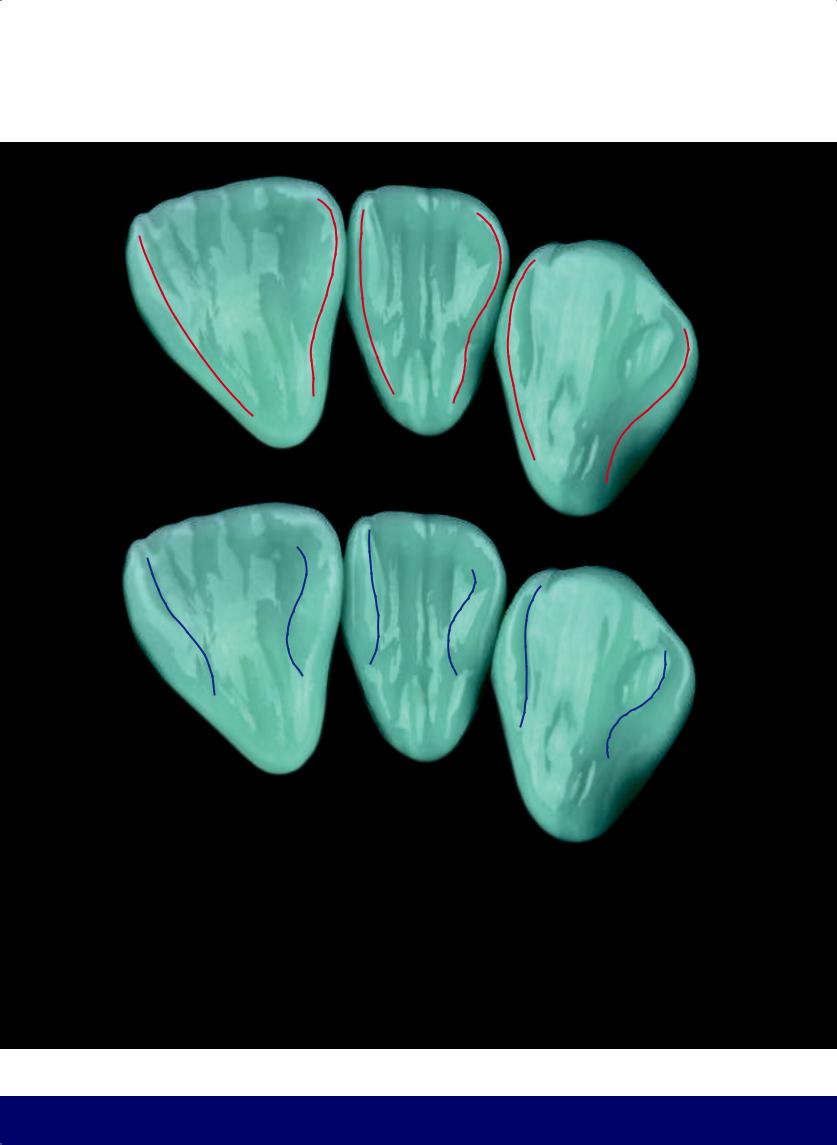
CHAPTER 12
—The lingual transitional line angles repeat what occurs on the labial surface, with the mesial border resembling a C and the distal border resembling an S. These lines and angles are the highest part of the mesial and distal marginal ridges, respectively.
—Lines separate the mesial and distal marginal ridges from the lingual depression in the lateral and central incisors and from the central lobe in the canines.
266
@dentistinfo стоматологический телеграм канал
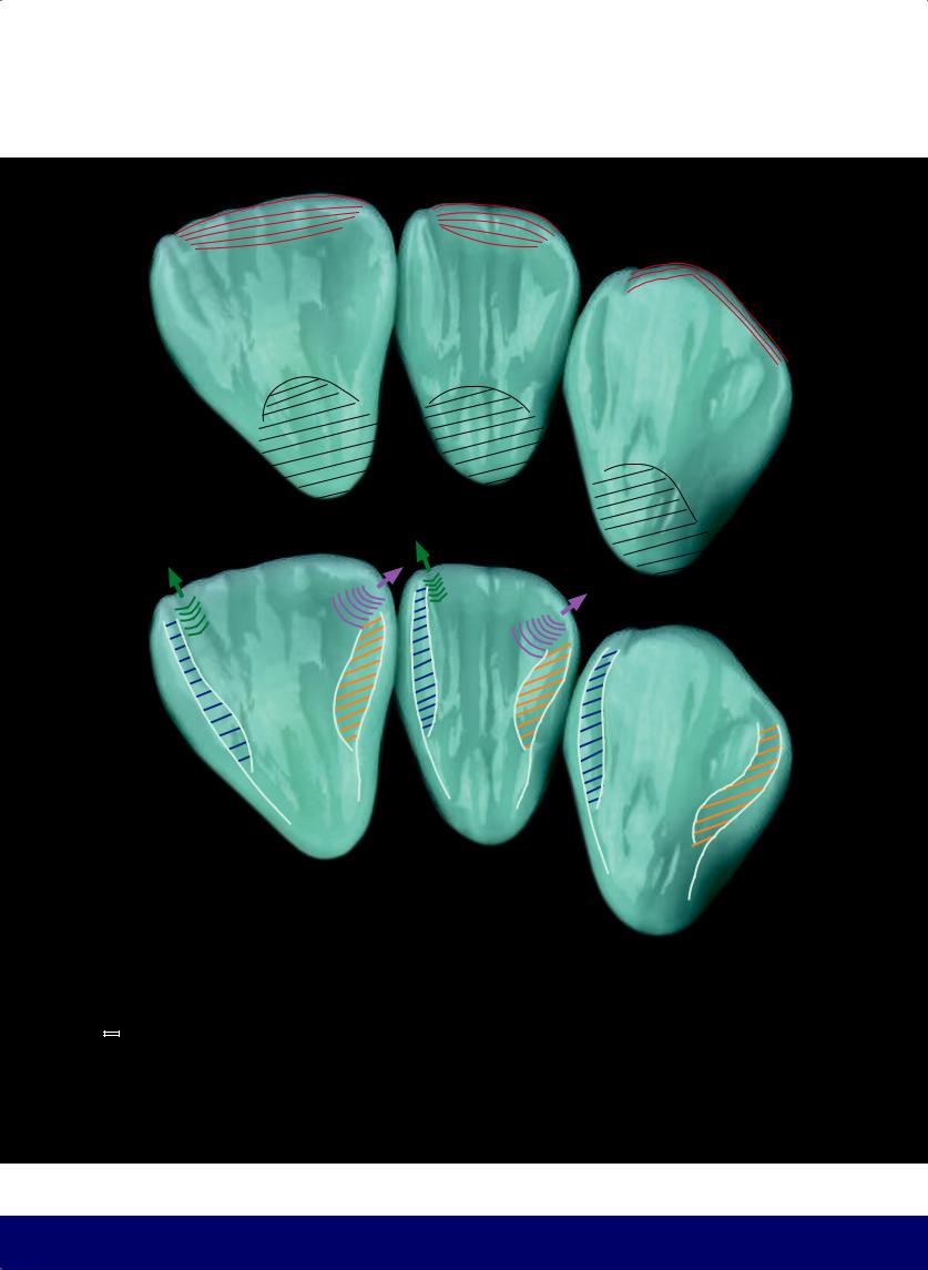
THE ESTHETIC AND FUNCTIONAL PARAMETERS OF ANTERIOR TEETH
—
—
Incisal edges of the central and lateral incisors and the cusp tip of the canine.
Cingulum.
The MESIAL MARGINAL CREST is narrower and longer because the MESIAL ESCAPE ROUTE is directed toward the incisal edge, while the DISTAL MARGINAL RIDGE is wider and shorter, as the DISTAL ESCAPE ROUTE lies between it and the incisal edge.
267
@dentistinfo стоматологический телеграм канал
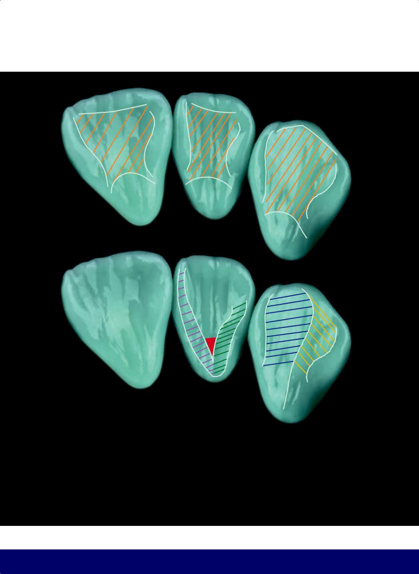
CHAPTER 12
—The lingual depression in the central and lateral incisors, and the central lobe in the canines.
—The central lobe on the lingual aspect of the canines is divided into two parts:
—The larger mesial side. (Note that for canine guidance, the mesial side slides laterally in the mandibular canine.)
—The smaller distal side.
268
FORAMEN CECUM
Pit resulting from the meeting of the mesial and distal marginal crests near the cingulum.
@dentistinfo стоматологический телеграм канал
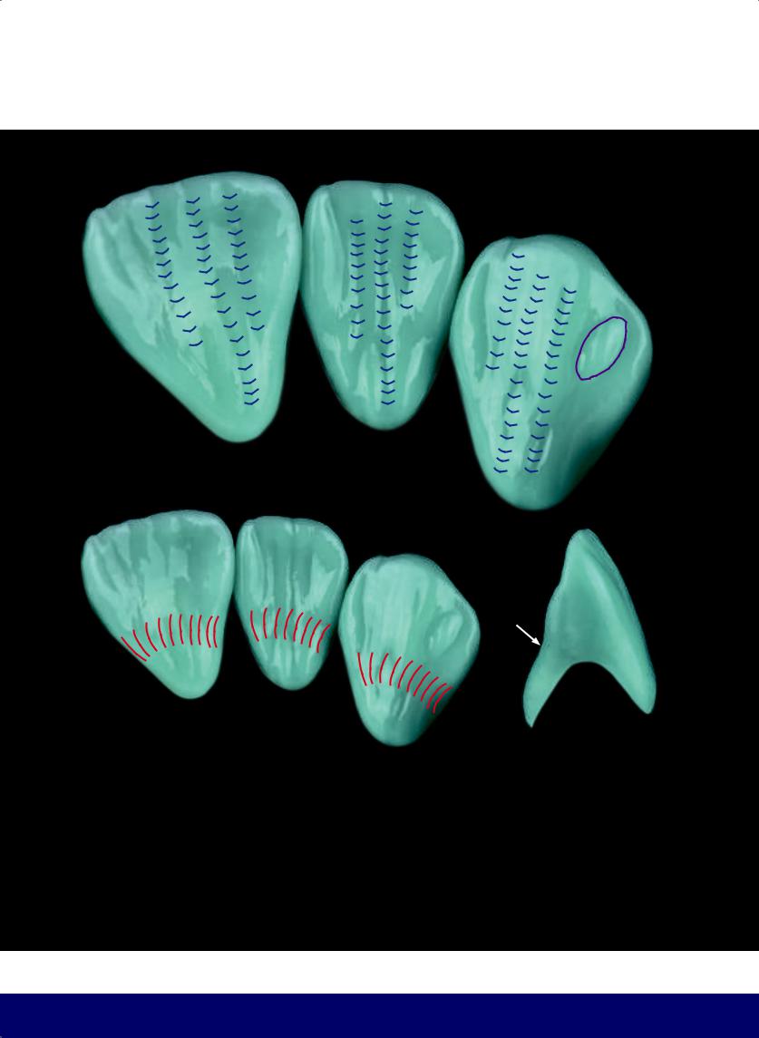
THE ESTHETIC AND FUNCTIONAL PARAMETERS OF ANTERIOR TEETH
—Lobe of the distal marginal crest, where the the buccal cusp of the mandibular first premolar touches on protrusive movement.
—Minor vertical grooves.
—Cingulum depression: separates the cingulum from the marginal ridges.
269
@dentistinfo стоматологический телеграм канал
CHAPTER 12
270
@dentistinfo стоматологический телеграм канал
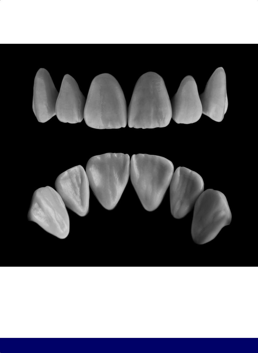
THE ESTHETIC AND FUNCTIONAL PARAMETERS OF ANTERIOR TEETH
271
@dentistinfo стоматологический телеграм канал

Incisor
Maxillary Central
13
272
@dentistinfo стоматологический телеграм канал
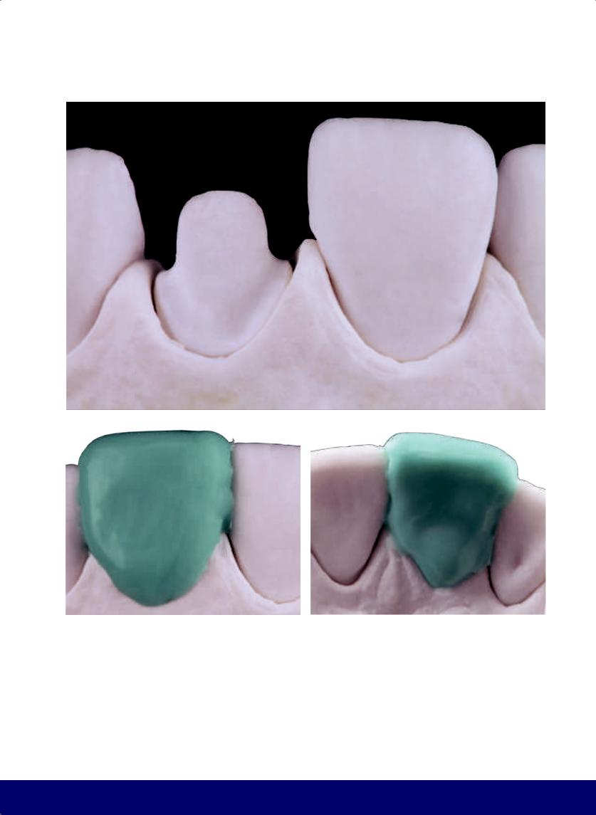
MAXILLARY CENTRAL INCISOR
a
b |
|
c |
|
|
|
Fig 13-1 | (a to c) With the proper position of the tooth preparation, fill out the tooth form with labial, lingual, and incisal wax and allow the wax to harden.
273
@dentistinfo стоматологический телеграм канал
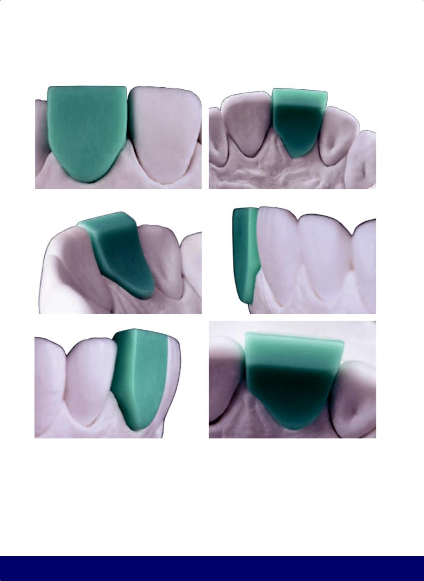
CHAPTER 13
a |
|
b |
|
|
|
c |
|
d |
|
|
|
e |
|
f |
|
|
|
Fig 13-2 | (a to f) Labial, lingual, and incisal perspectives of the wax block after the excess has been trimmed. The wax block still presents oversized dimensions ready for reduction.
274
@dentistinfo стоматологический телеграм канал
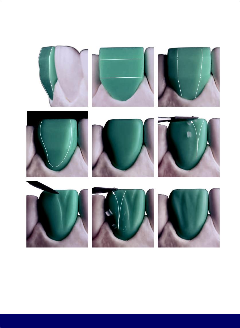
MAXILLARY CENTRAL INCISOR
a |
|
b |
|
c |
|
|
|
|
|
d |
e |
|
f |
|
|
|
|
g |
|
h |
|
i |
|
|
|
|
|
Fig 13-3 | (a and b) Carve the incisal and cervical planes (horizontal planes). (c) Carve the mesial and distal planes (vertical planes) while keeping horizontal planes. Schematically, teeth present three inclinations in the mesiodistal and cervico-incisal directions.
(d) Further reduce the wax block and delimit the flat area (or face of the tooth; define the C on the mesial border, the S on the distal border), the cervical line that follows the gingival zenith, and the incisal border. (e) Round the angles and finalize the form of the flat
area. Note the lines of brightness. (f to i) Vertical macro texture. Create the mesial and distal developmental grooves (resembling two Ys) that divide the labial face into three smaller structures: the mesial, central, and distal lobes. They begin between the cervical and middle thirds, open between the middle and incisal thirds, and diverge in the incisal third. (White arrows indicate the direction of instrumentation.)
275
@dentistinfo стоматологический телеграм канал
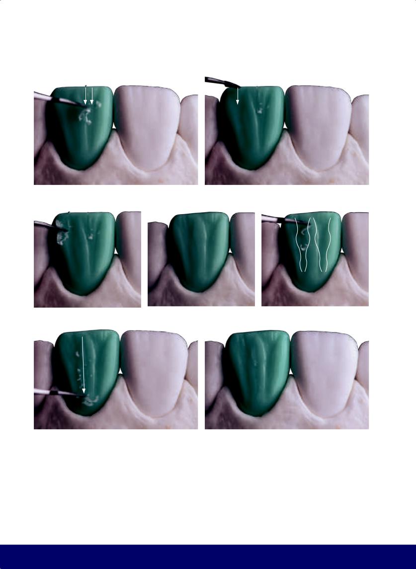
CHAPTER 13
a |
|
b |
|
|
|
c |
|
d |
|
e |
|
|
|
|
|
f |
|
g |
|
|
|
Fig 13-4 | (a to g) Smaller vertical grooves in and out of the Ys.
276
@dentistinfo стоматологический телеграм канал
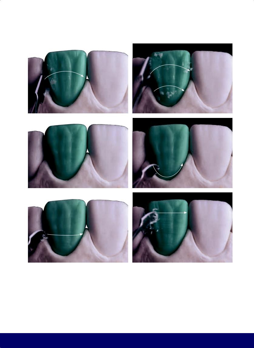
a |
b |
|
|
c |
d |
|
|
e |
f |
|
|
Fig 13-5 | (a to c) The horizontal macro texture involves three broad and subtle depressions with a trajectory opposite the crown/root line. (d to f) The horizontal micro texture mimics the striae of Retzius that parallel the crown/root line and become less curved, and finally straighten, as they approach the incisal border.
MAXILLARY CENTRAL INCISOR
277
@dentistinfo стоматологический телеграм канал
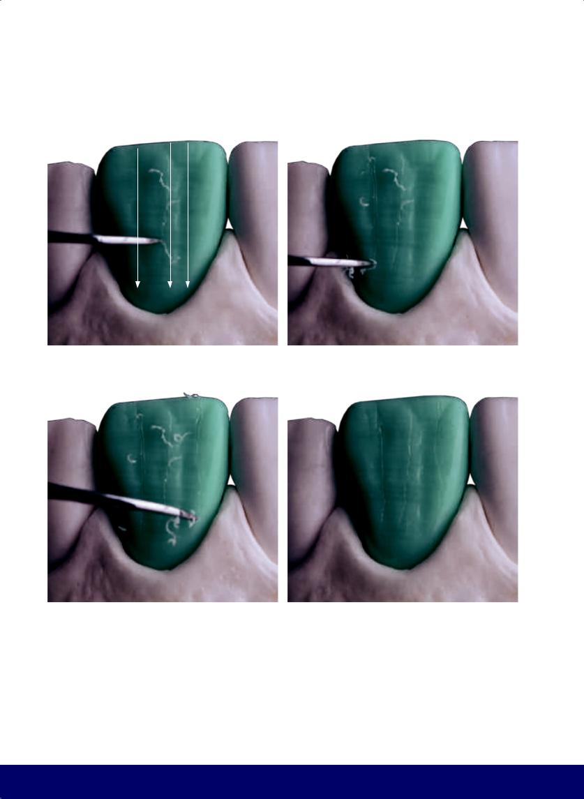
CHAPTER 13
a |
|
b |
|
|
|
c |
|
d |
|
|
|
Fig 13-6 | (a to d) The vertical micro texture includes cracks and grooves (“Wolverine claws”).
278
@dentistinfo стоматологический телеграм канал
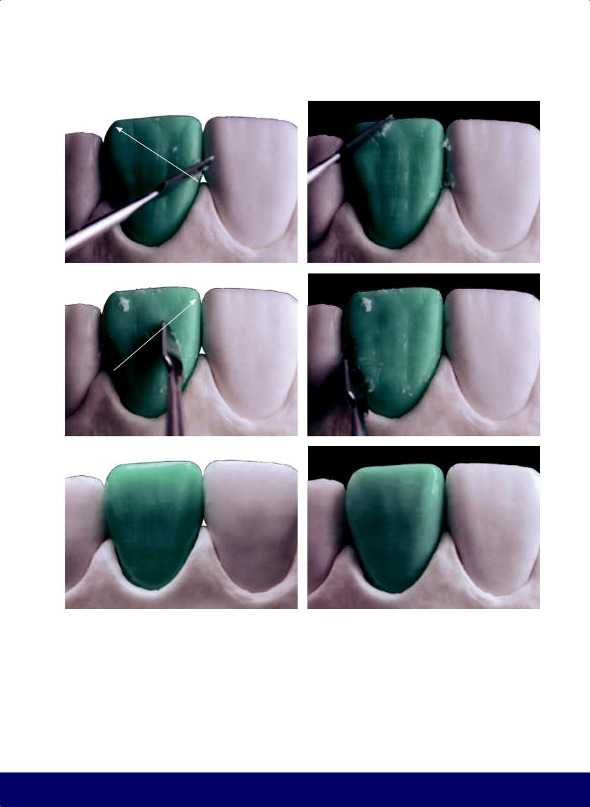
a |
b |
|
|
c |
d |
|
|
e |
f |
|
|
Fig 13-7 | (a to e) Control the labial texture by cross-cutting. Slide the carving instrument over the wax surface diagonally, top to bottom, mesial to distal, and then distal to mesial. Slide it smoothly and repeatedly. (f) Finished external labial contour.
MAXILLARY CENTRAL INCISOR
279
@dentistinfo стоматологический телеграм канал
CHAPTER 13
a |
|
b |
|
|
|
c |
|
d |
|
|
|
e |
|
f |
|
|
|
Fig 13-8 | (a) Concave lingual face. (b) Note the borders of the lingual surface with the mesial C, the subtle distal S, the cervical line that follows the gingival zenith, and the incisal edge. (c) Create the mesial marginal crest, which is narrow and long, by adding a depression directed toward the incisal edge to create a low relief area or the “mesial escape route.” (d) Make the distal marginal crest slightly wider and shorter than the mesial crest; the depression should exit diagonally between the crest and the incisal edge,
280
creating the “distal escape route.” (e and f) The cingulum depression is a low relief that marks the separation between the mesial and distal marginal ridges and the cingulum (according to Jan Hatjó).
@dentistinfo стоматологический телеграм канал
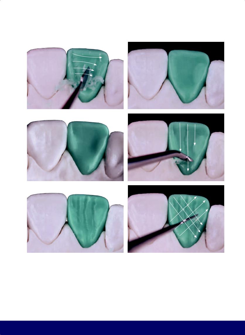
a |
b |
|
|
c |
d |
|
|
e |
f |
|
|
Fig 13-9 | (a to c) Remove the excess from the lingual aspect and consequently create the lingual fossa. (d to f) Create the lingual vertical grooves that run from the cingulum to the incisal edge.
MAXILLARY CENTRAL INCISOR
281
@dentistinfo стоматологический телеграм канал
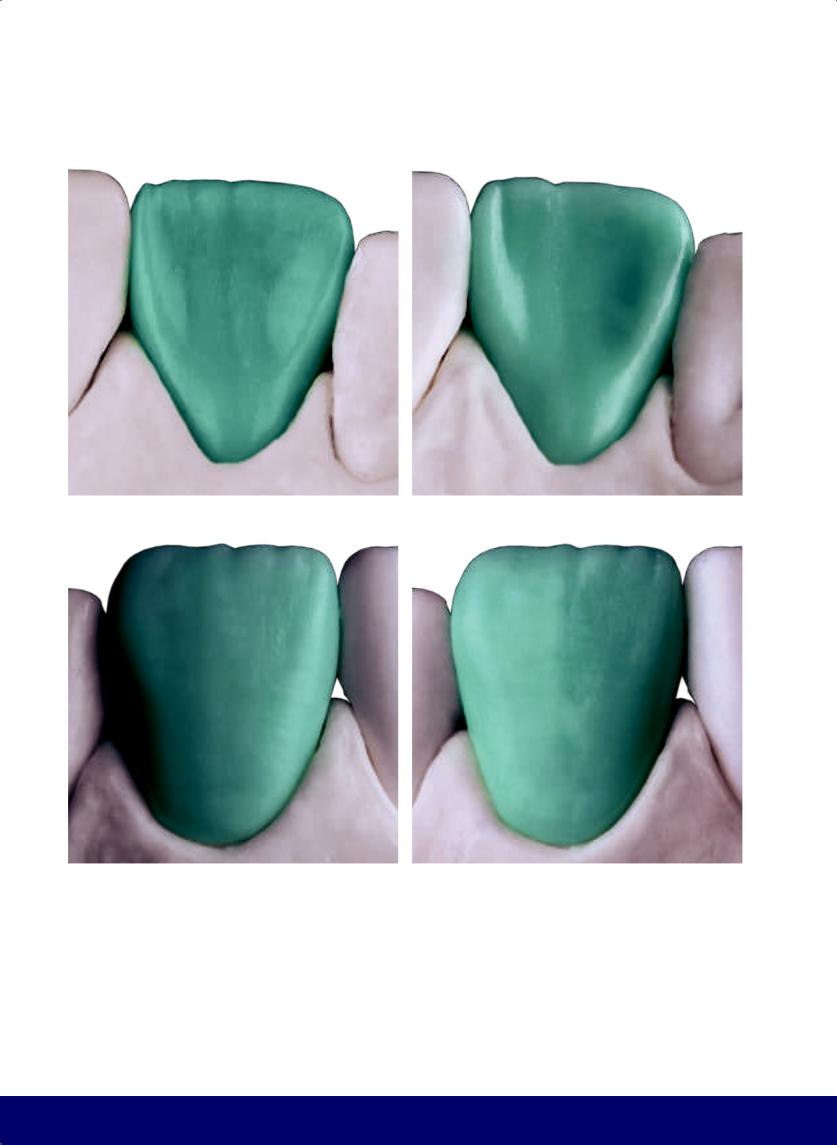
CHAPTER 13
a |
|
b |
|
|
|
c |
|
d |
|
|
|
Fig 13-10 | (a and b) Completed lingual face. (c and d) Completed labial face.
282
@dentistinfo стоматологический телеграм канал
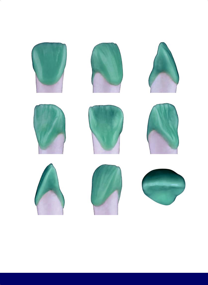
MAXILLARY CENTRAL INCISOR
a |
|
b |
|
c |
|
|
|
|
|
d |
|
e |
|
f |
|
|
|
|
|
g |
|
h |
|
i |
|
|
|
|
|
Fig 13-11 | (a to i) Completed maxillary central incisor.
283
@dentistinfo стоматологический телеграм канал

Incisor
Maxillary Lateral
14
284
@dentistinfo стоматологический телеграм канал
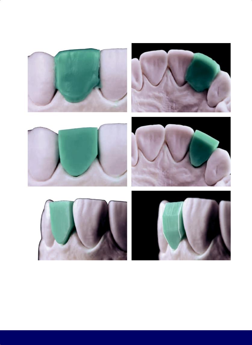
a |
b |
|
|
c |
d |
|
|
e |
f |
|
|
Fig 14-1 | (a and b) Fill out the wax block on the tooth preparation with excess labial, lingual, and incisal wax. (c to e) Lingual, labial, and incisal perspectives after trimming excess. The wax block still presents oversized dimensions ready for reduction. (f) Carve the incisal and cervical planes (horizontal planes).
MAXILLARY LATERAL INCISOR
285
@dentistinfo стоматологический телеграм канал
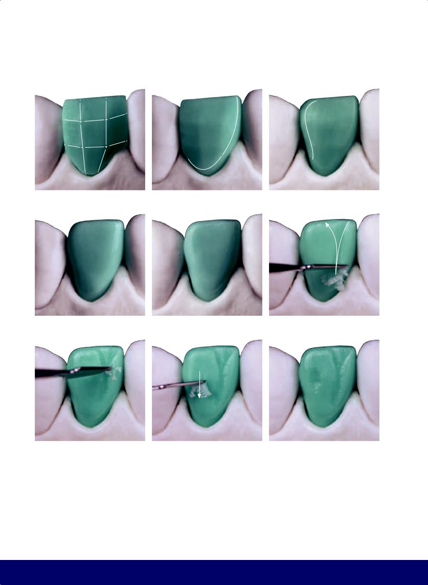
CHAPTER 14
a |
|
b |
|
c |
|
|
|
|
|
d |
|
e |
|
f |
|
|
|
|
|
g |
|
h |
|
i |
|
|
|
|
|
Fig 14-2 | (a) Carve the mesial and distal planes (vertical planes) in keeping with the horizontal planes. The labial surface should have three planes in the mesiodistal and cervico-incisal directions. (b and c) Reduce the overall wax block and delineate the flat area of the tooth face by defining the C on the mesial border, the S on the distal border, the cervical border, and the incisal border. (d and e) Round the angles and finalize the form of the flat area. Notice the transitional line angles. (f to i) For the vertical macro texture,
286
create the mesial and distal developmental grooves, dividing the labial face into two lobes: a narrower mesial lobe and a wider distal lobe. The mesial groove should resemble a Y and the distal groove is a slight depression. The mesial groove begins between the cervical and middle thirds and opens between the middle and incisal thirds, toward the incisal. (White arrows indicate the direction of instrumentation.)
@dentistinfo стоматологический телеграм канал
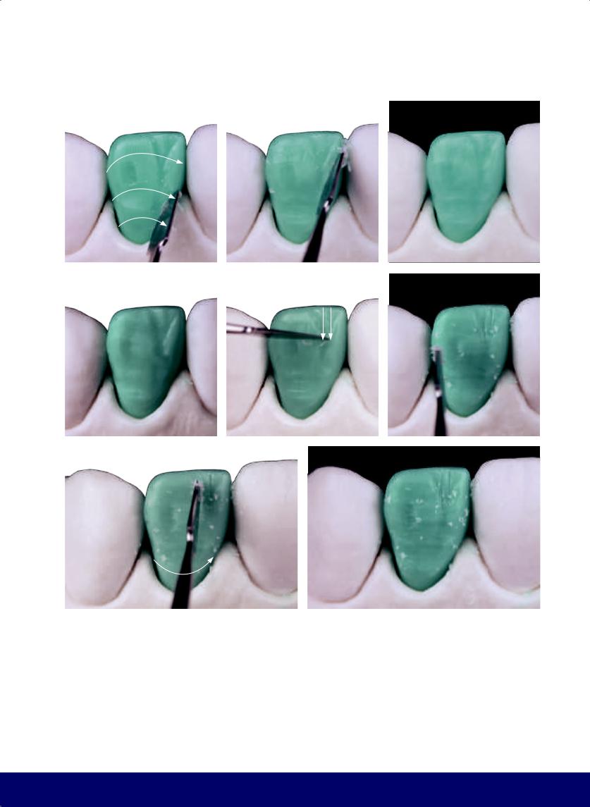
a |
|
b |
|
|
|
d |
|
e |
|
|
|
g |
h |
|
|
Fig 14-3 | (a to d) Horizontal macro texture is composed of three subtle broad depressions that follow a trajectory opposite the crown/root line. (e) Minor vertical grooves within and around the Y. (f to h) Horizontal micro texture. To create the striae of Retzius, gently slide the tip of the instrument parallel to the crown/root line.
MAXILLARY LATERAL INCISOR
c
f
287
@dentistinfo стоматологический телеграм канал
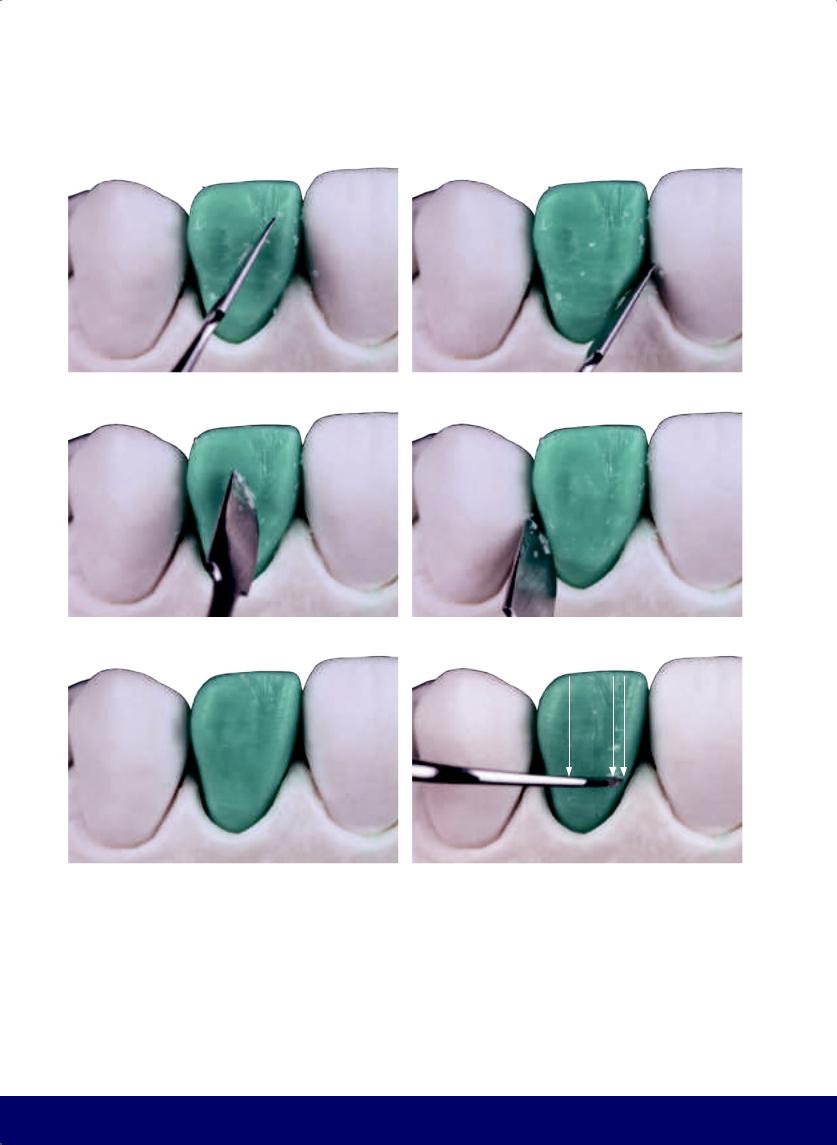
CHAPTER 14
a |
|
b |
|
|
|
c |
|
d |
|
|
|
e |
|
f |
|
|
|
Fig 14-4 | (a to d) Control the labial texture by cross-cutting: Slide the instrument diagonally over the surface of the wax, from top to bottom, from mesial to distal, and then from distal to mesial. (e and f) Add vertical micro texture of cracks or grooves or “Wolverine claws.”
288
@dentistinfo стоматологический телеграм канал
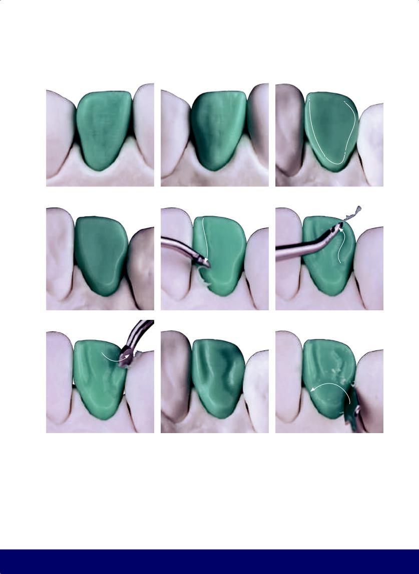
MAXILLARY LATERAL INCISOR
a |
|
b |
|
c |
|
|
|
|
|
d |
|
e |
|
f |
|
|
|
|
|
g |
|
h |
|
i |
|
|
|
|
|
Fig 14-5 | (a and b) Completed external labial surface. (c and d) Note the borders of the lingual surface with the mesial C, the subtle distal S, the cervical line that follows the gingival zenith, and the incisal edge. (e) Make the mesial marginal crest narrow and long by adding a depression directed toward the incisal border; this low relief is the “mesial escape route.” (f to h) Create the distal marginal ridge, which is slightly wider and shorter than the mesial
crest. The depression should exit diagonally between the crest and the incisal edge to create the “distal escape route.” (i) The cingulum depression is a low relief that marks the separation between the marginal mesial and distal ridges and the cingulum (according to Jan Hatjó).
289
@dentistinfo стоматологический телеграм канал
CHAPTER 14
a |
|
b |
|
c |
|
|
|
|
|
d |
|
e |
|
f |
|
|
|
|
|
g |
|
h |
|
i |
|
|
|
|
|
Fig 14-6 | (a to c) Remove lingual excess to reveal the lingual fossa. (d to g) Create the vertical lingual grooves, running from cingulum to incisal edge. (h and i) Completed lingual face.
290
@dentistinfo стоматологический телеграм канал
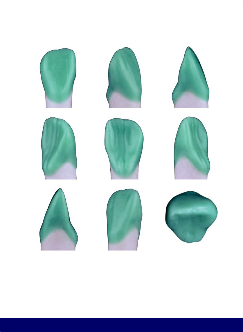
MAXILLARY LATERAL INCISOR
a |
|
b |
|
c |
|
|
|
|
|
d |
|
e |
|
f |
|
|
|
|
|
g |
|
h |
|
i |
|
|
|
|
|
Fig 14-7 | (a to i) Completed maxillary lateral incisor.
291
@dentistinfo стоматологический телеграм канал

Maxillary Canine
15
292
@dentistinfo стоматологический телеграм канал
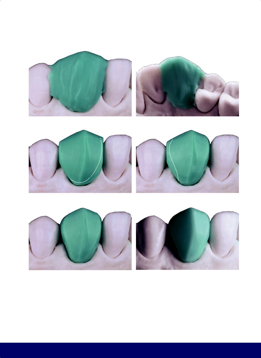
MAXILLARY CANINE
a |
|
b |
|
|
|
c |
|
d |
|
|
|
e |
|
f |
|
|
|
Fig 15-1 | (a and b) Construct the wax block with excess on the buccal, lingual, and incisal dimensions. Allow the wax to harden.
(c) Buccal perspective of the wax block after trimming excess. Note that the buccal face is divided in two parts by a curved line that goes from the cusp tip to the gingival zenith. The mesial side is smaller than the distal side. The mesial segment of the
longitudinal ridge of the cusp is also shorter than the distal segment. As a result, the cusp tip is slightly toward the mesial. (d) Reduce the buccal shape by defining the mesial C. (e) Form the subtle distal S. (f) Note that the mesial and distal planes already have, from the outset, a cervico-incisal bulge that emphasizes the slightly mesialized curve.
293
@dentistinfo стоматологический телеграм канал
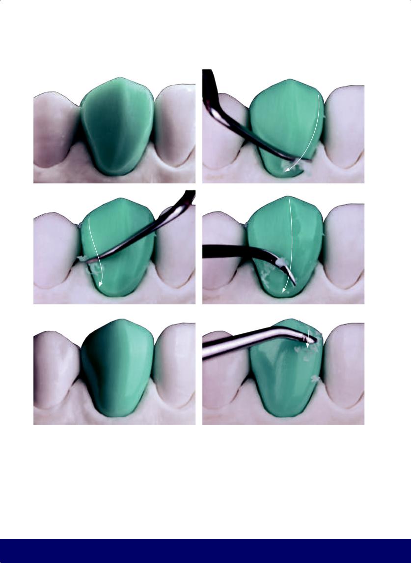
CHAPTER 15
a |
|
b |
|
|
|
c |
|
d |
|
|
|
e |
|
f |
|
|
|
Fig 15-2 | (a) Reduce the buccal shape and delineate the flat planes through the definition of the mesial C, the distal S, the cervical line, and the cusp tip. (b to e) Round the angles and finalize the form of the flat areas. Note the transitional line angles. (f to j) For the vertical macro texture, create developmental grooves. The mesial groove is very short given the structural reinforcement in the buccal “belly” of the canine. The distal groove is a wide and subtle depression that extends from the distal segment of the cusp to the distal papilla. (White arrows indicate the direction of instrumentation.)
294
@dentistinfo стоматологический телеграм канал
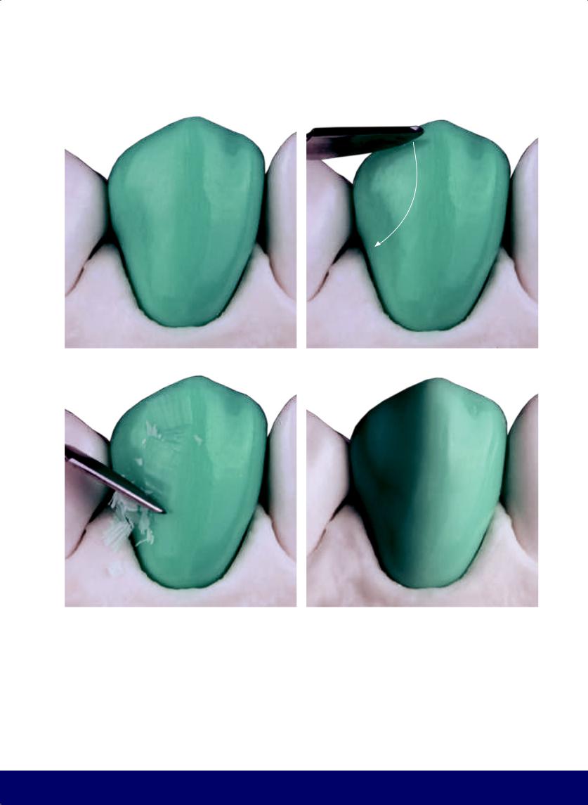
MAXILLARY CANINE
g |
|
h |
|
|
|
i |
|
j |
|
|
|
295
@dentistinfo стоматологический телеграм канал
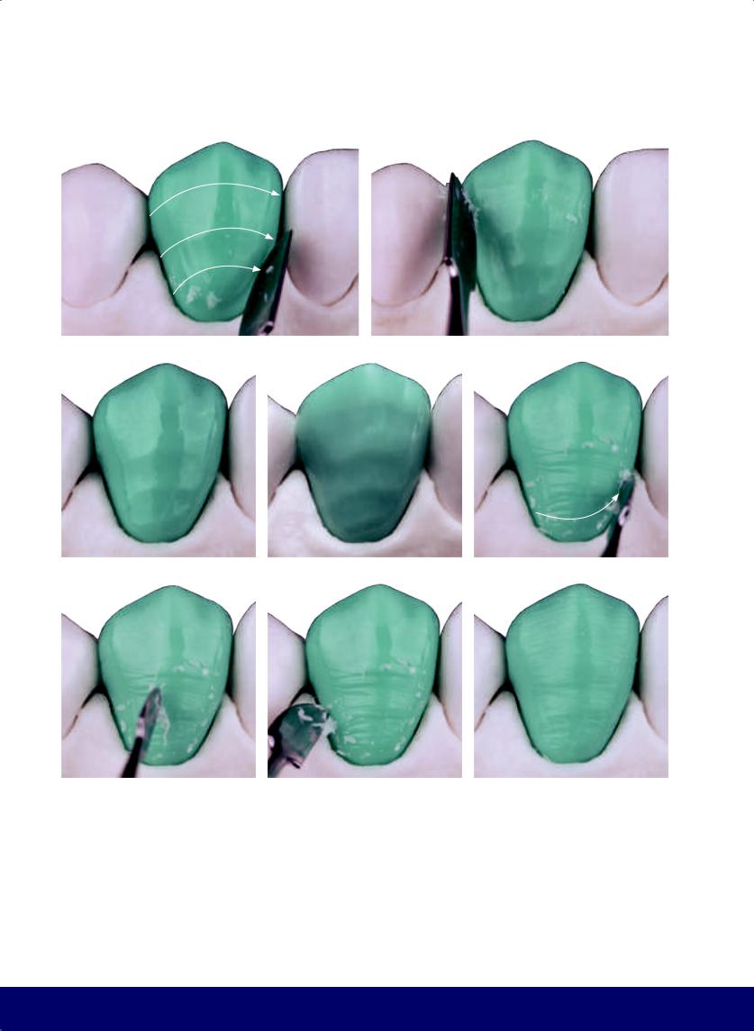
CHAPTER 15
a |
|
b |
|
|
|
c |
|
d |
|
e |
|
|
|
|
|
f |
|
g |
|
h |
|
|
|
|
|
Fig 15-3 | (a to d) The horizontal macro texture includes three broad and subtle depressions following a trajectory opposite the crown/root line. (e to h) For horizontal micro texture, the striae of Retzius follow a curved trajectory parallel to the crown/root line and become less curved and straighten before the curvature reverses in the approach to the cusp tip.
296
@dentistinfo стоматологический телеграм канал
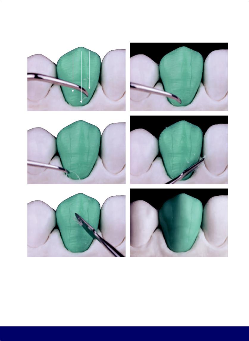
a |
b |
|
|
c |
d |
|
|
e |
f |
|
|
Fig 15-4 | (a to c) Vertical micro texture includes cracks or grooves (“Wolverine claws”). (d to f) Control the buccal texture by cross cuts; slide the carving instrument diagonally across the wax surface, from top to bottom, from mesial to distal, and then from distal to mesial.
MAXILLARY CANINE
297
@dentistinfo стоматологический телеграм канал

CHAPTER 15
a |
|
b |
|
c |
|
|
|
|
|
d |
|
e |
|
|
|
f |
|
g |
|
h |
|
|
|
|
|
Fig 15-5 | (a) Completed external buccal contour. (b) Concave lingual face. (c and d) Note the reduction of the distal plane. Observe the curved line, facing distally, that divides the lingual aspect into two parts: a larger mesial side and a smaller distal side. (This is where the mandibular canine slides in canine guidance.) (e to h) Create the lingual marginal ridges by adding the mesial and distal depressions directed toward the cusp tip.
298
@dentistinfo стоматологический телеграм канал
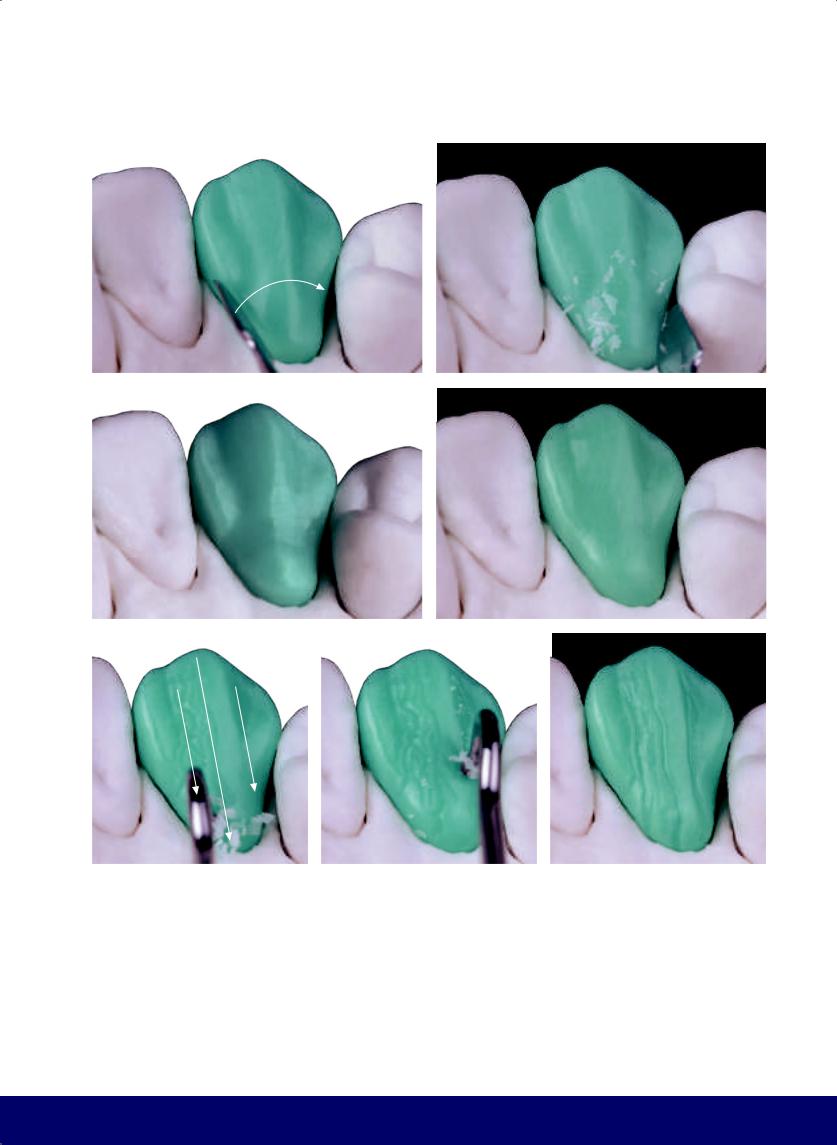
a |
b |
|
|
c |
d |
|
|
e |
|
f |
|
|
|
Fig 15-6 | (a to c) Form the cingulum depression, a low relief that marks the separation between the mesial and distal marginal ridges, the central lobe, and the cingulum. (d to g) Develop the vertical lingual grooves.
MAXILLARY CANINE
g
299
@dentistinfo стоматологический телеграм канал
CHAPTER 15
a |
|
b |
|
c |
|
|
|
|
|
d |
|
e |
|
|
|
Fig 15-7 | (a and b) Finalize the lingual face by softening the vertical texture. Slide the instrument diagonally from top to bottom, from mesial to distal, and vice versa. (c and d) Create a distal lobe within the distal groove. (e) The final definition of the lingual face.
300
@dentistinfo стоматологический телеграм канал
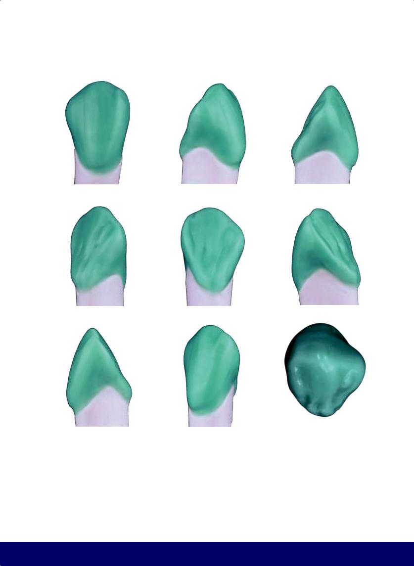
MAXILLARY CANINE
a |
|
b |
|
c |
|
|
|
|
|
c |
|
d |
|
e |
|
|
|
|
|
f |
|
g |
|
h |
|
|
|
|
|
Fig 15-8 | (a to i) Completed external canine contour.
301
@dentistinfo стоматологический телеграм канал

Progressive Technique for Maxillary Anterior Teeth
16
302
@dentistinfo стоматологический телеграм канал
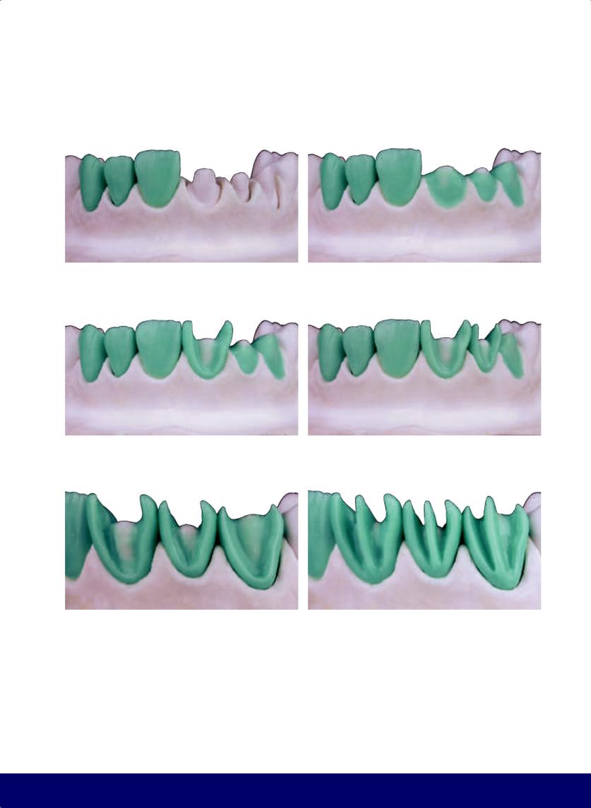
PROGRESSIVE TECHNIQUE FOR MAXILLARY ANTERIOR TEETH
a |
|
b |
|
|
|
c |
|
d |
|
|
|
e |
|
f |
|
|
|
Fig 16-1 | (a) Place the preparations. (b) Seat a wax base. (c to e) Define the labial plane first with the placement of the cervical support and the transitional line angles (the mesial C and distal S). These are three of the four “frames” of the crown. (f) Place the central support column. Note that in the canine this column curves toward the mesial, while in the central and the lateral incisors it is positioned more to the center.
303
@dentistinfo стоматологический телеграм канал
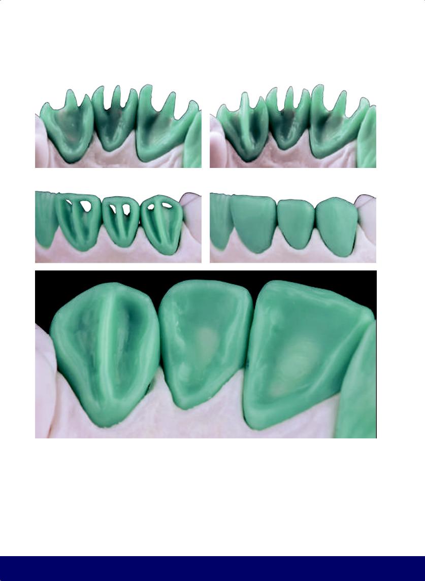
CHAPTER 16
a |
|
b |
|
|
|
c |
|
d |
|
|
|
e
Fig 16-2 | (a) Lingual view. (b) The canine is divided by a column curved to the distal. (c) Connect the vertical columns by means of the incisal “beam” in the central and lateral incisors and of the cusp tip in the canine. The frame of the crowns is complete. (d) Close the spaces between the columns and frames to complete the flat labial face. (e) Create the mesial and distal marginal ridges and “escape areas” by filling in the lingual surface.
304
@dentistinfo стоматологический телеграм канал
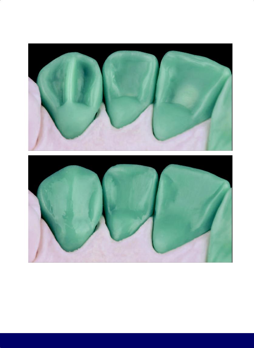
PROGRESSIVE TECHNIQUE FOR MAXILLARY ANTERIOR TEETH
a
b
Fig 16-3 | (a) Create the cingulum by filling in the cervical area.
(b) Fill in the lingual surface.
305
@dentistinfo стоматологический телеграм канал
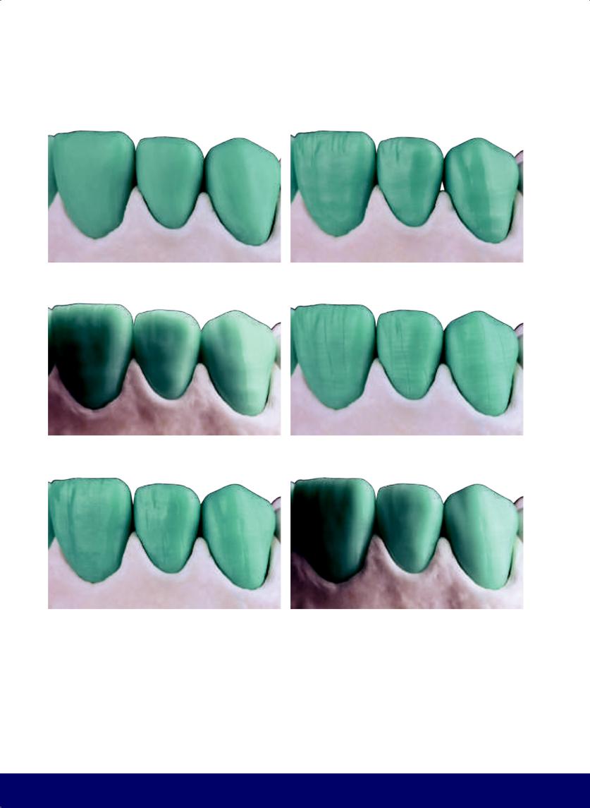
CHAPTER 16
a |
|
b |
|
|
|
c |
|
d |
|
|
|
e |
|
f |
|
|
|
Fig 16-4 | (a to l) Finalize the labial/buccal and lingual surfaces. Add macro and micro texture to elucidate the incidence of light at different angles.
306
@dentistinfo стоматологический телеграм канал
PROGRESSIVE TECHNIQUE FOR MAXILLARY ANTERIOR TEETH
g |
|
h |
|
|
|
i |
|
j |
|
|
|
k |
|
l |
|
|
|
307
@dentistinfo стоматологический телеграм канал

Anterior Segment and Full Arch Wax-ups
17
308
@dentistinfo стоматологический телеграм канал
ANTERIOR SEGMENT AND FULL ARCH WAX-UPS
a
b
c
Fig 17-1 | (a to c) Labial views of the anterior teeth.
309
@dentistinfo стоматологический телеграм канал
CHAPTER 17
a |
|
b |
|
|
|
c |
|
d |
|
|
|
e
Fig 17-2 | (a) Craniocaudal view. (b) Surface texture visible with a 45-degree profile view. (c to g) Lingual views of the anterior teeth.
310
@dentistinfo стоматологический телеграм канал
ANTERIOR SEGMENT AND FULL ARCH WAX-UPS
f
g
311
@dentistinfo стоматологический телеграм канал
CHAPTER 17
a
b
Fig 17-3 | (a and b) Lingual views of the right and left sides in color. (c and d) Same lingual views of the right and left sides in black and white.
312
@dentistinfo стоматологический телеграм канал
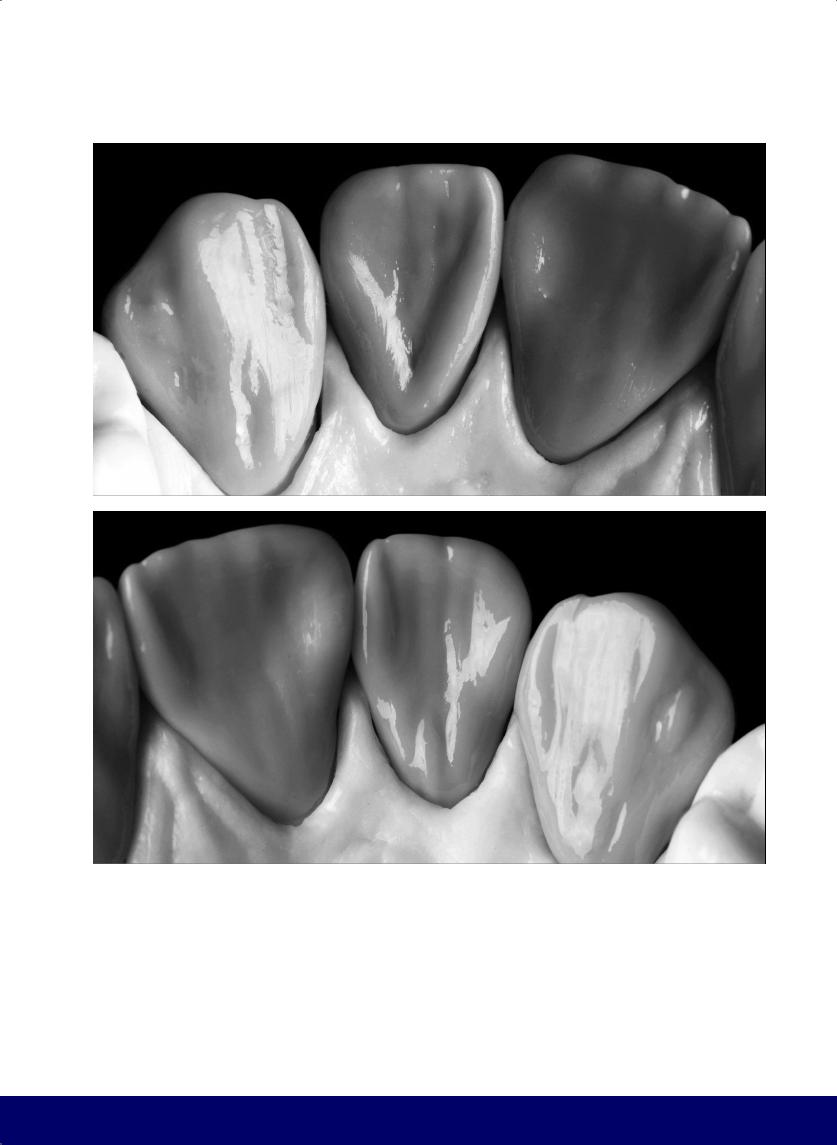
ANTERIOR SEGMENT AND FULL ARCH WAX-UPS
c
d
313
@dentistinfo стоматологический телеграм канал
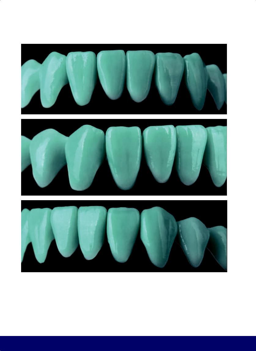
CHAPTER 17
a
b
c
Fig 17-4 | (a to f) The technique for constructing the mandibular teeth follows the same steps as those used for the maxillary teeth but with much more subtle macro and micro texture.
314
@dentistinfo стоматологический телеграм канал
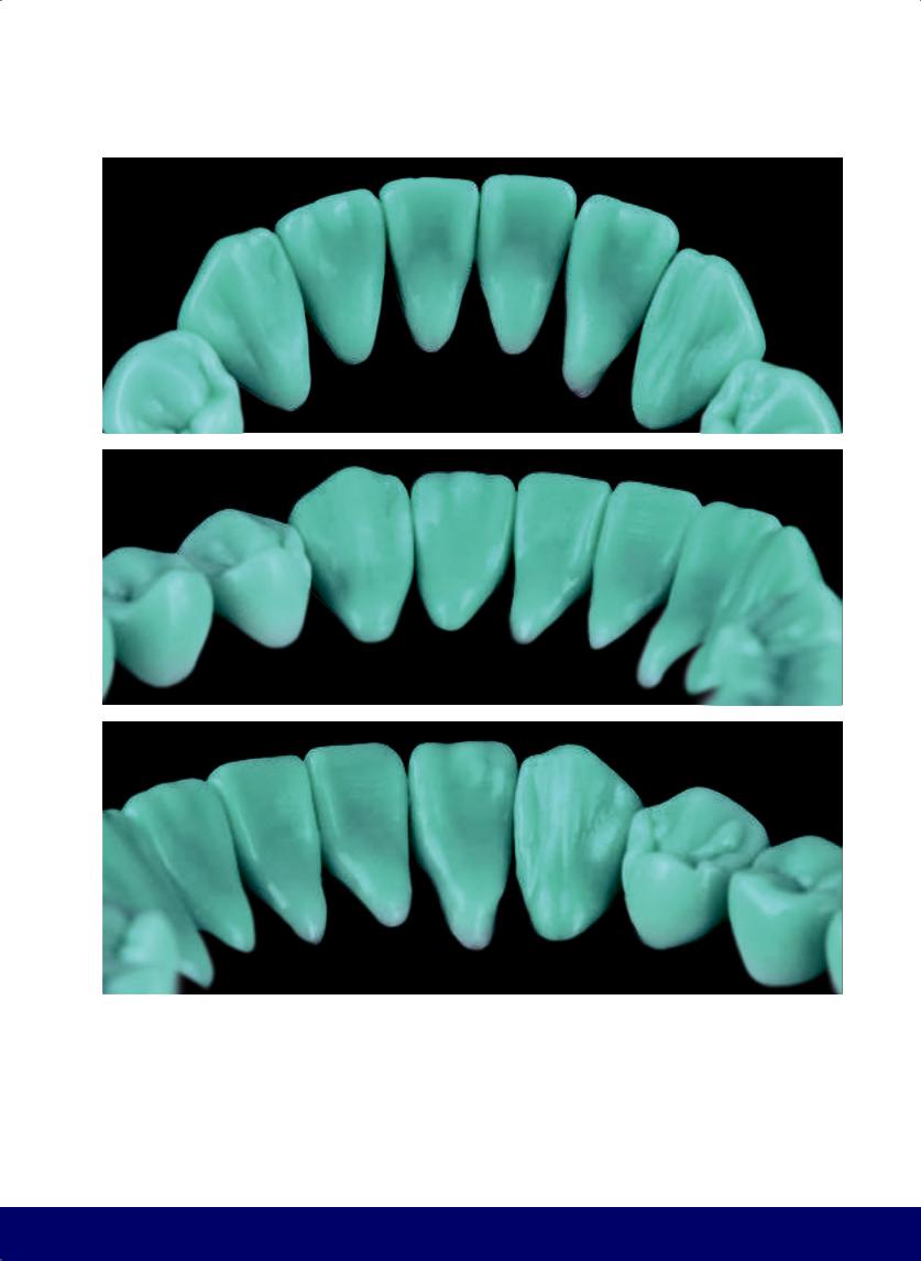
ANTERIOR SEGMENT AND FULL ARCH WAX-UPS
d
e
f
315
@dentistinfo стоматологический телеграм канал
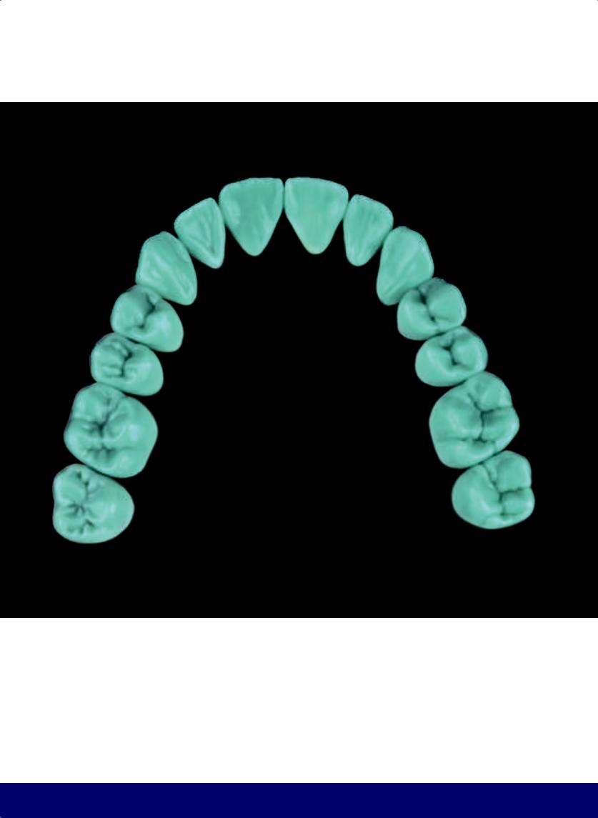
CHAPTER 17
316
@dentistinfo стоматологический телеграм канал
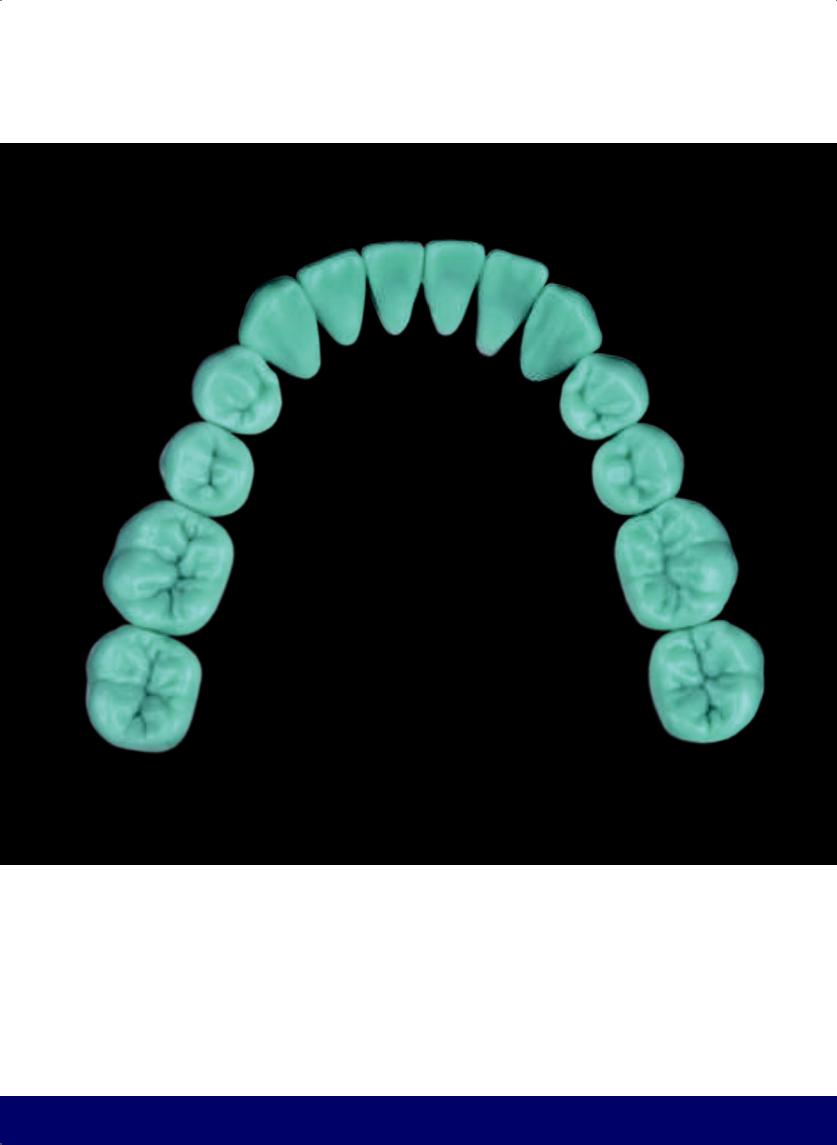
ANTERIOR SEGMENT AND FULL ARCH WAX-UPS
317
@dentistinfo стоматологический телеграм канал
CHAPTER 17
318
@dentistinfo стоматологический телеграм канал
ANTERIOR SEGMENT AND FULL ARCH WAX-UPS
319
@dentistinfo стоматологический телеграм канал
@dentistinfo стоматологический телеграм канал

RECOMMENDED READING
1.Angle EH. Classification of malocclusion. Dent Cosmos 1899; 41:248–264.
2.Adolfi D. Natural Esthetics. Carlos dos Reis (trans). Chicago: Quintessence, 2002.
3.Adolfi D, Scopin de Andrade O, Vernazza GJ, Adolfi MC. Functional and esthetic protocol for oral rehabilitation. In: Duarte S Jr (ed). Quintessence of Dental Technology 2013, vol 36. Chicago: Quintessence, 2013:139–161.
4.Adolfi D, Adolfi M. Use of different laboratory technologies and techniques in the reconstruction of total rehabilitations. Prosthesis News 2015;2:409–426.
5.Battistelli A, Severino D, La Manna O. Anatomic Functional Geometry Modeling: Modellazione Dentale Naturale Codificata. Villa Carcina, Italy: Teamwork Media, 2010.
6.Bottino MA, Faria R, do Amaral JMBL, Saavedra GSFA. Tecnologia CAD/CAM na reabilitação protética. In: Neto AA, de Castellucci Barbosa L, Barreto MA, Moreira AA. Estética do Sorriso em Reabilitação Protética. Nova Odessa, Brazil: Editora Napoleão, 2014: 158–203.
7.Bottino MA, Faria R, Valandro LF. Percepção Estética em Próteses Livres de Metal em Dentes Naturais e Implantes. São Paulo: Artes Médicas, 2009.
8.Dawson PE. Avaliação, Diagnóstico e Tratamento dos Problemas Oclusais. São Paulo: Artes Médicas, 1980.
9.Edwards B. The New Drawing on the Right Side of the Brain, ed 3. London: Harper, 2008.
10.Fradeani M. Esthetic Rehabilitation in Fixed Prosthodontics. London: Quintessence, 2004.
11.Francci CE, Riquieri H, Nishida AC, Saavedra GSFA. Harmonização do Sorriso—Do planejamento digital à cimentação de laminados com preparo dental: Parte I. Int J Esthet Dentedição 2016;1:14– 33.
12.Gürel G, Bichacho N. Permanent diagnostic provisional restorations for predictable results when redesigning the smile. Pract Proced Aesthet Dent 2006;18:281–286.
13.Hatjó J. Anteriores: A Beleza Natural dos Dentes Anteriores. São Paulo: Santos, 2008.
14.Kano P. Challenging Nature: Wax-Up Techniques in Aesthetics and Functional Occlusion. London: Quintessence, 2011.
15.Kano P, Xavier C, Ferencz JL, Van Dooren E, Silva NRFA. The anatomical shell technique: An approach to improve the esthetic predictability of CAD/ CAM restorations. In: Duarte S Jr (ed). Quintessence of Dental Technology 2013, vol 36. Chicago: Quintessence, 2013:27–37.
16.Kataoka S, Nishimura Y. Nature’s Morphology: An Atlas of Tooth Shape and Form. Sadan A (ed). Chicago: Quintessence, 2002.
17.Kyrillos M, Moreira M, Calicchio L. A Arquitetura do Sorriso. São Paulo: Quintessence, 2012.
18.Lundeen HC. Introduction to Occlusal Anatomy. Lexington, Kentucky: The Author, 1969.
19.Magne P, Belser U. Bonded Porcelain Restoration in the Anterior Dentition: A Biomimetic Approach. London: Quintessence, 2002.
20.McLaren EA, Cao PT. Smile analysis and esthetic design: “In the zone.” Inside Dentistry 2009;5:46–48.
21.Motsch A. Ajuste Oclusal em Dentes Naturais. São Paulo: Santos, 1990.
22.Nunes LJ. Princípios de Oclusão e Técnicas de Enceramento Progressivo e Escultura. Rio de Janeiro: Guanabara Koogan, 1980.
23.Okeson JP. Fundamentos de Oclusão e Desordens Temporo-Man- dibulares, ed 2. São Paulo: Artes Médicas, 1992.
24.Pagani C, da Rocha DM, Saavedra GSFA, de Carvalho RF. Previsibilidade e estética: A utilização do ensaio restaurador (mock-up) na construção da beleza do sorriso. In: Callegari A, Brito e Dias R (eds). Especialidade em Foco: Beleza do Sorriso, vol 1. Nova Odessa, Brazil: Editora Napoleão, 2013:66–108.
25.Ramfjord S, Ash MM. Occlusion, ed 3. Philadelphia: Saunders, 1983.
26.Riquieri H. Enceramento e Escultura Passo a Passo in Reabilitação Estética. Nova Odessa, Brazil: Napoleão, 2012:150–177.
27.Rufenacht CR. Fundamentals of Esthetics. Chicago: Quintessence, 1990.
28.Saavedra GSFA, Riquieri H, Nishida AC, Luz JN, Francci CE. Harmonização do sorriso—Do planejamento digital à cimentação de laminados com preparo dental—Parte II. Int J Esthet Dent 2016;1:206–225.
29.Scolavino S, Paolone G, Orsini G, Devoto W, Putignano A. The simultaneous modeling technique: Closing gaps in posteriors [in Portuguese]. Int J Esthet Dent 2016;11:58–81.
30.Simon H, Magne P. Clinically based diagnostic wax-up for optimal esthetics: The diagnostic mock-up. J Calif Dent Assoc 2008;36:355–362.
31.Shillingburg HT Jr, Hobo S, Whitsett LD, Jacobi R, Brackett SE. Fundamentals of Fixed Prosthodontics, ed 3. Chicago: Quintessence, 1997.
32.Soares C, Santana da Silva J. Fotografia odontológica clínica e laboratorial. In: Aranha C, Soares C, Santana da Silva J. Administração e Fotografia, vol 7, APDESP series. São Paulo: Napoleão, 2015:34–113.
33.Turano JC, Turano LM. Fundamentos de Protese Total. Rio de Janeiro: Quintessence, 1989.
34.Ubassy G. Analysis: The New Way in Dental Communication. Villa Carcina, Italy: Teamwork Media, 2000.
35.Vieira GF. Atlas of Anatomy of Permanent Teeth. São Paulo: Santos, 2005.
321
@dentistinfo стоматологический телеграм канал

This beautiful atlas conveys not only the practical knowledge of dental anatomy but also the art of sculpting it in wax. The ideal anatomy of each dental structure is described in detail and the waxing techniques are beautifully illustrated step by step for visual reference. The author demonstrates that for every morphologic feature there is an explanation in nature, assigning significance to every minute feature of dental morphology. Organized by tooth and arch, this book views morphology through a clinical lens and repeatedly draws connections between anatomical features and clinical concepts. The fundamental knowledge presented in this text is essential for improving waxing and sculpting techniques and will be useful for students and specialists alike.
C O N T E N TS
1The Esthetic and Functional Parameters of Posterior Teeth
2Maxillary First Premolar
3Maxillary Second Premolar
4Maxillary First Molar
5Maxillary Second Molar
6Maxillary Posterior Quadrant
7Mandibular First Premolar
8Mandibular Second Premolar
9Mandibular First Molar
10Mandibular Second Molar
11Mandibular Posterior Quadrant
12The Esthetic and Functional Parameters of Anterior Teeth
13Maxillary Central Incisor
14Maxillary Lateral Incisor
15Maxillary Canine
16Progressive Technique for Maxillary Anterior Teeth
17Anterior Segment and Full Arch Wax-ups
ISBN 978-0-86715-770-3
90000
9 780867 157703
@dentistinfo стоматологический телеграм канал