

CHAPTER 1 Evaluation of the Urologic Patient 39
A  B C
B C
F
D E
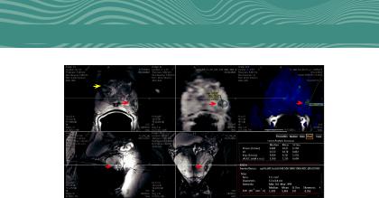
FIG. 1.20 A 66-year-old African American man with a prostate-specific antigen (PSA) of 7.0 and two prior negative biopsies. A 3T mpMRI with an endorectal coil of the prostate was obtained. There were two suspicious areas. (A), (D), and (E) are the triplanar axial, sagittal, and coronal plane images, respectively. The peripheral zone (blue line) and the central gland (yellow arrow) are well visualized. The red arrow represents a well-circumscribed heterogeneous benign prostatic hypertrophy nodule (11 mm 3 11 mm 3 14 mm) within the peripheral zone with no communication to the central gland. (B) The corresponding apparent diffusion coefficient map demonstrates areas of heterogeneous restriction (761 3 1026 mm2/s). (C) The lesion on the dynamic con- trast-enhanced (DCE) image exhibits focal type 2 and 3 enhancement curves. (F) The DCE quantitative analysis is listed. The patient underwent a fusion biopsy. The lesion was also appreciated on ultrasonography, and no cancer was detected.
is not specific for cancer and may require plain film radiography, CT, or MRI to confirm as well as correlation with prior history of bone fractures, trauma, surgery, or arthritis.
Positron Emission Tomography (PET). PET/CT and PET/MRI offer diagnostic information based on glucose, choline, or amino acid metabolism depending on the radiotracer used. Molecular imaging of cancer is most commonly performed using PET radiotracer
2-[18 F]fluoro-2-deoxy-D-glucose(18 F-FDG). It has a well-established role in the detection of residual seminomatous germ cell tumors following chemotherapy (Fig. 1.21).
While it is important to select the study that provides the most useful information, it is also important to consider radiation exposure (Table 1.6) to the patient to be “As Low as reasonably achievable” (ALARA).
HEMATURIA
Hematuria, or presence of blood in the urine, is a concerning urologic sign and it must be thoroughly evaluated because it signals
40 CHAPTER 1 Evaluation of the Urologic Patient
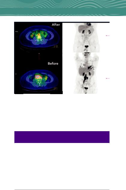
FIG. 1.21 Fluorine-18 fluorodeoxyglucose (18 F-FDG) PET/CT is useful for staging and restaging of seminoma in patients treated with chemotherapy. This patient presented with a right-sided seminoma with bulky right-sided retroperitoneal lymph nodes. Positron emission tomography/computed tomography after chemotherapy shows no uptake in the previously positive nodal region.
Table 1.6 Radiation Exposure From Common Urologic Imaging
Procedures
RELATIVE |
|
|
RADIATION |
EFFECTIVE DOSE |
|
LEVEL (RRL) |
ESTIMATED RANGE |
EXAMPLE EXAMINATIONS |
None |
0 |
Ultrasound, MRI |
Minimal |
,0.1 mSv |
Chest radiographs |
Low |
0.1–1.0 mSv |
Lumbar spine radiographs, |
|
|
pelvic radiographs |
Medium |
1–10 mSv |
Abdomen CT without contrast, |
|
|
nuclear medicine, bone scan, |
|
|
99m Tc-DMSA renal scan, IVP, |
|
|
retrograde pyelograms, KUB, |
|
|
chest CT with contrast |
High |
10–100 mSv |
Abdomen CT without and with |
|
|
contrast, whole-body PET |
IVP, Intravenous pyelogram; KUB, kidney, ureters, bladder
Modified from American College of Radiology: ACR Appropriateness Criteria Radiation Dose Assessment Introduction. http://www.acr.org/SecondaryMainMenuCategories/quality_safety/ app_criteria/RRLInformation.aspx, 2008.
CHAPTER 1 Evaluation of the Urologic Patient 41
the presence of a genitourinary (GU) malignancy in up to 25% of patients. Hematuria is classified as gross or microscopic. Gross hematuria (GH) is often alarming to patients, whereas microscopic hematuria (MH) goes unnoticed by the patient until it is detected by urinalysis. Per the American Urological Association (AUA), MH is defined as three or more RBCs/HPF and a single positive urinalysis is sufficient to prompt evaluation.
Causes of Microscopic Hematuria
In most studies, one-thirds to two-thirds of patients evaluated for MH have a demonstrable cause. These include urinary calculus, benign prostatic enlargement (BPH), urethral stricture, and various other conditions (Table 1.7). Malignancy is found in 0.68% to 4.3% of patients being evaluated for MH. The likelihood of identifying malignancy is greater with higher levels of MH (.25 RBCs/ HPF), GH, or with risk factors for malignancy (Table 1.8).
Selecting Patients for Evaluation
The AUA guidelines recommend evaluating all adults with asymp- tomatic MH or GH. A thorough history and exam is required as GH must be distinguished from pigmenturia, which may be due to exogenous sources (e.g., bilirubin, myoglobin), foods (e.g., beets and rhubarb), drugs (e.g., phenazopyridine), and simple dehydration. In women, GH must also be distinguished from vaginal bleeding, which usually can be achieved by obtaining a careful menstrual history.
In order to prevent a misdiagnosis or delay in diagnosis of a more concerning etiology for hematuria, patients who are suspected to have hematuria due to benign causes must have that benign cause substantiated by clinical evidence. They should also be further evaluated once the suspected benign cause has resolved. Patients who develop hematuria in the setting of anticoagulation (e.g., warfarin, enoxaparin, heparin, aspirin, clopidogrel) should still undergo a complete evaluation as the risk of underlying GU malignancy is similar to the patients not on anticoagulation.
Guideline-Based Evaluation of Microscopic and Gross
Hematuria
The AUA guidelines has an evaluation algorithm for MH
(Fig. 1.22). Again, all patients with gross hematuria should un-

42 CHAPTER 1 Evaluation of the Urologic Patient
Table 1.7 Differential Diagnosis of Asymptomatic Microhematuriaa
|
|
COMMON CLINICAL |
|
|
PRESENTATION AND RISK |
CATEGORY |
EXAMPLES |
FACTORS |
Neoplasm |
Any |
Pelvic irradiation, chronic |
|
|
urinary tract infections, |
|
|
indwelling foreign body |
|
Bladder cancer |
Older age, male predominance, |
|
|
tobacco use, occupational |
|
|
exposures, irritative voiding |
|
|
symptoms |
|
Ureteral or renal |
Family history of early colon |
|
pelvis cancer |
cancers or upper tract |
|
|
tumors, flank pain |
|
Renal cortical tumor |
Family history of early kidney |
|
|
tumors, flank pain, flank |
|
|
mass |
|
Prostate cancer |
Older age, family history, |
|
|
African-American |
|
Urethral cancer |
Obstructive symptoms, pain, |
|
|
bloody discharge |
Infection/ |
Any |
History of infection |
inflammation |
|
|
|
Cystitis |
Female predominance, dysuria |
|
Pyelonephritis |
Fever, flank pain, diabetes, |
|
|
female predominance |
|
Urethritis |
Exposure to sexually trans- |
|
|
mitted infections, urethral |
|
|
discharge, dysuria |
|
Tuberculosis |
Travel to endemic areas |
|
Schistosomiasis |
Travel to endemic areas |
|
Hemorrhagic cystitis |
Infections, pelvic radiation, |
|
|
certain medications, |
|
|
chemical exposures |
Calculus |
Any |
|
|
Nephroureterolithiasis |
Flank pain, family history, |
|
|
prior stone |
|
Bladder stones |
Bladder outlet obstruction |
Benign prostatic |
|
Male, older age, obstructive |
enlargement |
|
symptoms |
Medical renal |
Any |
Hypertension, azotemia, |
diseaseb |
|
dysmorphic erythrocytes, |
|
|
cellular casts, proteinuria |
|
Nephritis |
|
|
IgA nephropathy |
|

CHAPTER 1 Evaluation of the Urologic Patient 43
Table 1.7 Differential Diagnosis of Asymptomatic
Microhematuriaa—cont’d
|
|
COMMON CLINICAL |
|
|
PRESENTATION AND RISK |
CATEGORY |
EXAMPLES |
FACTORS |
Congenital or |
Polycystic kidney |
Family history of renal cystic |
acquired |
disease |
disease |
anatomic |
Ureteropelvic junction |
History of UTI, stone, flank |
abnormality |
obstruction |
pain |
|
Ureteral stricture |
History of surgery or radia- |
|
Urethral diverticulum |
tion, flank pain, |
|
Fistula |
hydronephrosis; stranguria, |
|
|
spraying urine |
|
|
Discharge, dribbling, |
|
|
dyspareunia, history of UTI, |
|
|
female predominance |
|
|
Pneumaturia, fecaluria, |
|
|
abdominal pain, recurrent |
|
|
UTI, history of diverticulitis |
|
|
or colon cancer |
Other |
Exercise-induced |
Recent vigorous exercise |
|
hematuriac |
|
|
Endometriosis |
Cyclic hematuria in a |
|
|
menstruating woman |
|
Hematologic or |
Family history or personal |
|
thrombotic disease |
history of bleeding or |
|
|
thrombosis |
|
Papillary necrosis |
African-American, sickle cell |
|
|
disease, diabetes, analgesic |
|
|
abuse |
|
Arteriovenous |
|
|
malformation |
|
|
Renal vein thrombosis |
|
|
Interstitial cystitis |
Voiding symptoms |
|
Trauma |
History |
|
Recent genitourinary |
History |
|
surgery or instru- |
|
|
mentation |
|
aDifferential diagnosis, having ruled out obvious benign causes, such as menstruation, recent instrumentation, uncomplicated cystitis, etc.
bPresence of hematologic illness, medical renal illness or use of anticoagulants or antiplatelet agents does not preclude the need for a hematuria evaluation.
cExercise-induced hematuria is a diagnosis of exclusion. Absence of hematuria after abstinence from exercise must be confirmed.
IgA, Immunoglobulin A; UTI, urinary tract infection.

Table 1.8 Selected Guidelines for Evaluation of Asymptomatic Microhematuria
|
REFERENCE, |
|
CRITERIA FOR |
|
|
|
GUIDELINE BODY |
YEAR |
AGE/SEX |
EVALUATION |
CYSTOSCOPY |
IMAGING |
BIOMARKERS |
American Urolog- |
Davis et al., |
Adults |
3 or more RBCs/HPF |
35 years old or |
CT urogram |
Not |
ical Association |
2012 |
|
on a single UA |
with risk factors |
preferred |
recommended |
Canadian Consen- |
Kassouf et al., |
Adults |
3 or more RBCs/HPF |
35 years old |
Provider discre- |
Not |
sus Document |
2016 |
|
on 2 out of 3 UAs |
|
tion |
recommended |
Kaiser Permanente |
Loo et al., 2009 |
Adults |
More than 3 RBCs/ |
Urologist discre- |
CT urogram or |
No consensus on |
|
|
|
HPF on 2 out of |
tion |
IVP 1 RUS |
cytology |
|
|
|
3 UAs |
|
|
|
American College |
Committee |
Adults |
Not stated |
Avoid in never- |
Avoid in never- |
Not stated |
of Obstetricians |
Opinion, 2017 |
Women |
|
smoking |
smoking |
|
and |
|
|
|
women age |
women age |
|
Gynecologists |
|
|
|
35–50; only |
35–50; only |
|
and American |
|
|
|
evaluate if .25 |
evaluate if .25 |
|
Urogynecologic |
|
|
|
RBCs/HPF |
RBCs/HPF |
|
Society |
|
|
|
|
|
|
Japanese |
Horie et al., |
Adults |
5 or more RBCs/HPF |
40 years old, |
US |
1/2 Cytology |
|
2014 |
40 years |
on a single UA |
with risk factors |
|
|
Dutch |
van der Molen |
Adults |
3 or more RBCs/HPF |
.50 years; pro- |
US if .50 years |
Cytology |
|
and Hovius, |
50 years |
on 2 out of 3 UAs |
vider discretion |
old; provider |
recommended |
|
2012 |
|
|
in younger |
discretion in |
in patients with |
|
|
|
|
patients |
younger |
negative |
|
|
|
|
|
patients |
evaluation |
Patient Urologic the of Evaluation 1 CHAPTER 44

American College |
Nielsen and |
Adults |
3 or more RBCs/HPF |
Not stated |
Not stated |
Not recom- |
of Physicians |
Qaseem, 2016 |
|
on a single UA |
|
|
mended |
UK National Insti- |
NICE, 2016 |
Patients |
11 blood or more |
Not stated |
Not stated |
Not stated |
tute for Health |
Anderson et al., |
over age |
on urine dip stick |
|
|
|
and Care Excel- |
2008 |
60 |
test PLUS |
|
|
|
lence (NICE) |
|
|
Dysuria or elevated |
|
|
|
|
|
|
leukocytosis |
|
|
|
CT, Computed tomography; HPF, high-power field; IVP, intravenous pyelogram; RBC, red blood cell; RUS, renal ultrasound; UA, urinalysis; US, ultrasound.
Patient Urologic the of Evaluation 1 CHAPTER
45
Patient with microhematuria
≥ 3 RBC/HPF on UA with microscopy
Repeat urinalysis positive
History and physical exam
Focus on risk factors for urothelial cancer and non-malignant causes1
Risk stratification1,3
|
Non-malignant or |
|
|
|
|
|
|
|
|
gynecologic source |
Evaluation directed by signs/symptoms |
|
|
||||
|
|
|
Include urine culture if infection is suspected |
|
|
|||
|
|
|
|
|
||||
|
|
|
|
|
|
|
|
|
|
Non-malignant or |
|
Non-malignant or gynecologic |
|
|
|||
|
|
|
|
|||||
|
gynecologic source ruled out |
|
source identified |
|
|
|||
|
Treat non-malignant or |
|||||||
|
|
|
|
|
|
|
||
|
|
|
|
|
|
|
|
gynecologic source4 |
Low Risk |
|
Intermediate Risk |
|
High Risk |
|||
All of the following: |
|
Any of the following: |
|
Any of the following: |
|||
Women age < 50; Men age < 40 yrs |
|
Women age 50–59; Men age 40–59 yrs |
|
Women and men age > 60 yrs |
|||
Never smoker or < 10 pack-years |
|
10–30 Pack-years smoking |
|
>30 Pack-years smoking |
|||
3–10 RBC/HPF on one UA |
|
11–25 RBC/HPF on one UA |
|
> 25 RBC/HPF on one UA |
|||
No additional risk factors for |
|
One or more additional risk factors for |
|
History of gross hematuria |
|||
urothelial cancer1 |
|
urothelial cancer1 |
|
Previously low-risk, no prior |
|||
No prior episodes of MH3 |
|
Previously low-risk, no prior |
|
evaluation and > 25 RBC/HPF on |
|||
|
|
|
evaluation and 3–25 RBC/HPF on |
|
repeat UA |
||
|
|
|
repeat UA |
|
|
|
|
|
|
|
|
|
|
|
|
Shared |
|
|
|
|
|
|
|
decision-making |
|
|
|
|
|
|
|
Repeat urinalysis positive
Repeat Urinalysis
Repeat urinalysis negative
Release from care
|
Repeat Urinalysis within 6 |
|
Cystoscopy and Renal |
|
|
|
||
|
months OR Cystoscopy and Renal |
|
|
Cystoscopy and CT Urogram6 |
||||
|
|
Ultrasound5 |
|
|||||
|
Ultrasound5 |
|
|
|
|
|
|
|
|
|
|
|
|
|
|
|
|
Patient Urologic the of Evaluation 1 CHAPTER 46
|
|
|
|
|
|
|
|
|
|
|
|
Repeat urinalysis |
Evaluation performed |
|
|
|
|
|
|||||
negative |
|
|
|
|
|
|
|
|
|||
|
|
|
|
|
|
|
|
||||
|
|
|
|
|
|
|
Evaluation negative |
Evaluation positive |
|
Treat as indicated |
|
|
|
|
Consider Repeat Urinalysis |
|
|
||||||
|
|
|
|
|
If urologic diagnosis is non-malignant, |
||||||
|
|
|
|
|
|
|
|||||
|
|
|
within 12 months |
|
|
|
repeat urinalysis after treatment |
||||
 Repeat urinalysis Repeat urinalysis positive
  negative
Release from urologic care
Shared decision-making regarding repeat evaluation vs. observation
Consider cross-sectional imaging with urography or retrograde pyelograms if not performed previously
Re-evaluate
If patient develops gross hematuria, increase in degree of microhematuria or new urologic symptoms
Repeat urinalysis
positive Repeat urinalysis negative
Release from urologic care
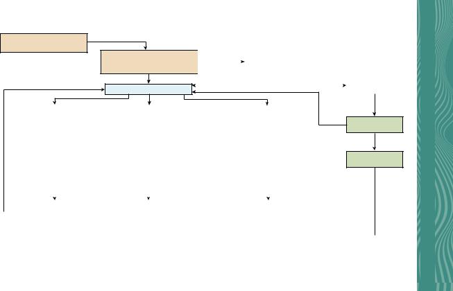
1.Main risk factors for urothelial cancer are those in the AUA risk stratification system (age, male sex, smoking, degree of microhematuria and history of gross hematuria). Additional risk factors for urothelial carcinoma include but are not limited to irritative lower urinary tract voiding symptoms, history of cyclophosphamide or ifosfamide chemotherapy, family history of urothelial carcinoma or Lynch Syndrome, occupational exposures to benzene chemicals or aromatic amines, history of chronic indwelling foreign body in the urinary tract
2.If medical renal disease is suspected, consider nephrologic evaluation, but pursue concurrent risk-based urological evaluation
3.Patients may be low-risk at first presentation with microhematuria, but may only be considered intermediateor high-risk if found to have persistent microhematuria
4.There are non-malignant and gynecologic sources of hematuria that do not require treatment and/or may confound the diagnosis of MH. Clinicians can consider catheterized urine specimen in women with vaginal atrophy or pelvic organ prolapse. Clinicians must use careful judgment and patient engagement to decide whether to pursue MH evaluation in the setting of chronic conditions that do not require treatment, such as the aforementioned gynecologic conditions, non-obstructing stones or BPH.
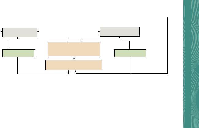
5.Clinician may perform cross-sectional imaging with urography or retrograde pyelograms if hematuria persists after negative renal ultrasound
6.MR Urogram or Non-contrast imaging plus retrograde pyelograms if contraindications to CT Urogram
FIG. 1.22 Microscopic (MH) Algorithm. https://www.auanet.org/documents/Guidelines/GUI-20-5442%20MH%20Algorithm.pdf.
Patient Urologic the of Evaluation 1 CHAPTER
47

48 CHAPTER 1 Evaluation of the Urologic Patient
dergo a complete evaluation of the upper and lower genitourinary tracts. For MH, patients meeting criteria should undergo a risk stratified evaluation, even if one phase of the evaluation shows a suspected cause for the MH. For example, if a patient is found to have nephrolithiasis or a renal tumor on upper tract imaging, they should still undergo cystoscopy for to evaluate for lower tract pathology.
•A thorough history and physical exam should be performed.
The goal is to identify causes that may warrant variation from the standard evaluation, such as infection, menstrual bleeding, known medical-renal disease, food/medications, trauma, or recent GU instrumentation. The history should also in- clude an assessment of symptoms, such as GH, voiding symp- toms, or flank pain. It should also include risk factors for hematuria. Current medications, including anticoagulants, should be elicited even though it would not preclude an evaluation for hematuria. The history should include questions about tobacco use as it is the number one risk factor for bladder cancer.
•Physical examination should focus on the GU system (e.g., flank tenderness, palpable masses in the flank, abdomen, su- prapubic region, urethra; prostate exam, meatal stenosis). If urethral stricture or BPH is suspected, urine flow rate and postvoid residual measurement may be helpful.
•Laboratory testing includes urinalysis (if not performed previ- ously) to assess for hematuria, dysmorphic RBCs, cellular casts, or proteinuria. Urine culture should be performed if the urinalysis is concerning for a urinary tract infection (UTI). Prostate-specific antigen should be checked in the appropriate setting and patient population.
•Urine cytologic examination is highly sensitive and specific for the detection of high-grade urothelial carcinoma. However, current evidence indicates that currently available urinary biomarkers, including cytology, is not sufficiently sensitive to replace cystoscopy or imaging, therefore, it should be not be performed in patients with MH. However, cytologic examination may be considered in patient with a negative initial workup in whom urothelial carcinoma is still suspected, as well as in patients with symptomatic MH or GH.