

187-2017
.pdfCHAPTER 33
neutrophils. GM-CSF acts together with interleukin-2 to stimulate T-cell proliferation and appears to be a locally active factor at the site of inflammation. GM-CSF mobilizes peripheral blood stem cells, but it is significantly less efficacious and more toxic than G-CSF in this regard.
Clinical Pharmacology
A. Cancer Chemotherapy-Induced Neutropenia
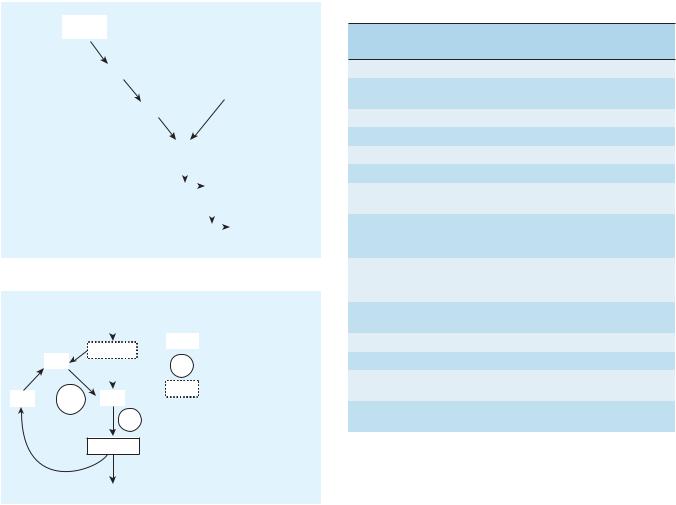
Neutropenia is a common adverse effect of the cytotoxic drugs used to treat cancer and increases the risk of serious infection in patients receiving chemotherapy. Unlike the treatment of anemia and thrombocytopenia, transfusion of neutropenic patients with granulocytes collected from donors is performed rarely and with limited success. The introduction of G-CSF in 1991 represented a milestone in the treatment of chemotherapy-induced neutropenia. This growth factor dramatically accelerates the rate of neutrophil recovery after dose-intensive myelosuppressive chemotherapy (Figure 33–5). It reduces the duration of neutropenia and usually raises the nadir count, the lowest neutrophil count seen following a cycle of chemotherapy.
The ability of G-CSF to increase neutrophil counts after myelosuppressive chemotherapy is nearly universal, but its impact on clinical outcomes is more variable. Many, but not all, clinical trials and meta-analyses have shown that G-CSF reduces episodes of febrile neutropenia, requirements for broad-spectrum antibiotics, infections, and days of hospitalization. Clinical trials have not shown improved survival in cancer patients treated with G-CSF. Clinical guidelines for the use of G-CSF after cytotoxic chemotherapy recommend reserving G-CSF for patients at high risk for febrile neutropenia based on age, medical history, and disease characteristics; patients receiving dose-intensive chemotherapy regimens that carry a greater than 20% risk of causing febrile
|
30.0 |
|
|
|
|
|
|
L |
25.0 |
|
|
|
|
|
|
/ |
|
|
|
|
|
|
|
9 |
20.0 |
|
|
|
|
|
|
× 10 |
|
|
|
|
|
|
|
15.0 |
|
|
|
|
|
|
|
ANC |
10.0 |
|
|
|
|
|
|
5.0 |
|
|
|
|
|
|
|
|
|
|
|
|
|
|
|
|
0 |
4 |
8 |
12 |
16 |
20 |
24 |
Study day
FIGURE 33 5 Effects of granulocyte colony-stimulating factor (G-CSF; red line) or placebo (green line) on absolute neutrophil count (ANC) after cytotoxic chemotherapy for lung cancer. Doses of chemotherapeutic drugs were administered on days 1 and 3. G-CSF or placebo injections were started on day 4 and continued daily through day 12 or 16. The first peak in ANC reflects the recruitment of mature cells by G-CSF. The second peak reflects a
marked increase in new neutrophil production by the bone marrow under stimulation by G-CSF. (Normal ANC is 2.2–8.6 × 10⁹/L.)
(Reproduced, with permission, from Crawford J et al: Reduction by granulocyte colony-stimulating factor of fever and neutropenia induced by chemotherapy in patients with small-cell lung cancer. N Engl J Med 1991;325:164. Copyright © 1991 Massachusetts Medical Society. Reprinted with permission from Massachusetts Medical Society.)
Agents Used in Cytopenias; Hematopoietic Growth Factors |
603 |
neutropenia; patients with a prior episode of febrile neutropenia after cytotoxic chemotherapy; patients at high risk for febrile neutropenia; and patients who are unlikely to survive an episode of febrile neutropenia. Pegfilgrastim is an alternative to G-CSF for prevention of chemotherapy-induced febrile neutropenia. Pegfilgrastim can be administered once per chemotherapy cycle, and it may shorten the period of severe neutropenia slightly more than G-CSF.
Like G-CSF and pegfilgrastim, GM-CSF also reduces the duration of neutropenia after cytotoxic chemotherapy. It has been more difficult to show that GM-CSF reduces the incidence of febrile neutropenia, probably because GM-CSF itself can induce fever. In the treatment of chemotherapy-induced neutropenia, G-CSF 5 mcg/kg daily or GM-CSF 250 mcg/m2 daily is usually started within 24–72 hours after completing chemotherapy and is continued until the absolute neutrophil count is greater than 10,000 cells/µL. Pegfilgrastim is given as a single dose of 6 mg.
The utility and safety of the myeloid growth factors in the postchemotherapy supportive care of patients with acute myeloid leukemia (AML) have been the subject of a number of clinical trials. Because leukemic cells arise from progenitors whose proliferation and differentiation are normally regulated by hematopoietic growth factors, including GM-CSF and G-CSF, there was concern that myeloid growth factors could stimulate leukemic cell growth and increase the rate of relapse. The results of randomized clinical trials suggest that both G-CSF and GM-CSF are safe following induction and consolidation treatment of myeloid and lymphoblastic leukemia. There has been no evidence that these growth factors reduce the rate of remission or increase relapse rate. On the contrary, the growth factors accelerate neutrophil recovery and reduce infection rates and days of hospitalization. Both G-CSF and GM-CSF have FDA approval for treatment of patients with AML.
B. Other Applications
G-CSF and GM-CSF have also proved to be effective in treating the neutropenia associated with congenital neutropenia, cyclic neutropenia, myelodysplasia, and aplastic anemia. Many patients with these disorders respond with a prompt and sometimes dramatic increase in neutrophil count. In some cases, this results in a decrease in the frequency of infections. Because neither G-CSF nor GM-CSF stimulates the formation of erythrocytes and platelets, they are sometimes combined with other growth factors for treatment of pancytopenia.
The myeloid growth factors play an important role in autologous stem cell transplantation for patients undergoing high-dose chemotherapy. High-dose chemotherapy with autologous stem cell support is increasingly used to treat patients with tumors that are resistant to standard doses of chemotherapeutic drugs. The high-dose regimens produce extreme myelosuppression; the myelosuppression is then counteracted by reinfusion of the patient’s own hematopoietic stem cells (which are collected prior to chemotherapy). The administration of G-CSF or GM-CSF early after autologous stem cell transplantation reduces the time to engraftment and to recovery from neutropenia in patients receiving stem cells obtained either from bone marrow or from

604 |
SECTION VI Drugs Used to Treat Diseases of the Blood, Inflammation, & Gout |
peripheral blood. These effects are seen in patients being treated for lymphoma or for solid tumors. G-CSF and GM-CSF are also used to support patients who have received allogeneic bone marrow transplantation for treatment of hematologic malignancies or bone marrow failure states. In this setting, the growth factors speed the recovery from neutropenia without increasing the incidence of acute graft-versus-host disease.
Perhaps the most important role of the myeloid growth factors in transplantation is for mobilization of PBSCs. Stem cells collected from peripheral blood have nearly replaced bone marrow as the hematopoietic preparation used for autologous and allogeneic transplantation. The cells can be collected in an outpatient setting with a procedure that avoids much of the risk and discomfort of bone marrow collection, including the need for general anesthesia. In addition, there is evidence that PBSC transplantation results in more rapid engraftment of all hematopoietic cell lineages and in reduced rates of graft failure or delayed platelet recovery.
G-CSF is the cytokine most commonly used for PBSC mobilization because of its increased efficacy and reduced toxicity compared with GM-CSF. To mobilize stem cells for autologous transplantation, donors are given 5–10 mcg/kg daily subcutaneously for 4 days. On the fifth day, they undergo leukapheresis. The success of PBSC transplantation depends on transfusion of adequate numbers of stem cells. CD34, an antigen present on early progenitor cells and absent from later, committed, cells, is used as a marker for the requisite stem cells. The goal is to infuse at least 5 × 10⁶ CD34 cells/kg; this number of CD34 cells usually results in prompt and durable engraftment of all cell lineages. It may take several separate leukaphereses to collect enough CD34 cells, especially from older patients and patients who have been exposed to radiation therapy or chemotherapy.
For patients with multiple myeloma or non-Hodgkin’s lymphoma who respond suboptimally to G-CSF alone, the novel hematopoietic stem cell mobilizer plerixafor can be added to G-CSF. Plerixafor is a bicyclam molecule originally developed as an anti-HIV drug because of its ability to inhibit the CXC chemokine receptor 4 (CXCR4), a co-receptor for HIV entry into CD4+ T lymphocytes (see Chapter 49). Early clinical trials of plerixafor revealed a remarkable ability to increase CD34 cells in peripheral blood. Plerixafor mobilizes CD34 cells by preventing chemokine stromal cell-derived factor-1α (CXCL12) from binding to CXCR4 and directing the CD34 cells to “home” to the bone marrow. Plerixafor is administered by subcutaneous injection after 4 days of G-CSF treatment and 11 hours prior to leukapheresis; it can be used with G-CSF for up to 4 continuous days. Plerixafor is eliminated primarily by the renal route and must be dose-adjusted for patients with renal impairment. The drug is well tolerated; the most common adverse effects associated with its use are injection site reactions, gastrointestinal disturbances, dizziness, fatigue, and headache.
Toxicity
Although the three growth factors have similar effects on neutrophil counts, G-CSF and pegfilgrastim are used more frequently than GM-CSF because they are better tolerated. G-CSF and
pegfilgrastim can cause bone pain, which clears when the drugs are discontinued. GM-CSF can cause more severe side effects, particularly at higher doses. These include fever, malaise, arthralgias, myalgias, and a capillary leak syndrome characterized by peripheral edema and pleural or pericardial effusions. Allergic reactions may occur but are infrequent. Splenic rupture is a rare but serious complication of the use of G-CSF for PBSC mobilization.
MEGAKARYOCYTE GROWTH FACTORS
Patients with thrombocytopenia have a high risk of hemorrhage. Although platelet transfusion is commonly used to treat thrombocytopenia, this procedure can cause adverse reactions in the recipient; furthermore, a significant number of patients fail to exhibit the expected increase in platelet count. Thrombopoietin (TPO) and IL-11 both appear to be key endogenous regulators of platelet production. A recombinant form of IL-11 was the first agent to gain FDA approval for treatment of thrombocytopenia. Recombinant human thrombopoietin and a pegylated form of a shortened human thrombopoietin protein underwent extensive clinical investigation in the 1990s. However, further development was abandoned after autoantibodies to the native thrombopoietin formed in healthy human subjects and caused thrombocytopenia. Efforts shifted to investigation of novel, nonimmunogenic agonists of the thrombopoietin receptor, which is known as Mpl. Two thrombopoietin agonists (romiplostim and eltrombopag) are approved for treatment of thrombocytopenia.
Chemistry & Pharmacokinetics
Interleukin-11 is a 65to 85-kDa protein produced by fibroblasts and stromal cells in the bone marrow. Oprelvekin, the recombinant form of IL-11 approved for clinical use (Table 33–4), is produced by expression in Escherichia coli. The half-life of IL-11 is 7–8 hours when the drug is injected subcutaneously.
Romiplostim is a thrombopoietin agonist peptide covalently linked to antibody fragments that serve to extend the peptide’s half-life. The Mpl-binding peptide has no sequence homology with human thrombopoietin, and there is no evidence in animal or human studies that the Mpl-binding peptide or romiplostim induces antibodies to thrombopoietin. After subcutaneous administration, romiplostim is eliminated by the reticuloendothelial system with an average half-life of 3–4 days. Its half-life is inversely related to the serum platelet count; it has a longer half-life in patients with thrombocytopenia and a shorter half-life in patients whose platelet counts have recovered to normal levels. Romiplostim is approved for therapy of patients with chronic immune thrombocytopenia who have had an inadequate response to other therapies.
Eltrombopag is an orally active small nonpeptide thrombopoietin agonist molecule approved for therapy of patients with chronic immune thrombocytopenia who have had an inadequate response to other therapies, and for treatment of thrombocytopenia in patients with hepatitis C to allow initiation of interferon therapy. Following oral administration, peak eltrombopag levels

CHAPTER 33
are observed in 2–6 hours and the half-life is 26–35 hours. Eltrombopag is excreted primarily in the feces.
Pharmacodynamics
Interleukin-11 acts through a specific cell surface cytokine receptor to stimulate the growth of multiple lymphoid and myeloid cells. It acts synergistically with other growth factors to stimulate the growth of primitive megakaryocytic progenitors and, most importantly, increases the number of peripheral platelets and neutrophils.
Romiplostim has high affinity for the human Mpl receptor. Eltrombopag interacts with the transmembrane domain of the Mpl receptor. Both drugs induce signaling through the Mpl receptor pathway and cause a dose-dependent increase in platelet count. Romiplostim is administered once weekly by subcutaneous injection. Eltrombopag is an oral drug. For both drugs, peak platelet count responses are observed in approximately 2 weeks.
Clinical Pharmacology
Interleukin-11 is approved for the secondary prevention of thrombocytopenia in patients receiving cytotoxic chemotherapy for treatment of nonmyeloid cancers. Clinical trials show that it reduces the number of platelet transfusions required by patients who experience severe thrombocytopenia after a previous cycle of chemotherapy. Although IL-11 has broad stimulatory effects on hematopoietic cell lineages in vitro, it does not appear to have significant effects on the leukopenia caused by myelosuppressive
Agents Used in Cytopenias; Hematopoietic Growth Factors |
605 |
chemotherapy. Interleukin-11 is given by subcutaneous injection at a dose of 50 mcg/kg daily. It is started 6–24 hours after completion of chemotherapy and continued for 14–21 days or until the platelet count passes the nadir and rises to more than 50,000 cells/µL.
In patients with chronic immune thrombocytopenia who failed to respond adequately to previous treatment with steroids, immunoglobulins, or splenectomy, romiplostim and eltrombopag significantly increase platelet count in most patients. Both drugs are used at the minimal dose required to maintain platelet counts of greater than 50,000 cells/µL.
Toxicity
The most common adverse effects of IL-11 are fatigue, headache, dizziness, and cardiovascular effects. The cardiovascular effects include anemia (due to hemodilution), dyspnea (due to fluid accumulation in the lungs), and transient atrial arrhythmias. Hypokalemia also has been seen in some patients. All of these adverse effects appear to be reversible.
Eltrombopag is potentially hepatotoxic and liver function must be monitored, particularly when used in patients with hepatitis C. Portal vein thrombosis also has been reported with eltrombopag and romiplostim in the setting of chronic liver disease. In patients with myelodysplastic syndromes, romiplostim increases the blast count and risk of progression to acute myeloid leukemia. Marrow fibrosis has been observed with thrombopoietin agonists but is generally reversible when the drug is discontinued. Rebound thrombocytopenia has been observed following discontinuation of TPO agonists.
SUMMARY Agents Used in Anemias and Hematopoietic Growth Factors
|
Mechanism of |
|
Clinical |
Pharmacokinetics, Toxicities, |
Subclass, Drug |
Action |
Effects |
Applications |
Interactions |
|
|
|
|
|
|
|
|
|
|
|
|
|
|
|
|
|
|
|
|
|
|
|
|
|
|
|
|
|
|
|
|
|
|
|

606 |
SECTION VI |
Drugs Used to Treat Diseases of the Blood, Inflammation, & Gout |
|
|
||
|
|
Mechanism of |
|
Clinical |
|
Pharmacokinetics, Toxicities, |
|
|
|
|
|||
Subclass, Drug |
Action |
Effects |
Applications |
|
Interactions |
|
|
|
|
|
|
|
|
|
|
|
|
|
|
|
|
|
|
|
|
|
|
|
|
|
|
|
|
|
|
|
|
|
|
|
|
|
|
|
|
|
|
|
|
|
|
|
|
|
|
|
|
|
|
|
|
|
|
|
|
|
|
|
|
|
|
|
|
|
|
|
|
|
|
|
|
|
|
|
|
|
|
|
|
|
|
|
|
|
|
|
|
|
|
|
|
|
|
|

CHAPTER 33 Agents Used in Cytopenias; Hematopoietic Growth Factors 607
P R E P A R A T I O N S
A V A I L A B L E
GENERIC NAME |
AVAILABLE AS |
Darbepoetin alfa |
Aranesp |
Deferasirox |
Exjade |
Deferoxamine |
Generic, Desferal |
Eltrombopag |
Promacta |
Epoetin alfa |
Erythropoietin (EPO), Epogen, |
|
Procrit |
Epoetin beta (Methoxy |
Mircera |
polyethylene glycol–epoetin beta) |
|
Filgrastim (G-CSF) |
Neupogen, Granix |
Folic acid (folacin, pteroylglutamic |
Generic |
acid) |
|
Iron |
|
Oral: See Table 33–3. |
|
Iron dextran (parenteral) |
INFeD, Dexferrum |
Sodium ferric gluconate |
Ferrlecit |
complex (parenteral) |
|
Iron sucrose (parenteral) |
Venofer |
Ferric carboxymaltose |
Injectafer |
(parenteral) |
|
Ferumoxytol (parenteral) |
Feraheme |
Oprelvekin (IL-11) |
Neumega |
Pegfilgrastim |
Neulasta |
Plerixafor |
Mozobil |
Romiplostim |
Nplate |
Sargramostim (GM-CSF) |
Leukine |
Vitamin B12 |
|
Oral, parenteral |
Generic cyanocobalamin or |
|
hydroxocobalamin |
Nasal |
Nascobal, CaloMist |
|
|
REFERENCES
Aapro MS et al, European Organisation for Research and Treatment of Cancer: 2010 update of EORTC guidelines for the use of granulocyte-colony stimulating factor to reduce the incidence of chemotherapy-induced febrile neutropenia in adult patients with lymphoproliferative disorders and solid tumours. Eur J Cancer 2011;47:8.
Albaramki J et al: Parenteral versus oral iron therapy for adults and children with chronic kidney disease. Cochrane Database Syst Rev 2012;(1):CD007857.
Auerbach M, Al Talib K: Low-molecular weight iron dextran and iron sucrose have similar comparative safety profiles in chronic kidney disease. Kidney Int 2008;73:528.
Barzi A, Sekeres MA: Myelodysplastic syndromes: A practical approach to diagnosis and treatment. Cleve Clin J Med 2010;77:37.
Brittenham GM: Iron-chelating therapy for transfusional iron overload. N Engl J Med 2011;364:146.
Clark SF: Iron deficiency anemia: diagnosis and management. Curr Opin Gastroenterol 2009;25:122.
Darshan D, Fraer DM, Anderson GJ: Molecular basis of iron-loading disorders. Expert Rev Mol Med 2010;12:e36.
Gertz MA: Current status of stem cell mobilization. Br J Haematol 2010;150:647.
Kessans MR, Gatesman ML, Kockler DR: Plerixafor: A peripheral blood stem cell mobilizer. Pharmacotherapy 2010;30:485.
McKoy JM et al: Epoetin-associated pure red cell aplasia: Past, present, and future considerations. Transfusion 2008;48:1754.
Rees DC, Williams TN, Gladwin MT: Sickle-cell disease. Lancet 2010;376:2018.
Rizzo JD et al: American Society of Clinical Oncology/American Society of Hematology clinical practice guideline update on the use of epoetin and darbepoetin in adult patients with cancer. J Clin Oncol 2010;28:4996.
Sauer J, Mason JB, Choi SW: Too much folate: A risk factor for cancer and cardiovascular disease? Curr Opin Clin Nutr Metab Care 2009;12:30.
Solomon LR: Disorders of cobalamin (vitamin B12) metabolism: Emerging concepts in pathophysiology, diagnosis and treatment. Blood Rev 2007;21:113.
Stasi R et al: Thrombopoietic agents. Blood Rev 2010;24:179.
Wolff T et al: Folic acid supplementation for the prevention of neural tube defects: An update of the evidence for the U.S. Preventive Services Task Force. Ann Intern Med 2009;150:632.
C A S E S T U D Y A N S W E R
This patient’s megaloblastic anemia appears to be due to vitamin B12 (cobalamin) deficiency secondary to inadequate dietary B12. It is important to measure serum concentrations of both folic acid and cobalamin because megaloblastic anemia can result from deficiency of either nutrient. It is especially important to diagnose vitamin B12 deficiency because this deficiency, if untreated, can lead to irreversible
neurologic damage. Folate supplementation, which can compensate for vitamin B12–derived anemia, does not prevent B12-deficiency neurologic damage. To correct this patient’s vitamin B12 deficiency, she would probably be treated parenterally with cobalamin because of her neurologic symptoms, followed by oral supplementation to maintain her body stores of vitamin B12.

C H A P T E R
Drugs Used in Disorders
of Coagulation
James L. Zehnder, MD
C A S E S T U D Y
A 25-year-old woman presents to the emergency department complaining of acute onset of shortness of breath and pleuritic pain. She had been in her usual state of health until 2 days prior when she noted that her left leg was swollen and red. Her only medication was oral contraceptives. Family history was significant for a history of “blood clots” in multiple members of the maternal side of her family. Physical examination demonstrates an anxious woman with stable vital signs. The left lower extremity demonstrates erythema
Hemostasis refers to the finely regulated dynamic process of maintaining fluidity of the blood, repairing vascular injury, and limiting blood loss while avoiding vessel occlusion (thrombosis) and inadequate perfusion of vital organs. Either extreme—excessive bleeding or thrombosis—represents a breakdown of the hemostatic mechanism. Common causes of dysregulated hemostasis include hereditary or acquired defects in the clotting mechanism and secondary effects of infection or cancer. Atrial fibrillation is associated with stasis of blood in the atria, formation of clots, and increased risk of occlusive stroke. Because of the high prevalence of chronic atrial fibrillation, especially in the older population, use of anticoagulants is common. Guidelines for the use of oral anticoagulants (CHA2DS2-VASC score, see January C et al reference) are based on various risk factors (congestive heart failure, hypertension, age, diabetes, history of stroke, vascular disease, and sex). The drugs used to inhibit thrombosis and to limit abnormal bleeding are the subjects of this chapter.
and edema and is tender to touch. Oxygen saturation by fingertip pulse oximeter while breathing room air is 87% (normal > 90%). Ultrasound reveals a deep vein thrombosis in the left lower extremity; chest computed tomography scan confirms the presence of pulmonary emboli. Laboratory blood tests indicate elevated -dimer levels. What therapy is indicated acutely? What are the long-term therapy options? How long should she be treated? Should this individual use oral contraceptives?
MECHANISMS OF BLOOD
COAGULATION
The vascular endothelial cell layer lining blood vessels has an anticoagulant phenotype, and circulating blood platelets and clotting factors do not normally adhere to it to an appreciable extent. In the setting of vascular injury, the endothelial cell layer rapidly undergoes a series of changes resulting in a more procoagulant phenotype. Injury exposes reactive subendothelial matrix proteins such as collagen and von Willebrand factor, which results in platelet adherence and activation, and secretion and synthesis of vasoconstrictors and platelet-recruiting and activating molecules. Thus, thromboxane A2 (TXA2) is synthesized from arachidonic acid within platelets and is a platelet activator and potent vasoconstrictor. Products secreted from platelet granules include adenosine diphosphate (ADP), a powerful inducer of platelet aggregation, and serotonin (5-HT), which stimulates
608

CHAPTER 34 Drugs Used in Disorders of Coagulation |
609 |
+
+
+
Wall defect |
|
|
||
C |
|
vWF |
EC |
|
|
|
|
||
ADP |
|
|
PGI2 |
|
TXA2 |
|
|
|
|
|
– |
|
|
|
5-HT |
|
Intrinsic |
Extrinsic |
|
GP |
|
|
||
IIb/IIIa |
|
|
|
|
Platelets |
Degranulation |
|
Xa |
|
|
|
|
||
GP |
|
+ |
+ |
|
IIb/IIIa |
|
Activation |
|
|
|
|
|
||
GP |
|
|
|
+ |
IIb/IIIa |
Fibrin |
|
|
|
|
|
|
|
|
|
|
+ |
Thrombin |
Prothrombin |
|
|
|
|
|
 Fibrinogen
Thrombus formation at the site of the damaged vascular wall (EC, endothelial cell) and the role of platelets and clotting factors. Platelet membrane receptors include the glycoprotein (GP) Ia receptor, binding to collagen (C); GP Ib receptor, binding von Willebrand factor (vWF); and GP IIb/IIIa, which binds fibrinogen and other macromolecules. Antiplatelet prostacyclin (PGI2) is released from the endothelium. Aggregating substances released from the degranulating platelet include adenosine diphosphate (ADP), thromboxane A2 (TXA2), and serotonin (5-HT). Production of factor Xa by intrinsic and extrinsic pathways is detailed in Figure 34–2.
Decker JW: New directions in anticoagulant and antiplatelet treatment. [Editorial.] Br Heart J 1995;74:337.)
aggregation and vasoconstriction. Activation of platelets results in a conformational change in the αIIbβIII integrin (IIb/IIIa) receptor, enabling it to bind fibrinogen, which cross-links adjacent platelets, resulting in aggregation and formation of a platelet plug (Figure 34–1). Simultaneously, the coagulation system cascade is activated, resulting in thrombin generation and a fibrin clot, which stabilizes the platelet plug (see below). Knowledge of the hemostatic mechanism is important for diagnosis of bleeding disorders. Patients with defects in the formation of the primary platelet plug (defects in primary hemostasis, eg, platelet function defects, von Willebrand disease) typically bleed from surface sites (gingiva, skin, heavy menses) with injury. In contrast, patients with defects in the clotting mechanism (secondary hemostasis, eg, hemophilia A) tend to bleed into deep tissues (joints, muscle, retroperitoneum), often with no apparent inciting event, and bleeding may recur unpredictably.
The platelet is central to normal hemostasis and thromboembolic disease, and is the target of many therapies discussed in this chapter. Platelet-rich thrombi (white thrombi) form in the high flow rate and high shear force environment of arteries. Occlusive arterial thrombi cause serious disease by producing downstream ischemia of extremities or vital organs, and they can result in limb amputation or organ failure. Venous clots tend to be more fibrinrich, contain large numbers of trapped red blood cells, and are recognized pathologically as red thrombi. Deep venous thrombi (DVT) can cause severe swelling and pain of the affected extremity,
but the most feared consequence is pulmonary embolism (PE). This occurs when part or all of the clot breaks off from its location in the deep venous system and travels as an embolus through the right side of the heart and into the pulmonary arterial circulation. Occlusion of a large pulmonary artery by an embolic clot can precipitate acute right heart failure and sudden death. In addition lung ischemia or infarction will occur distal to the occluded pulmonary arterial segment. Such emboli usually arise from the deep venous system of the proximal lower extremities or pelvis. Although all thrombi are mixed, the platelet nidus dominates the arterial thrombus and the fibrin tail dominates the venous thrombus.
BLOOD COAGULATION CASCADE
Blood coagulates due to the transformation of soluble fibrinogen into insoluble fibrin by the enzyme thrombin. Several circulating proteins interact in a cascading series of limited proteolytic reactions (Figure 34–2). At each step, a clotting factor zymogen undergoes limited proteolysis and becomes an active protease (eg, factor VII is converted to factor VIIa). Each protease factor activates the next clotting factor in the sequence, culminating in the formation of thrombin (factor IIa). Several of these factors are targets for drug therapy (Table 34–1).
Thrombin has a central role in hemostasis and has many functions. In clotting, thrombin proteolytically cleaves small peptides
610 |
SECTION VI Drugs Used to Treat Diseases of the Blood, Inflammation, & Gout |
|
|
|
|
|
|
|
|
|
|
Clotting in the Lab |
||||||||||
|
|
|
|
|
|
|
|
|
|
|
|
|
|
|
|
|
||||
|
|
|
|
|
Contact |
|
|
|
|
|
|
|
|
|
|
|||||
|
|
|
|
|
|
factors |
|
|
|
|
|
|
|
|
|
|
||||
|
|
|
|
|
|
|
|
|
XIa |
|
|
|
|
|
||||||
|
|
|
|
|
|
|
|
|
|
Tissue |
|
|||||||||
|
|
|
|
|
|
|
|
|
|
|
|
|
|
|
|
|
|
|||
|
|
|
|
|
|
|
|
|
|
|
|
|
|
|
|
|
|
factor, |
|
|
Intrinsic |
|
|
|
|
|
|
|
|
IXa |
|
|
VIIa |
Extrinsic |
|||||||
|
|
|
|
|
|
|
|
|
|
|
|
|||||||||
|
|
|
|
|
|
|
|
|
VIIIa |
|
|
|
|
|||||||
pathway |
|
|
|
|
|
|
|
|
|
|
|
|
|
pathway |
||||||
|
|
|
|
|
|
|
|
|
|
|
|
|
|
|
|
|
||||
(PTT) |
|
|
|
|
|
|
|
|
|
|
Xa |
|
|
|
|
(PT) |
||||
|
|
|
|
|
|
|
|
|
|
|
|
|
|
|
|
|
|
|||
|
|
|
|
|
|
|
|
|
|
|
|
|
|
Va |
|
|||||
|
|
|
|
|
|
|
|
|
|
|
II |
|
|
|
IIa |
|
||||
|
|
|
|
|
|
|
|
|
|
|
|
|
|
|||||||
|
|
|
|
|
|
|
|
|
|
|
|
|||||||||
|
|
|
|
|
|
|
|
|
|
|
fibrinogen |
|
|
|
fibrin |
|
||||
|
|
|
|
|
|
|
|
|
|
|
|
|
|
|
||||||
|
|
|
|
|
|
|
|
|
|
|
|
|
|
|
||||||
|
|
|
|
|
|
|
|
|
|
|
|
|
|
|||||||
|
|
|
|
|
|
|
|
|
|
|
|
|
|
|
|
|
|
clot |
|
|
|
|
|
|
|
|
|
|
|
|
Clotting in Vivo |
|
|
|
|
|
|||||
|
|
|
|
|
|
|
|
|
|
|
|
|
|
|
||||||
|
|
|
|
|
|
|
Wound |
|
|
|
|
|
||||||||
|
|
|
|
|
|
|
|
|
|
|
Natural Anticoagulant Systems |
|||||||||
|
|
|
|
|
|
|
|
|
|
|
|
|
|
|
|
Antithrombin III/heparin |
||||
|
|
|
|
|
|
|
TF/VIIa |
|
|
|
|
|||||||||
|
|
|
|
|
|
|
|
|
|
|
|
|
|
|
|
|||||
|
|
|
|
|
|
|
|
|
|
|
|
|
|
|
||||||
|
|
IXa |
|
|
|
|
|
|
|
|
|
|
Protein C/Protein S |
|||||||
|
|
|
|
|
|
|
|
|
|
|
|
|
|
|
|
Tissue factor pathway |
||||
|
|
|
|
|
|
|
|
|
|
|
|
|
|
|
|
|||||
XIa |
|
VIIIa |
|
Xa |
|
|
|
|
|
inhibitor |
||||||||||
Va
Thrombin
Fibrin
FIGURE 34 2 A model of blood coagulation. With tissue factor (TF), factor VII forms an activated complex (VIIa-TF) that catalyzes the activation of factor IX to factor IXa. Activated factor XIa also catalyzes this reaction. Tissue factor pathway inhibitor inhibits the catalytic action of the VIIa-TF complex. The cascade proceeds as shown, resulting ultimately in the conversion of fibrinogen to fibrin, an essential component of a functional clot. The two major anticoagulant drugs, heparin and warfarin, have very different actions. Heparin, acting
in the blood, directly activates anticlotting factors, specifically antithrombin, which inactivates the factors enclosed in rectangles. Warfarin, acting in the liver, inhibits the synthesis of the factors enclosed in circles. Proteins C and S exert anticlotting effects by inactivating activated factors Va and VIIIa.
from fibrinogen, allowing fibrinogen to polymerize and form a fibrin clot. Thrombin also activates many upstream clotting factors, leading to more thrombin generation, and activates factor XIII, a transaminase that cross-links the fibrin polymer and stabilizes the clot. Thrombin is a potent platelet activator and mitogen. Thrombin also exerts anticoagulant effects by activating the protein C pathway, which attenuates the clotting response (Figure 34–2). It should therefore be apparent that the response to vascular injury is a complex and precisely modulated process that
TABLE 34 1 Blood clotting factors and drugs that affect them.1
Component |
|
Target for the |
or Factor |
Common Synonym |
Action of: |
IFibrinogen
II |
Prothrombin |
Heparin, dabigatran (IIa); |
|
|
warfarin (synthesis) |
III |
Tissue thromboplastin |
|
IV |
Calcium |
|
VProaccelerin
VII |
Proconvertin |
Warfarin (synthesis) |
VIII |
Antihemophilic factor |
|
|
(AHF) |
|
IX |
Christmas factor, |
Warfarin (synthesis) |
|
plasma thromboplas- |
|
|
tin component (PTC) |
|
X |
Stuart-Prower factor |
Heparin, rivaroxiban, |
|
|
apixaban, edoxaban (Xa); |
|
|
warfarin (synthesis) |
XI |
Plasma thromboplas- |
|
|
tin antecedent (PTA) |
|
XII |
Hageman factor |
|
XIII |
Fibrin-stabilizing factor |
|
Proteins C |
|
Warfarin (synthesis) |
and S |
|
|
Plasminogen |
|
Thrombolytic enzymes, |
|
|
aminocaproic acid |
|
|
|
1See Figure 34–2 and text for additional details.
ensures that under normal circumstances, repair of vascular injury occurs without thrombosis and downstream ischemia—that is, the response is proportionate and reversible. Eventually vascular remodeling and repair occur with reversion to the quiescent resting anticoagulant endothelial cell phenotype.
Initiation of Clotting: The Tissue Factor-VIIa Complex
The main initiator of blood coagulation in vivo is the tissue factor (TF)–factor VIIa pathway (Figure 34–2). Tissue factor is a transmembrane protein ubiquitously expressed outside the vasculature but not normally expressed in an active form within vessels. The exposure of TF on damaged endothelium or to blood that has extravasated into tissue binds TF to factor VIIa. This complex, in turn, activates factors X and IX. Factor Xa along with factor Va forms the prothrombinase complex on activated cell surfaces, which catalyzes the conversion of prothrombin (factor II) to thrombin (factor IIa). Thrombin, in turn, activates upstream clotting factors, primarily factors V, VIII, and XI, resulting in amplification of thrombin generation. The TF-factor VIIa-catalyzed activation of factor Xa is regulated by tissue factor pathway inhibitor (TFPI). Thus after initial activation of factor X to Xa by TF-VIIa, further propagation of the clot occurs by feedback amplification of thrombin through the intrinsic pathway factors

VIII and IX. (This provides an explanation for why patients with deficiency of factor VIII or IX—hemophilia A and hemophilia B, respectively—have a severe bleeding disorder.)
It is also important to note that the coagulation mechanism in vivo does not occur in solution, but is localized to activated cell surfaces expressing anionic phospholipids such as phosphatidylserine, and is mediated by Ca2+ bridging between the anionic phospholipids and γ-carboxyglutamic acid residues of the clotting factors. This is the basis for using calcium chelators such as ethylenediamine tetraacetic acid (EDTA) or citrate to prevent blood from clotting in a test tube.
Antithrombin (AT) is an endogenous anticoagulant and a member of the serine protease inhibitor (serpin) family; it inactivates the serine proteases IIa, IXa, Xa, XIa, and XIIa. The endogenous anticoagulants protein C and protein S attenuate the blood clotting cascade by proteolysis of the two cofactors Va and VIIIa. From an evolutionary perspective, it is of interest that factors V and VIII have an identical overall domain structure and considerable homology, consistent with a common ancestor gene; likewise the serine proteases are descendants of a trypsin-like common ancestor. Thus, the TF-VIIa initiating complex, serine proteases, and cofactors each have their own lineage-specific attenuation mechanism (Figure 34–2). Defects in natural anticoagulants result in an increased risk of venous thrombosis. The most common defect in the natural anticoagulant system is a mutation in factor V (factor V Leiden), which results in resistance to inactivation by the protein C/protein S mechanism.
Fibrinolysis
Fibrinolysis refers to the process of fibrin digestion by the fibrinspecific protease, plasmin. The fibrinolytic system is similar to the coagulation system in that the precursor form of the serine protease plasmin circulates in an inactive form as plasminogen. In response to injury, endothelial cells synthesize and release tissue plasminogen activator (t-PA), which converts plasminogen to plasmin (Figure 34–3). Plasmin remodels the thrombus and limits its extension by proteolytic digestion of fibrin.
Both plasminogen and plasmin have specialized protein domains (kringles) that bind to exposed lysines on the fibrin clot and impart clot specificity to the fibrinolytic process. It should be noted that this clot specificity is only observed at physiologic levels of t-PA. At the pharmacologic levels of t-PA used in thrombolytic therapy, clot specificity is lost and a systemic lytic state is created, with attendant increase in bleeding risk. As in the coagulation cascade, there are negative regulators of fibrinolysis: endothelial cells synthesize and release plasminogen activator inhibitor (PAI), which inhibits t-PA; in addition α2 antiplasmin circulates in the blood at high concentrations and under physiologic conditions will rapidly inactivate any plasmin that is not clot-bound. However, this regulatory system is overwhelmed by therapeutic doses of plasminogen activators.
If the coagulation and fibrinolytic systems are pathologically activated, the hemostatic system may careen out of control, leading to generalized intravascular clotting and bleeding. This process is called disseminated intravascular coagulation (DIC) and may
CHAPTER 34 Drugs Used in Disorders of Coagulation |
611 |
||||||||
|
|
|
|
|
|
Plasminogen |
|
|
|
Activation |
|
|
Inhibition |
|
|||||
Various stimuli |
|
|
|
|
|||||
|
|
+ |
|
|
|
|
|
|
|
Blood |
|
|
|
Blood |
|
+ |
− |
Antiactivators |
|
|
|
|
|
|
|||||
proactivator |
|
|
activator |
|
|
|
|
||
t-PA, urokinase |
+ |
− |
Aminocaproic |
|
|||||
Streptokinase |
|
|
acid |
|
|||||
|
|
|
|
||||||
|
|
|
|
|
|
+ |
|
|
|
|
|
|
Activator |
|
|
|
|
||
Proactivator
Plasmin
Anistreplase
++
Fibrinogen |
|
|
Thrombin |
|
Fibrin |
||
|
Fibrinogen |
|
|
Fibrin clot |
|
degradation |
|
degradation |
|
|
|
|
|||
|
|
|
|
products, |
|||
products |
|
|
Factor XIII |
|
|||
|
|
|
D-dimer |
||||
|
|
|
|
||||
FIGURE 34 3 Schematic representation of the fibrinolytic system. Plasmin is the active fibrinolytic enzyme. Several clinically useful activators are shown on the left in bold. Anistreplase is a combination of streptokinase and the proactivator plasminogen. Aminocaproic acid (right) inhibits the activation of plasminogen to plasmin and is useful in some bleeding disorders. t-PA, tissue plasminogen activator.
follow massive tissue injury, advanced cancers, obstetric emergencies such as abruptio placentae or retained products of conception, or bacterial sepsis. The treatment of DIC is to control the underlying disease process; if this is not possible, DIC is often fatal.
Regulation of the fibrinolytic system is useful in therapeutics. Increased fibrinolysis is effective therapy for thrombotic disease.
Tissue plasminogen activator, urokinase, and streptokinase all activate the fibrinolytic system (Figure 34–3). Conversely, decreased fibrinolysis protects clots from lysis and reduces the bleeding of hemostatic failure. Aminocaproic acid is a clinically useful inhibitor of fibrinolysis. Heparin and the oral anticoagulant drugs do not affect the fibrinolytic mechanism.
■ BASIC PHARMACOLOGY OF THE ANTICOAGULANT DRUGS
The ideal anticoagulant drug would prevent pathologic thrombosis and limit reperfusion injury yet allow a normal response to vascular injury and limit bleeding. Theoretically this could be accomplished by preservation of the TF-VIIa initiation phase of the clotting mechanism with attenuation of the secondary intrinsic pathway propagation phase of clot development. At this time such a drug does not exist; all anticoagulants and fibrinolytic drugs have an increased bleeding risk as their principle toxicity.

612 |
SECTION VI Drugs Used to Treat Diseases of the Blood, Inflammation, & Gout |
INDIRECT THROMBIN INHIBITORS
The indirect thrombin inhibitors are so-named because their antithrombotic effect is exerted by their interaction with a separate protein, antithrombin. Unfractionated heparin (UFH), also known as high-molecular-weight (HMW) heparin, low- molecular-weight (LMW) heparin, and the synthetic pentasaccharide fondaparinux bind to antithrombin and enhance its inactivation of factor Xa (Figure 34–4). Unfractionated heparin and to a lesser extent LMW heparin also enhance antithrombin’s inactivation of thrombin.
HEPARIN
Chemistry & Mechanism of Action
Heparin is a heterogeneous mixture of sulfated mucopolysaccharides. It binds to endothelial cell surfaces and a variety of plasma proteins. Its biologic activity is dependent upon the endogenous anticoagulant antithrombin. Antithrombin inhibits clotting factor proteases, especially thrombin (IIa), IXa, and Xa, by forming equimolar stable complexes with them. In the absence of heparin, these reactions are slow; in the presence of heparin, they are accelerated 1000-fold. Only about a third of the molecules in commercial heparin preparations have an accelerating effect because the remainder lack the unique pentasaccharide sequence needed for high-affinity binding to antithrombin. The active heparin molecules bind tightly to antithrombin and cause a conformational change in this inhibitor. The conformational change of antithrombin exposes its active site for more rapid interaction with the proteases (the activated clotting factors). Heparin functions as a cofactor for the antithrombin-protease reaction without being consumed. Once the antithrombin-protease complex is formed, heparin is released intact for renewed binding to more antithrombin.
The antithrombin binding region of commercial unfractionated heparin consists of repeating sulfated disaccharide units
composed of -glucosamine- -iduronic acid and -glucosamine- -glucuronic acid. High-molecular-weight fractions of heparin with high affinity for antithrombin markedly inhibit blood coagulation by inhibiting all three factors, especially thrombin and factor Xa. Unfractionated heparin has a molecular weight range of 5000–30,000 Da. In contrast, the shorter-chain, low-molecular- weight fractions of heparin inhibit activated factor X but have less effect on thrombin than the HMW species. Nevertheless, numerous studies have demonstrated that LMW heparins such as enoxaparin, dalteparin, and tinzaparin are effective in several thromboembolic conditions. In fact, these LMW heparins—in comparison with UFH—have equal efficacy, increased bioavailability from the subcutaneous site of injection, and less frequent dosing requirements (once or twice daily is sufficient).
USP heparin is harmonized to the World Health Organization International Standard (IS) unit dose. Enoxaparin is obtained from the same sources as regular UFH, but doses are specified in milligrams. Fondaparinux also is specified in milligrams. Dalteparin, tinzaparin, and danaparoid (an LMW heparinoid containing heparan sulfate, dermatan sulfate, and chondroitin sulfate), on the other hand, are specified in anti-factor Xa units.
Monitoring of Heparin Effect
Close monitoring of the activated partial thromboplastin time (aPTT or PTT) is necessary in patients receiving UFH. Levels of UFH may also be determined by protamine titration (therapeutic levels 0.2–0.4 unit/mL) or anti-Xa units (therapeutic levels 0.3–0.7 unit/mL). Weight-based dosing of the LMW heparins results in predictable pharmacokinetics and plasma levels in patients with normal renal function. Therefore, LMW heparin levels are not generally measured except in the setting of renal insufficiency, obesity, and pregnancy. LMW heparin levels can be determined by anti-Xa units. For enoxaparin, peak therapeutic levels should be 0.5–1 unit/mL for twice-daily dosing, determined 4 hours after administration, and approximately 1.5 units/mL for once-daily dosing.
Antithrombin III
(inactive)
|
|
|
Thrombin |
|
Antithrombin III |
Thrombin |
Antithrombin III |
Factor |
|
(active) |
(active) |
Xa |
|
|
|
Factor |
|||
|
|
|
|
|
|
|
|
Factor |
IXa |
Unfractionated Heparin |
LMW Heparin |
XIa |
|
|
|
|
|
|
|
FIGURE 34 4 Differences between low-molecular-weight (LMW) heparins and high-molecular-weight heparin (unfractionated heparin). Fondaparinux is a small pentasaccharide fragment of heparin. Activated antithrombin III (AT III) degrades thrombin, factor X, and several other factors. Binding of these drugs to AT III can increase the catalytic action of AT III 1000-fold. The combination of AT III with unfractionated heparin increases degradation of both factor Xa and thrombin. Combination with fondaparinux or LMW heparin more selectively increases degradation of Xa.