

МРТ - в диагностике болезней суставов
.pdfAnatomy and MRI of the Joints |
242 |
Superior
Lateral Medial
Inferior
243 |
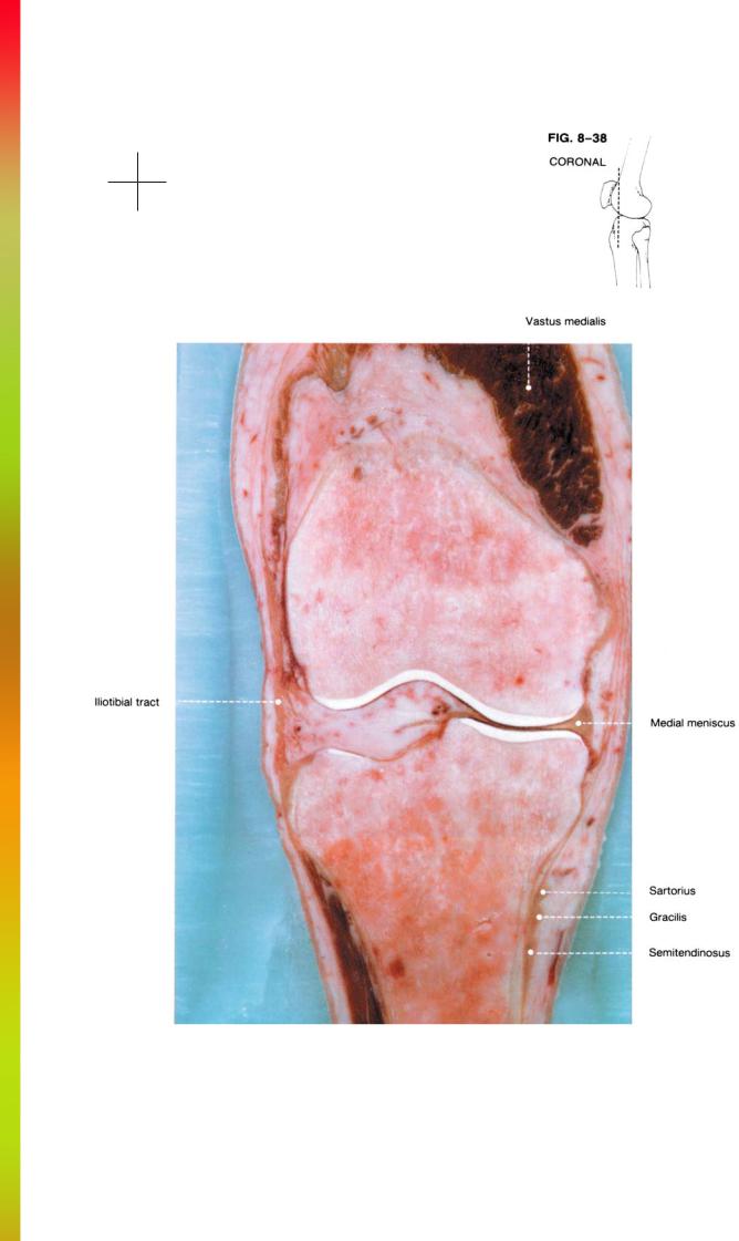
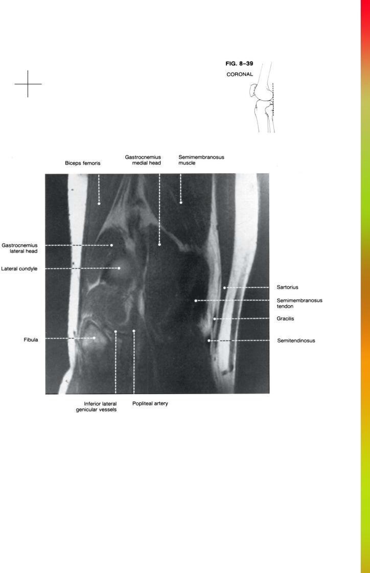
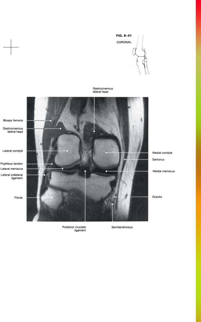
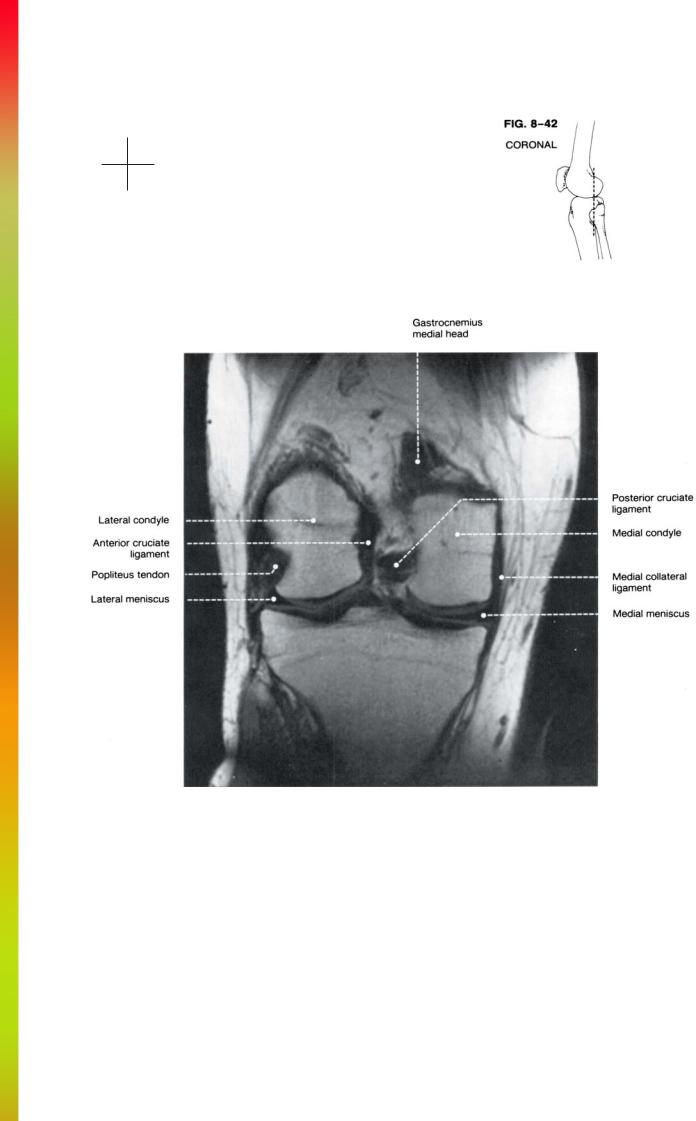
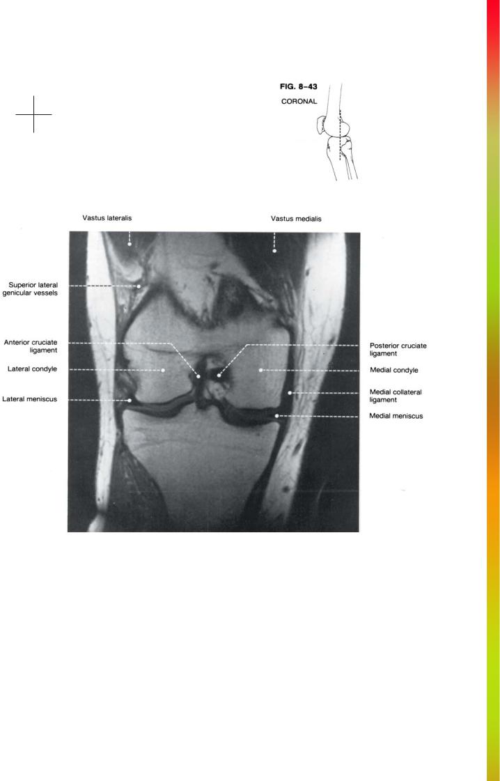
THE KNEE |
Superior
Lateral Medial
Inferior
Anatomy and MRI of the Joints |
244 |
Superior
Lateral Medial
Inferior

245 |
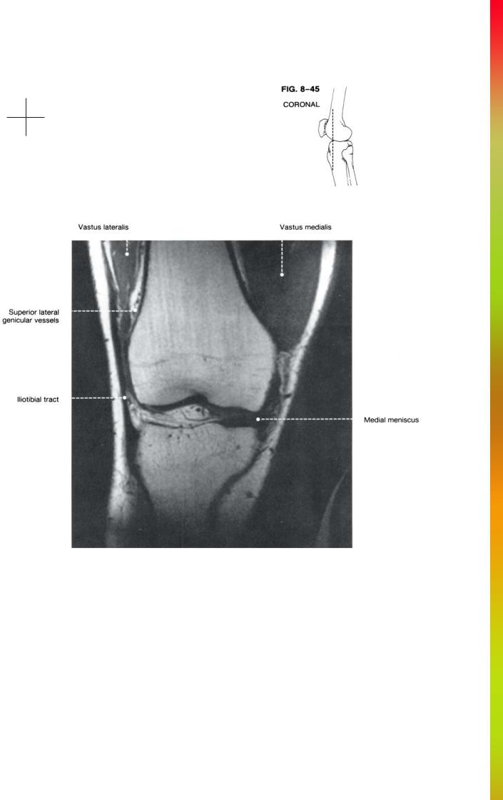
THE KNEE |
Superior
Lateral Medial
Inferior
Anatomy and MRI of the Joints |
246 |
Superior
Lateral Medial
Inferior
247 |
THE KNEE |
Superior
Lateral Medial
Inferior
Anatomy and MRI of the Joints |
248 |
Superior
Lateral Medial
Inferior

249 |
THE KNEE |
Superior
Lateral Medial
Inferior
Anatomy and MRI of the Joints |
250 |
Superior
Lateral Medial
Inferior

CHAPTER 9
THE ANKLE
William D. Middleton, M.D.
Like the wrist, the ankle has a complex bony architecture with multiple ligaments, muscles, and tendons oriented in a variety of planes. Understanding the cross-sectional relationships of these various anatomic structures can be difficult if the ankle is described in isolation. Therefore, this chapter will also include discussion of the subtalar region as well as portions of the mid-foot.
The ankle joint itself is composed of the articulation between the tibia and fibula and the talus. The convex talar dome rests in the concave surface of the distal tibia and is secured medially and laterally by the medial and lateral malleoli. This arrangement allows for dorsiflexion and plantarflexion in the sagittal plane while restricting motion in the coronal and axial planes.
The subtalar joint between the talus and calcaneus consists of three separate facets. The largest is the posterior facet. The middle and anterior facets are smaller, variable in shape, and may be fused into a single unit with a continuous cartilaginous covering. The mid-subtalar facet is formed below by a ledge of bone projecting off the medial surface of the calcaneus called the sustentaculum tali. The sustentaculum tali is necessary for bony support of the talus because the talus projects medially at approximately a 15-degree angle from the long axis of the calcaneus.
Anteriorly the head of the talus articulates with the navicular and the calcaneus articulates with the cuboid. The talonavicular and calcaneocuboid joints together are known as the transverse tarsal joint. The navicular articulates anteriorly with the three cuneiforms and the cuboid articulates anteriorly with the base of the fourth and fifth metatarsals.
A number of important ligaments support the ankle. The strongest is the deltoid ligament which arises from the medial malleolus. Its strength is necessary since the medial malleolus is short and provides relatively little bony support to the medial aspect of the ankle. The deltoid ligament has many fibers. The deepest run between the medial malleolus and the talus. These fibers fan out to reach both anterior and
posterior aspect of the talus. Superficial fibers also extend to the sustentaculum tali and the navicular. These superficial fibers also attach to and blend with the plantar calcaneonavicular (spring) ligament. Figure 9-1 illustrates the fibers of the deltoid ligament.
Laterally the ankle is supported by several separate ligaments. The calcaneofibular ligament extends posteriorly from the lateral malleolus to the calcaneus. The anterior talofibular ligament runs from the anterior aspect of the lateral malleolus to the neck of the talus while the posterior talofibular ligament connects these bones posteriorly. These ligaments are shown in Fig. 9-2.
The subtalar joint is supported by the interosseous ligament running between the posterior and mid-subtalar joints from the superior aspect of the calcaneus to the inferior aspect of the talus (Fig. 9-2). The space in which the interosseous ligament runs is called the tarsal sinus.
Another important ligament arising from the calcaneus is the spring ligament (also known as the plantar calcaneonavicular ligament). The spring ligament extends from the sustentaculum tali to the inferior aspect of the navicular and provides important support for the head of the talus which is lacking in significant bony support. The spring ligament blends superiorly with the tibionavicular fibers of the deltoid ligament and inferiorly with the plantar calcaneocuboid (short plantar) ligament.
The musculotendinous structures in the ankle can be divided into medial, lateral, anterior, and posterior groups. The posterior group consists of a single tendon-the Achilles tendon. This tendon arises from the soleus and gastrocnemius muscles and is the largest in the body. It attaches to the posterior surface of the calcaneus.
The medial group includes the flexor hallucis longus, flexor digitorum longus, and the tibialis posterior. The relationships of these three tendons to each other and adjacent structures is shown in Figs. 9-1 and 9-3. The flexor hallucis longus is the most posterior of the three,
251