

МРТ - в диагностике болезней суставов
.pdf
CHAPTER 7
THE HIP
Thomas L. Lawson, M.D. and William D. Middleton, M.D.
The function of the bony pelvis is to aid in the distribution and transmission of body weight to the limbs and to help absorb the stress of the muscular activity in the erect posture. The articulations of the bony pelvis include the hip joint, sacroiliac joint, and the pubic symphysis. In these latter two joints, free motion has been sacrificed in favor of stability and strength of union. The hip joint is the major joint of motion associated with the pelvis. It must be a strong joint and able to withstand great stresses and weight and also provide flexibility for motion of the lower extremity. The hip is structurally complex and reinforced by ligaments and bony struts to aid in this function. The hip joint allows for extensive motion: flexion, extension, adduction, abduction, circumduction, and rotation. The hip joint is well designed to allow not only for this wide range of motion, but also to provide the stability and strength to carry the weight of the body in an erect posture.
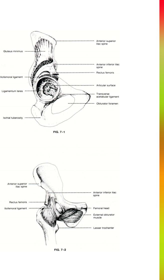
The hip is a ball and socket or an enarthrodial joint (Fig. 7-1). It is formed by the spherical-shaped head of the femur and the cup-shaped cavity of the acetabulum. The wall of the acetabulum is thick and heavily reinforced to support the weight of the body. The semilunar articular portion is open below and has a deep central nonarticular portion. The acetabulum is contributed to and is formed by the three component bones of the pelvis. One-fifth of the smooth lunate surface of the articular portion of the acetabulum is formed from the body of the pubis, twofifths from the ilium, and two-fifths from the body of the ischium. The prominent osseous rim of the acetabulum forms the attachment of the labrum or cotyloid ligament of the hip joint. The uneven internal edge provides an attachment for the synovial membrane of the joint.
The articulating surfaces of the hip are covered with cartilage and lubricated by synovial fluid. The cartilage on the head of the femur covers the entire surface of the femoral head with the exception of a depression just below its center for the attachment of the ligamentum
teres.Conversely, the cartilage of the acetabulum is an incomplete cartilage ring of a horseshoe shape, with the opening of the horseshoe directed caudally.
The central acetabular fossa is a deep, irregular nonarticular portion of the joint. It is formed mainly from the ischium. In the fossa a small mass of fat can be found as well as the attachment of the ligamentum teres, one of the principle ligaments of the joint.
The acetabular fossa is open inferiorly as the acetabular notch. This notch is bridged by the transverse ligament. Just caudal to the acetabular notch is the obturator foramen, a large apperture surrounded by the bodies and rami of the ischium and the pubis.
The second component of the hip joint is the femoral head. The head of the femur is smooth and forms an approximate two-thirds of a sphere. The articular surface of the head of the femur is largest in its craniad and anterior portion, where the greatest weight is directed. Along its medial surface there is a small irregular depression, the fovea capitis or centralis, which forms the site of attachment of the ligamentum teres.
The head of the femur is attached to the body of the bone via the neck. This is a strong buttress of bone of about 5 cm in length. Its function is to displace the femoral shaft away from the pelvis to allow freedom of motion and to minimize contact of the leg with the pelvis. The neck forms an angle with the shaft of the femur which varies from approximately 115° to 140°. The head also points somewhat forward or anterior and as a result an anterior angle between the neck and shaft of the femur is formed that averages about 8°.
The greater trochanter is a large bony prominence along the lateral and craniad aspect of the base of the femoral neck. Along the medial and caudal aspect of the femoral neck is a smaller prominence called the lesser trochanter. Between the two bony masses there is an irregular ridge along the anterior aspect of the femur called the intertrochanteric line. Posteriorly, the two trochanters are joined by a thick intertrochanteric
153
Anatomy and MRI of the Joints |
154 |

crest. Both trochanters as well as the intertrochanteric line and crest are important muscle attachment sites.
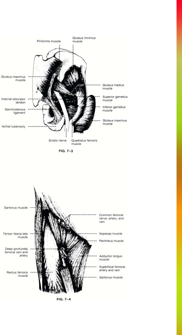
The femoral head is held to the acetabulum by five major ligaments: the capsular ligament, ligamentum teres, iliofemoral ligament, transverse ligament, and cotyloid ligament or acetabular labrum (Fig. 7-2). The capsular ligament is a strong, dense, fibrous band that extends from the margin of the acetabulum craniad over and surrounding the neck of the femur caudally. Its upper circumference is attached to the acetabulum, just external to the cotyloid ligament or labrum. Along the lower circumference of the acetabulum it is attached to the transverse ligament, as this ligament bridges the acetabular notch. The capsular ligament surrounds the neck of the femur and is attached to it. It is attached anteriorly to the anterior intertrochanteric line, posteriorly to the intertrochanteric crest, and craniad to the base of the neck of the femur. From these insertions the fibers are reflected upward over the neck of the femur forming a tubular sheath and a dense ligamentous joint capsule. While the external surface of the capsular ligament is rough and covered by numerous muscles, its inner surface is smooth, glistening, and lubricated by synovial fluid.
The iliofemoral ligament is a thick fibrous band that extends obliquely across the front or anterior aspect of the joint. It is intimately associated with the capsular ligament and serves to strengthen it. It is attached above to the lower portion of the anterior inferior spine of the ilium. Caudally it diverges to form two bands, one of which passes downward to insert on the anterior intertrochanteric line. The second band passes caudally and laterally and inserts on the upper portion of the intertrochanteric line adjacent to the neck and greater tuberosity of the femur.
The ligamentum teres, also called the ligamentum capitis femoris, is a strong triangular fibrous band that crosses the joint. The narrow apex of the ligament is attached to the femoral head at the fovea capitis femoris. The broad base of the ligament inserts into the acetabular fossa. Along its inferior portion the ligamentum teres blends into the cotyloid and transverse ligaments. The ligamentum teres is formed by connective tissue, but is surrounded by a tubular sheath of synovial membrane.
The cotyloid ligament or acetabular labrum is the fibrocartilaginous rim attached to the margin of the acetabulum. The function of the ligament is to deepen the acetabular fossa as well as to protect the edges of the acetabulum and to smooth the irregularities of its surface. On cross-section this ligament is prismoidal. The base which is attached to the margin of the acetabulum is thick, while its free edge is thin and sharp.
The transverse ligament bridges the caudal opening of the acetabulum between the acetabular notch. It forms a
155 |
THE HIP |
portion of the cotyloid ligament or acetabular labrum, although histologically it has a slightly different appearance. There is a small notch or opening at the lower portion of the acetabulum between the transverse ligament and the acetabulum itself. Through this small foramen passes nutrient vessels to the joint itself.
The synovial membrane of the joint is extensive. It covers all portions of the neck of the femur contained within the joint capsular ligament. From the neck it is reflected on the internal surface of the capsular ligament and covers both surfaces of the cotyloid ligament as well as the ligamentum teres.
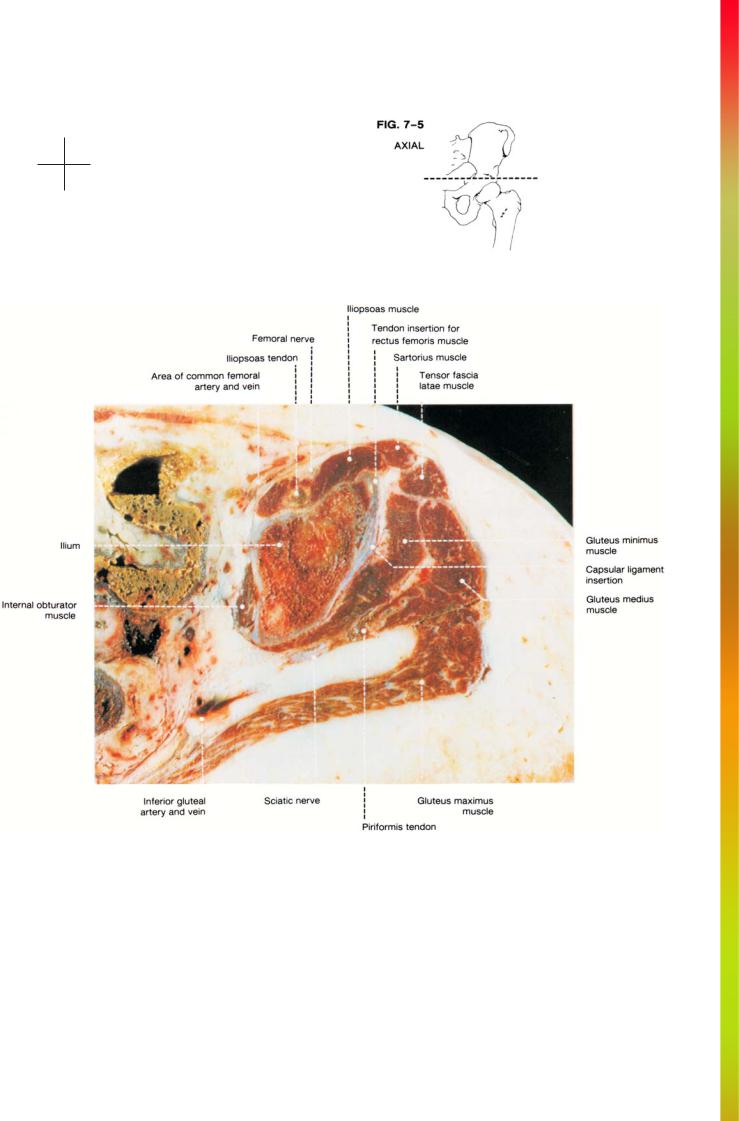
The anatomical relations of the muscle of the hip can be best understood by dividing them into posterior, anterior, medial, and lateral groups. The posterior group, also known as the short rotators of the hip, consist of the piriformis, the obturator internus and externus, the two gemelli, and the quadratus femoris muscles (Fig. 7-3). The piriformis muscle is the most superior of this group, arising from the lateral mass of the sacrum. It extends laterally and inferiorly through the greater sciatic foramen to insert via a rounded tendon onto the upper medial surface of the greater trochanter of the femur. The internal obturator muscle arises from the deep inner surface of the obturator foramen and passes posteriorly through the lesser sciatic foramen and around the ischium where it turns laterally and extends to insert onto the greater trochanter of the femur just posterior and caudal to the piriformis muscle. The superior and inferior gemelli muscles arise from the ischial spine and ischial tuberosity, respectively, and travel along the superior and inferior aspect of the internal obturator muscle, converging laterally with the insertion of the internal obturator muscle. The external obturator muscle originates from the superficial surface of the obturator foramen and passes posteriorly and laterally beneath the hip joint. It inserts onto the posterior aspect of the greater trochanter below the obturator internus. The quadratus femoris muscle is the most inferior of this group. It arises from the lateral border of the ischial tuberosity and passes laterally to insert onto the posterior intertrochanter crest.
The lateral muscle group consists of the three gluteus muscles and the tensor fascia latae muscle. The gluteus minimus muscle is the deepest and most anterior gluteal muscle arising along the anterior lateral surface of the ilium. It extends inferiorly to insert on the anterior superior aspect of the greater trochanter. The gluteus medius muscle originates posterior and superior to the gluteus minimus. As it extends inferiorly, it covers the minimus and inserts on the posterior lateral surface of the greater trochanter. The gluteus maximus muscle is the largest and most superficial of the gluteal muscles. It originates from the posterior-most aspect of the ischium, the sacrum, and the sacrotuberous ligament. The gluteus maximus muscle extends inferiorly and laterally, with the bulk of the muscle inserting into the iliotibial tract. Approximately one quarter of the deep portion of the muscle inserts directly on the posterior superior aspect of the proximal femur. The tensor fascia latae
Anatomy and MRI of the Joints |
156 |

muscle is a short strap-like muscle that originates anterior to the gluteus medius near the anterior superior iliac spine. It extends inferiorly to insert on the anterior aspect of the iliotibial tract.
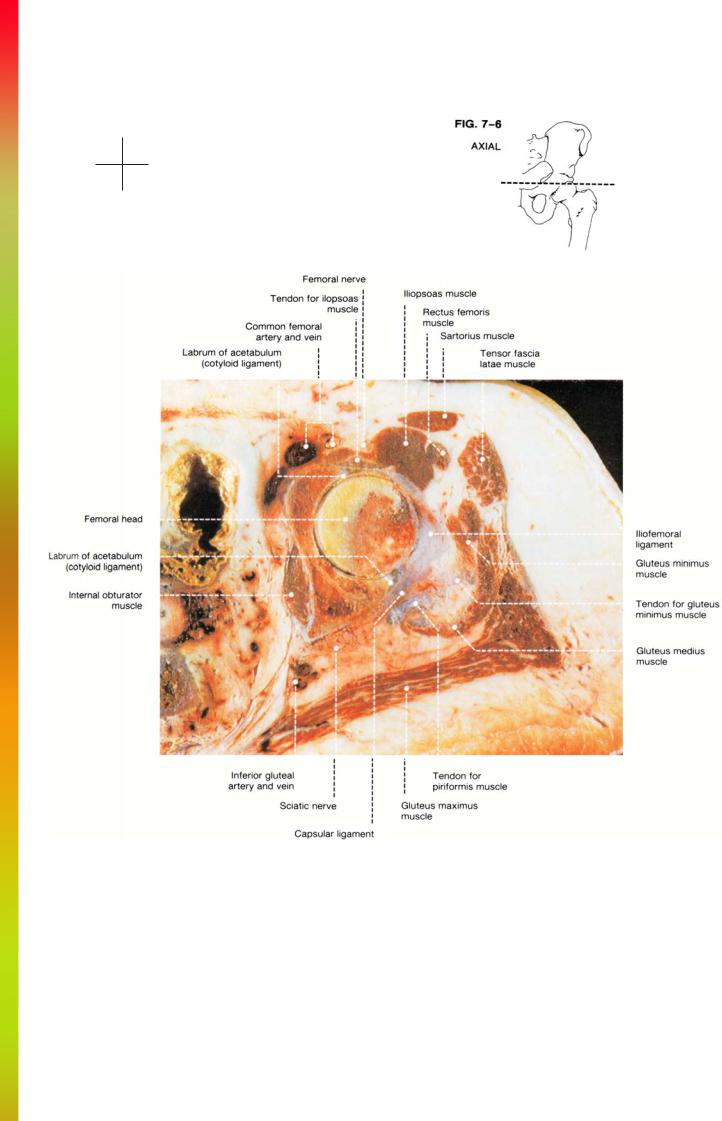
The anterior muscles include the iliopsoas, sartorius, and rectus femoris muscles (Fig. 7-4). The iliopsoas muscle is the strongest flexor of the hip. At the level of the hip it passes deep to the inguinal ligament and crosses the anterior aspect of the joint. It inserts onto the lesser trochanter. The rectus femoris muscle is the most superior and superficial muscle of the quadriceps group and is the only muscle of this group that crosses the hip joint. It arises primarily as a thick tendon from the anterior inferior iliac spine, although a smaller tendon arises from the ilium immediately above the acetabulum. The sartorius muscle is the longest muscle in the body and travels as the most superficial muscle of the anterior thigh. It arises from the anterior superior iliac spine and travels inferiorly and medially toward the knee.
The medial muscle group includes the pectineus, the adductors, and the gracilis muscles. Although all of these muscles act on the hip, only the pectineus muscle is well
157 |
THE HIP |
seen in images of the hip. It arises from the anterior aspect of the superior pubic ramus and extends inferiorly and laterally over the external obturator muscle to insert onto the posterior aspect of the upper femur.
Several large neurovascular structures can be identified in cross-sectional views of the hip. The femoral nerve, artery, and vein travel vertically along the anterior aspect of the hip. They are located superficial to the iliopsoas muscle and deep to the inguinal ligament. The common femoral artery bifurcates into the superficial femoral and deep (profunda) femoral arteries approximately 3 cm below the inguinal ligament. The medial and lateral femoral circumflex arteries are branches of the deep femoral artery which provide blood supply to the hip joint.
The sciatic nerve is the largest peripheral nerve in the body. It originates from the lumbosacral trunk and the sacral plexus anterior to the piriformis muscle. It exits the pelvis through the greater sciatic foramen between the piriformis muscle and the sacrospinous ligament. Outside the pelvis the sciatic nerve runs dorsal to the internal obturator and gemelli muscles and deep to the gluteus maximus muscle.


THE HIP
AXIAL |
|
|
Cryomicrotomes............................................................... |
FIGS. 7-5 to 7 |
-10 |
MR Images ....................................................................... |
FIGS. 7-11 to 7-18 |
|
SAGITTAL |
|
|
Cryomicrotomes .............................................................. |
FIGS. 7-19 to |
7-24 |
MR Images ....................................................................... |
FIGS. 7-25 to |
7-32 |
CORONAL |
|
|
Cryomicrotomes .............................................................. |
FIGS. 7-33 to |
7-40 |
MR Images ....................................................................... |
FIGS. 7-41 to |
7-48 |
Anatomy and MRI of the Joints |
160 |
Anterior
Medial Lateral
Posterior
161 |
THE HIP |
Anterior
Medial Lateral
Posterior