

4. Lung cavity syndrome
REASONS: The formation of a cavity in the lung is usually preceded by compaction of the lung tissue. Most often, this is inflammatory infiltration (abscesses, lung gangrene, staphylococcal destruction of the lungs, pneumonia, tuberculosis), tumor decay, pulmonary infarction. SYMPTOMS:
Shortness of breath, often a productive cough, possibly hemoptysis;
Lag in the act of breathing of the affected half of the chest;
Strengthening of voice trembling and bronchophony;
Percussion: a blunt-tympanic sound is determined. With a large cavity located on the periphery, tympanic sound is observed;
Auscultatory: bronchial, and sometimes amphoric breathing, sonorous medium and large bubble rales are revealed.
It should be emphasized that all these signs are determined in the presence of a smooth-walled cavity of at least 4 cm in diameter, located close enough to the surface of the chest containing air, connecting to the bronchus and surrounded by a densified lung tissue. In the absence of these conditions, the cavity in the lung remains "dumb" and is detected only during x-ray examination.
X-ray (linear tomography, CT) signs of a cavity syndrome in the lung are limited enlightenment of a round or oval shape, usually against a background of blackout. A horizontal level of fluid in the cavity is characteristic if it communicates with the bronchus and contains exudate and air.
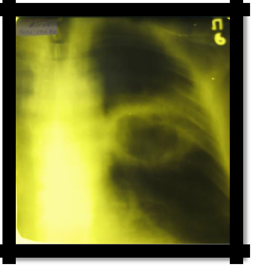
Pic 2.2 Linear tomography
5. The syndrome of fluid accumulation in the pleural cavity
REASONS: As a rule, a complication of another disease.
The causes of transudate are heart failure, kidney disease - nephrotic syndrome, hypo- and dysproteinemia.
The causes of exudate are inflammation of the pleura (pleurisy) of various etiologies (infectious exudate in pneumonia, abscess, tuberculosis; non-infectious exudate in malignant tumors, autoimmune diseases, uremia), wounds, trauma (hemothorax), chylous exudate (x). SYMPTOMS:
9
respiratory dyspnea;
On examination - protrusion and limitation of mobility of the corresponding side, smoothing of intercostal spaces;
Palpation - increased resistance of intercostal spaces, weakening or absence of voice trembling;
Percussion - dullness or absolute dullness over a liquid; directly above its level - a blunt-tympanic sound; the appearance of a dull sound on the healthy side due to displacement of the mediastinal organs (with large effusions); downward hepatic dullness. (Damuaso Line, Rauchfus-Grocco and Garland Triangles).
Auscultatory - the presence of fluid suggests the absence or weakening of breathing and bronchophony, and above the fluid level in the Garland triangle - a bronchial shade of breathing (on the effusion side).
X-ray homogeneous dimming is determined in the lower part of the pulmonary field with a characteristic oblique upper border for exudate and a more horizontal upper border for transudate. In the latter case, the process is often two-way. There is a shift in the mediastinal organs to a healthy side.
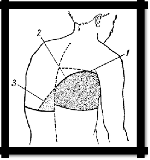
Pic 2.4 The line of Damoiso-Ellis-Sokolov (1); The Garland Triangle (2)
and the Rauchfus-Grocco Triangle (3) with exudative pleurisy.
Pic 2.5 Left-
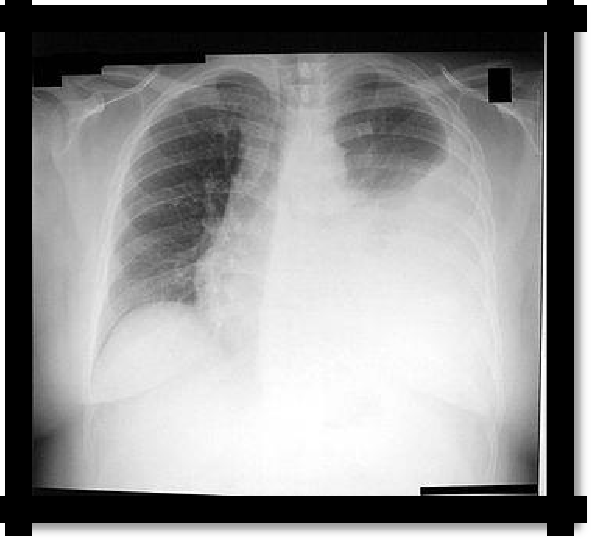
sided exudative pleuritis
10
Table 2.1 Differential diagnostic signs of exudates and transudates.
-
Type of effusion
Transudate
Exudate
Relative density
Usually
below
1.015;
rarely
Not
lower
than
1.015;
(with
compression of
large
usually 1.018
vessels by a tumor) - above
1.013-1.025
Coagulation
-
+
Color
and
Almost
transparent;
lemon
Serous
exudates
in
transparency
yellow or light yellow
appearance
do not
differ
from
transudates;
other
types
of
exudates
are
cloudy;цвет различен
Rivalt's reaction
-
+
Содержание белка
<30 g / l, usually 5-25 g / l
> 30 g / l, usually 30-50 g / l,
in purulent - up to 80 g / l
Cytological
There are few cellular elements;
There
are
more cellular
study
common
elements
than
in
mesothelial cells, red blood cells,
transudates. The number of
sometimes
lymphocytes
cellular elements,
their
predominate;
types and condition depend
after
repeated
punctures
on the etiology and phase
sometimes - eosinophils
of
the
inflammatory,
oncological processes